Last Updated on November 27, 2025 by Bilal Hasdemir

At Liv Hospital, we use the latest imaging technology to help patients with leg issues. A CT scan on legs provides clear pictures of bones, muscles, and blood vessels, allowing doctors to detect injuries or diseases accurately. This helps us plan the best and most effective treatment for each patient.
We focus on making sure you get the best care. If you’re in pain or hurt, a CT scan of your legs can help us find out what’s wrong. It guides us to the right tests or treatments and keeps an eye on your condition.
Key Takeaways
- CT scans provide detailed images of leg anatomy for accurate diagnosis.
- They help guide further tests or treatments for leg conditions.
- Our medical experts use CT scans to monitor conditions effectively.
- A CT scan of the legs is a key tool for diagnosing leg problems.
- Liv Hospital is dedicated to top-notch care and support.
Understanding CT Scans on Legs: The Basics

CT scans of the legs are key for seeing inside the lower legs. They help find many issues, like breaks or blood vessel problems.
What Is a CT Scan and How Does It Work?
A CT scan uses X-rays and a computer to show body parts in detail. When you get a CT scan of your legs, you lie on a table that moves into a big machine. This machine takes X-ray pictures from all sides.
Then, a computer makes these pictures into clear images. These images show bones, muscles, and soft tissues well. This helps doctors make accurate diagnoses.
Difference Between CT Scans and Other Imaging Methods
CT scans are different from X-rays and MRI scans. CT scans show detailed cross-sections, not just 2D images like X-rays. They are also faster and better for people with metal implants or who are scared of tight spaces.
| Imaging Method | Key Features | Typical Use Cases |
| CT Scan | Detailed cross-sectional images, quick and suitable for metal implants | Fractures, vascular diseases, and detailed soft tissue evaluation |
| X-ray | 2D images, quick, low radiation | Initial fracture assessment, bone alignment |
| MRI | High detail of soft tissues, no radiation | Soft tissue injuries, certain tumors, and detailed joint evaluation |
Knowing how CT scans work and their benefits helps us see their importance. They are key in diagnosing and treating leg problems.
Key Fact #1: CT Scans Provide Detailed Cross-Sectional Images

CT scans have changed how we diagnose and treat leg problems. They give detailed images of the lower extremities. This is key to understanding the complex anatomy.
Visualization of Bones, Muscles, and Soft Tissues
CT scans are great at showing bones, muscles, and soft tissues clearly. This is very helpful for finding issues like fractures, muscle tears, and soft tissue injuries.
Detailed imaging helps doctors see how bad an injury or disease is. This makes it easier to get the right treatment.
Advantages of Cross-Sectional Imaging
CT scans have many benefits over old imaging methods. They let doctors look at complex structures from different sides. This makes diagnoses more accurate.
| Advantages | Description |
| Detailed Visualization | Clear images of bones, muscles, and soft tissues |
| Multi-Angular Examination | Ability to examine structures from various angles |
| Enhanced Diagnostic Accuracy | Improved diagnosis due to high-resolution images |
CT scans of the lower extremity give detailed cross-sectional images. This helps doctors make better decisions for patient care. It’s a big reason why leg conditions are managed well.
Key Fact #2: Common Medical Reasons for a CT Scan on the Legs
CT scans are key for diagnosing leg injuries and conditions. They help us check on many medical issues in the lower legs.
Trauma and Suspected Fractures
CT scans are often used to check for leg trauma or fractures. They give clear images of bones. This helps us spot fractures or injuries that X-rays might miss.
Vascular Abnormalities and Blood Clots
CT scans also help find vascular problems and blood clots in the legs. They use contrast to show blood vessels. This helps us see blockages or clots causing pain or swelling.
Tumors and Infections
CT scans help diagnose tumors and infections in the legs. They give detailed images. This lets us see how big the problem is and plan treatment.
To show why CT scans are used on legs, let’s look at this table:
| Condition | Symptoms | CT Scan Findings |
| Trauma/Fracture | Pain, swelling, deformity | Fracture lines, bone displacement |
| Vascular Abnormality | Pain, swelling, coldness | Blood clots, vessel narrowing |
| Tumor/Infection | Pain, swelling, redness | Masses, abscesses, and bone destruction |
The table shows how CT scans help diagnose many leg issues. This helps doctors make better care plans for patients.
Key Fact #3: CT Lower Extremity Exams for Vascular Assessment
CT scans have changed how we diagnose vascular diseases in the legs. They give detailed images that help doctors treat vascular conditions.
Detecting Blood Clots and Deep Vein Thrombosis
CT scans are great for finding blood clots and deep vein thrombosis (DVT). They can see the venous system and spot clots that could cause big problems.
If someone has DVT symptoms, a CT scan can confirm if there’s a clot. Knowing this is key to choosing the right treatment.
Leg CT Angiogram Procedure
A leg CT angiogram focuses on the blood vessels in the legs. It uses contrast material to show the arteries and veins clearly.
The process includes:
- Preparation: The patient gets ready on the CT table, and an IV line is set up.
- Contrast Injection: Contrast material is given through the IV to make blood vessels stand out.
- Scanning: The CT scanner takes pictures of the leg’s blood vessels as the contrast moves through.
- Image Reconstruction: The raw data is turned into detailed images for doctors to study.
Assessing Arterial Disease
CT exams are also key for checking arterial disease in the legs. They show the arteries clearly, helping diagnose PAD.
| Condition | Symptoms | CT Scan Findings |
| Deep Vein Thrombosis (DVT) | Leg pain, swelling, warmth | Presence of a clot in the deep veins |
| Peripheral Arterial Disease (PAD) | Leg pain during walking, coldness | Narrowing or blockage of arteries |
A medical expert says, “CT scans are vital for vascular assessment. They’re non-invasive and very accurate for diagnosing vascular conditions.”
CT lower extremity exams help doctors give more accurate diagnoses and treatment plans for vascular conditions.
Key Fact #4: The Efficiency of Modern Leg Scanners
Modern leg scanners have changed medical imaging a lot. They are very efficient and accurate. This has helped doctors to diagnose and treat patients better.
Rapid Imaging Technology
The technology in modern leg scanners is fast and precise. It’s very important in emergencies when time is critical. This technology lets doctors make quick decisions for patient care.
Typical Duration: 10-15 Minutes
A CT scan of the leg usually takes 10 to 15 minutes. This is a big plus because it means patients don’t have to wait long. The can scan machine technology helps get great images fast, making the experience better for patients.
We use these advanced leg scanner technologies to give our patients the best care. The speed of modern leg scanners shows how far medical imaging has come.
Key Fact #5: CT Scans for Confirming Broken Bones
When X-rays don’t show enough, CT scans help us see bone injuries clearly. They are key to confirming fractures when X-rays can’t show the full injury.
When X-Rays Are Inconclusive
X-rays are usually the first choice for checking bone fractures. But, they might not always be clear, like if the fracture is complex or bones overlap. CT scans are better in these cases, giving us a detailed look to diagnose and assess fractures.
For example, in trauma or suspected fractures in tricky spots like the hip or ankle, CT scans are better. They show bone and soft tissues better than X-rays.
Detailed Fracture Assessment
CT scans are great for detailed fracture views because they show bones in 3D. This helps us see the fracture’s size, shape, and where it is. Accurate fracture assessment is key to treatment planning.
They let us check how the fracture pieces fit and if they’re out of place. Knowing this helps us plan the best treatment for each patient.
Key Fact #6: Visualizing Both Bone and Soft Tissue Pathology
We use CT scans to check leg conditions. They show both bone structures and soft tissue problems. This makes CT scans very useful in medicine.
Comprehensive Tissue Evaluation
CT scans look at all leg tissues, like bones, muscles, and soft tissues. They’re great when both bone and soft tissue injuries are considered. For example, in trauma, they spot fractures and check soft tissue damage.
These scans give doctors clear images. They help doctors understand the whole picture of a patient’s condition. This leads to better treatment plans.
Detecting Subtle Abnormalities
CT scans are good at finding small problems that other tests miss. This is key to catching leg issues early.
For soft tissue, they find inflammation, tumors, or infections. For bones, they check fractures, degeneration, or other problems.
CT scans give a detailed look at leg conditions. This helps doctors make better decisions for patients.
Key Fact #7: CT Scans with Metal Implants
Patients with metal implants can get CT scans. But we use special steps to get the best images.
Having a metal implant, like a hip replacement, can mess with CT scan images. Metal artifacts might show up as streaks or distortions. This can hide important details.
Modern Protocols for Metal Artifact Reduction
Today’s CT scanners have new ways to cut down on metal artifacts. One method is metal artifact reduction (MAR). It uses smart algorithms to fix the problems caused by metal.
These new steps really help improve image quality. Even with metal implants, we can make more accurate diagnoses. Our facilities have the latest tech for these cases.
Limitations and Considerations
Even with new CT scan methods, metal artifacts might not go away completely. How well these methods work depends on the metal type, its location, and the scanning tech used.
Some metal implants, like pacemakers, need extra care or other imaging methods. Our team looks at each case to find the best solution.
In short, CT scans with metal implants are tricky. But thanks to modern tech and methods, we can usually get clear images and accurate diagnoses.
The CT Scan of Leg Procedure: What to Expect
Getting ready for a CT scan on your leg involves a few steps. These steps help make the process smooth and successful. We know that getting a medical scan can make you nervous. So, we’re here to help you know what to expect during your leg CT scan.
Before Your Leg CT Scan
Before the scan, you’ll get instructions on how to prepare. This might include:
- Avoiding food and drink for a few hours before, depending on the use of contrast dye.
- Removing any metal objects, like jewelry or glasses, to avoid image problems.
- Telling your healthcare provider about any allergies or medical conditions, like kidney disease.
Table: Pre-CT Scan Preparations
| Preparation | Purpose |
| Avoid eating/drinking | Ensures clear images when contrast dye is used |
| Remove metal objects | Prevents image problems |
| Disclose allergies/medical conditions | Ensures safety with contrast dye and the scan |
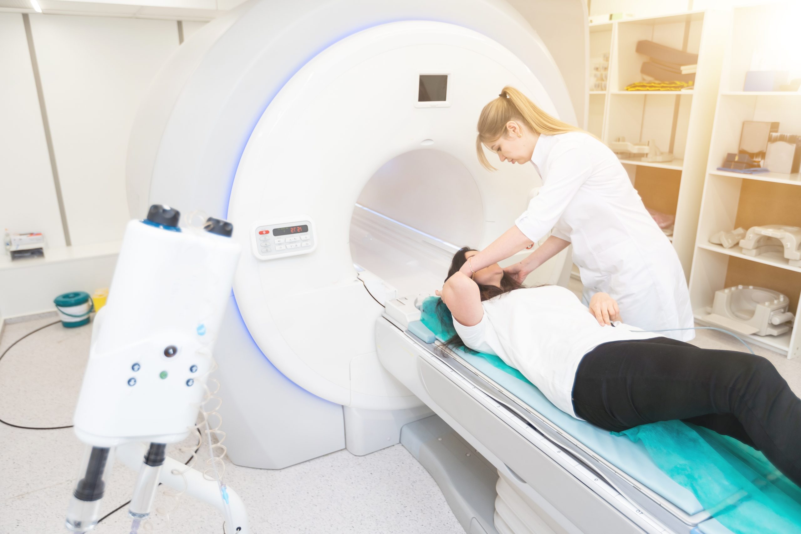
During the Procedure
During the CT scan, you’ll lie on a table that slides into a large machine. The scan is usually quick, lasting about 10-15 minutes. You might get contrast dye to make the images better. Our team is trained to keep you comfortable and safe.
After Your Scan
After the scan, you can go back to your normal activities unless told not to by your doctor. The images will be checked by a radiologist. Then, your doctor will get the results and decide what to do next.
We know getting a CT scan can be scary. But our team is here to make it as easy and stress-free as possible. If you have any questions or worries about your leg CT scan, please let us know.
3D Reconstruction and Image Processing
Advanced image processing makes CT scans show leg anatomy in 3D. This helps doctors see complex conditions more clearly. It makes CT scans more useful for diagnosis.
Creating Three-Dimensional Models
Creating 3D models from CT scans uses special software. It turns images into 3D models. This 3D reconstruction helps doctors see how different parts of the leg fit together.
For example, 3D models are key for complex fractures or big tumors. They show details that 2D images can’t. Doctors can look at these models from all sides, getting a better view.
Storage and Review of CT Images
Once CT scans are done, the images are saved digitally. They use DICOM standards for easy sharing. This makes it simple for different healthcare systems to work together.
These images can be seen on many devices. Doctors can use them on workstations, computers, or even phones. This makes it easier to discuss and diagnose patients, even from far away.
| Feature | 2D Imaging | 3D Reconstruction |
| Visualization | Limited to single-plane views | Multi-angle, spatial visualization |
| Diagnostic Accuracy | Good for straightforward cases | Enhanced for complex conditions |
| Data Storage | Typically, less storage is required | More storage is required due to detailed data |
Using 3D models and advanced image processing helps doctors. They can plan better treatments for leg problems with CT scans.
Radiation Exposure and Safety Considerations
It’s important to know the good and bad of CT scans on the legs. CT scans are great for finding problems, but they do use radiation.
Radiation can raise cancer risk. Yet, CT scans are often needed for accurate diagnosis and treatment plans. This makes them worth the risk.
Understanding Radiation Doses
A CT scan of the legs might use 2-3 mSv of radiation. This is less than the yearly background radiation most people get.
We try to use less radiation by adjusting the scanner. This way, we get good images without too much radiation.
| Procedure | Typical Radiation Dose (mSv) | Equivalent Background Radiation |
| CT Scan of Legs | 2-3 | 8-12 months |
| Chest X-Ray | 0.1 | 10 days |
| Background Radiation (annual) | 3 | 1 year |
Risk vs. Benefit Assessment
We look at the good and bad of CT scans. They help find problems and plan treatments. But there’s a risk of radiation harm.
The risk is higher for younger people. So, we’re extra careful with kids and young adults.
In short, CT scans of the legs have benefits and risks. Knowing about radiation helps us use it wisely. This way, we help patients while keeping radiation low.
When to Choose an MRI Scan of the leg vs. a CT Scan for Leg Imaging
Choosing between an MRI and a CT scan for leg imaging depends on the patient’s needs. We look at several factors. These include the type of tissue and the specific clinical scenario.
Comparative Advantages
MRI scans have some key advantages over CT scans for leg conditions. MRI shows soft tissues in great detail. This makes it perfect for checking muscles, tendons, and ligaments. Plus, MRI doesn’t use harmful radiation, which is good for patients needing many scans.
On the other hand, CT scans are better for seeing bones and finding fractures. But when soft tissue matters most, MRI is usually the better choice.
Specific Clinical Scenarios
There are times when MRI is better than CT scans for leg imaging:
- Checking soft tissue injuries like ligament sprains or muscle tears
- Finding conditions like osteonecrosis or stress fractures early with MRI
- Looking at tumors or infections in the leg soft tissues
In these situations, MRI’s detailed images help a lot with treatment plans and patient care.
The choice between MRI and CT scans for leg imaging depends on the situation and what’s needed for diagnosis and treatment.
Conclusion
CT scans of the legs play a key role in diagnosing and treating many medical issues. They provide detailed images of bones, muscles, and soft tissues. This helps doctors see problems like trauma, vascular issues, tumors, and infections clearly.
Modern CT scanners work fast, taking images in 10-15 minutes. This is great for patients who can’t stay in one place for long. New technology also makes CT scans better, even for people with metal implants.
When looking at leg problems, knowing the strengths and weaknesses of CT scans is important. It helps doctors pick the best tool for a diagnosis. As medical imaging gets better, CT scans will keep being a key part of patient care.
FAQ
What is a CT scan of the leg, and how does it work?
A CT scan of the leg uses X-rays and computer tech to show detailed images. It works by moving an X-ray machine around the leg. This captures images from different angles, then makes detailed pictures.
What are the common medical reasons for a CT scan of the legs?
CT scans of the legs help find and track issues like trauma, fractures, and tumors. They also check for vascular problems, blood clots, and infections.
How long does a CT scan of the leg typically take?
A CT scan of the leg usually takes 10-15 minutes. But it can vary based on the scan’s details and complexity.
Can I have a CT scan if I have metal implants in my leg?
Yes, you can get a CT scan with metal implants. New methods help reduce metal artifacts. But your doctor will weigh the risks and benefits first.
What is the difference between a CT scan and an MRI scan of the leg?
CT scans use X-rays, while MRI scans use magnetic fields and radio waves. CT scans are better for bones. MRI scans are better for soft tissues.
How is radiation exposure managed during a CT scan?
Radiation exposure is kept low by using the least amount needed for clear images. Modern scanners and protocols help keep it safe.
Can a CT scan detect broken bones that an X-ray can’t?
Yes, CT scans can find broken bones that X-rays miss. This is true for complex or fragmented fractures.
What is a leg CT angiogram, and when is it used?
A leg CT angiogram uses dye to see blood vessels in the leg. It’s for diagnosing blood clots, deep vein thrombosis, and arterial disease.
How are CT images stored and reviewed?
CT images are stored digitally and reviewed on computers or devices. They can also be made into 3D models for better understanding.
What should I expect during a CT scan of my leg?
During a CT scan, you’ll lie on a table that slides into the scanner. You’ll need to stay very quiet. The scan is painless and lasts about 10-15 minutes.
References
- Scheschenja, M., et al. (2024). CT angiography of the lower limbs: combining small field reconstruction with iterative reconstruction to improve image quality. Acta Radiologica, 65(7), 725-732. https://journals.sagepub.com/doi/abs/10.1177/02841851241258655
- Hwang, J. Y., et al. (2017). Doppler ultrasonography of the lower extremity arteries: basic scanning techniques and clinical applications. Ultrasonography, 36(1), 3-13. https://pmc.ncbi.nlm.nih.gov/articles/PMC5381852/


