Understanding CT scan results is key for both patients and doctors. At Liv Hospital, we help our patients understand their diagnosis and treatment choices.
A CT scan is a critical tool for diagnosing diseases and injuries. It shows detailed images of the body’s inside. With over 80 million CT scans done every year, knowing what the results mean is vital.
We’ll show you how to understand your CT scan results. We’ll cover important terms and concepts. This will help you make better health decisions.
Key Takeaways
- CT scans provide detailed images of internal structures, aiding in accurate diagnosis.
- Understanding CT scan results is key for patients and healthcare providers.
- Key components of a CT scan report include patient information, clinical history, and findings.
- Common terminology includes Hounsfield Units (HU) and density descriptions.
- CT scans can detect various abnormalities, including tumors, internal bleeding, and infections.
The Fundamentals of CT Scan Technology

To understand CT scan results, knowing how CT scans work is key. CT scans use X-rays to make detailed images of the body. These images are more detailed than regular X-rays.
What is a CT Scan and How Does It Work?
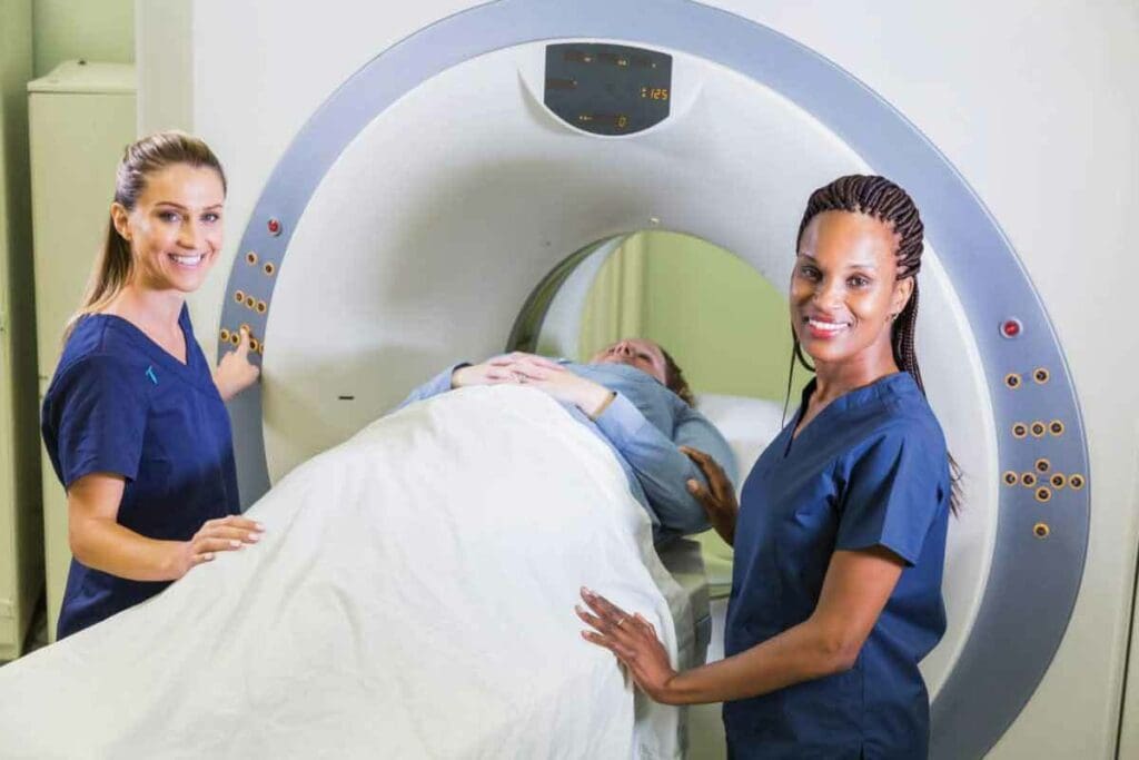
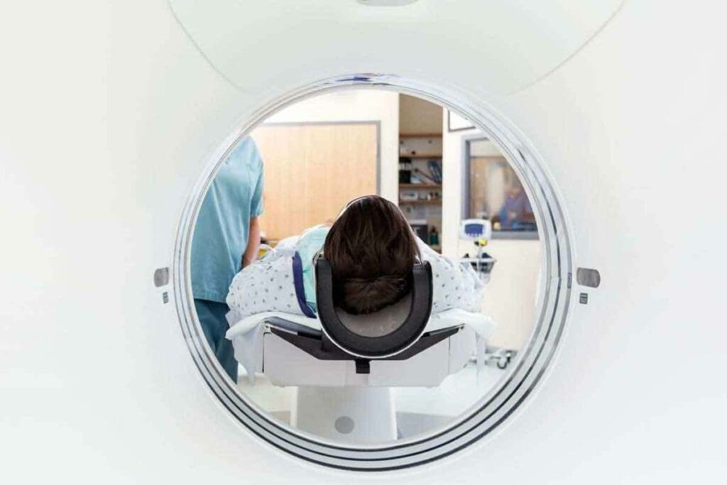
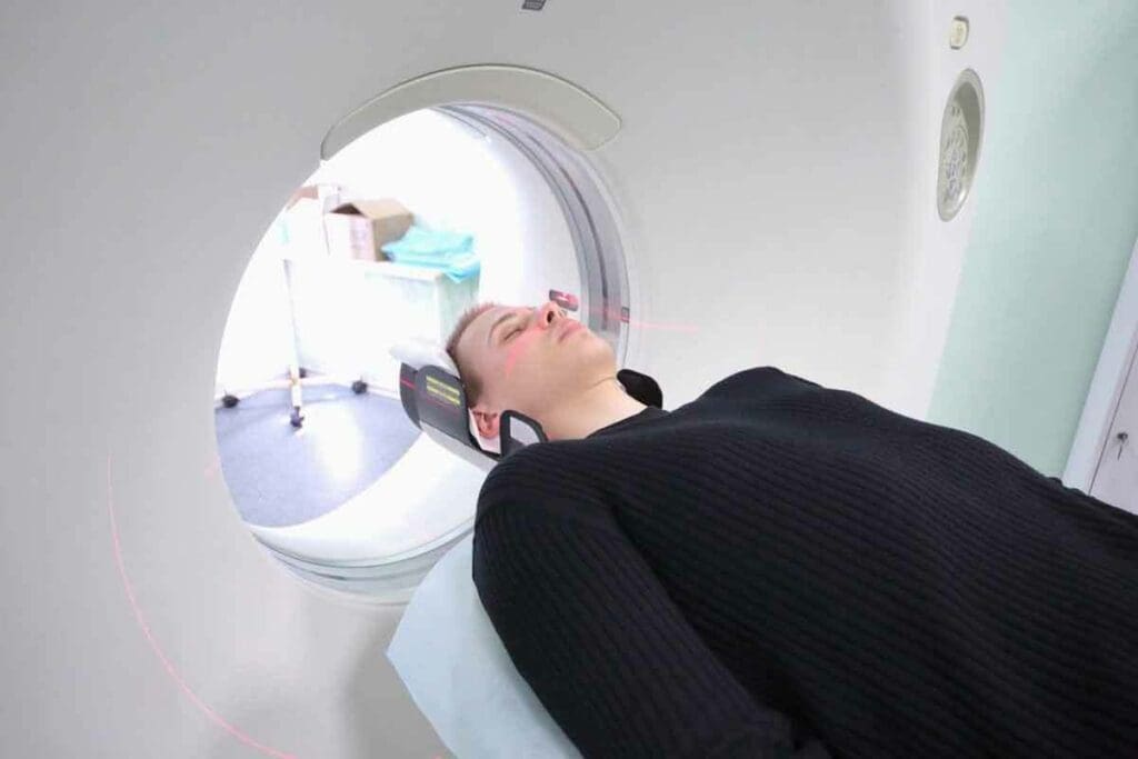
A CT (Computed Tomography) scan is a way to see inside the body. It uses computer-processed X-rays to make detailed images. The scanner is a big, doughnut-shaped machine that moves around the patient.
It takes X-ray measurements from many angles. Then, a computer turns this data into detailed images. These images can be seen one by one or together to show the body in 3D.
Comparing CT Scans to Other Imaging Modalities
CT scans are just one of many imaging tools used in medicine. Others include MRI, ultrasound, and PET scans. Each tool is good for different things.
| Imaging Modality | Primary Use | Key Benefits |
| CT Scan | Detailed images of internal structures, cancer detection, and trauma assessment | Quick, detailed, and versatile |
| MRI | Soft tissue evaluation, neurological disorders, and joint injuries | High contrast resolution, no radiation |
| Ultrasound | Pregnancy monitoring, gallbladder disease, and vascular evaluation | Non-invasive, real-time imaging, and no radiation |
| PET Scan | Cancer staging, metabolic activity assessment, and neurological disorders | Functional information, high sensitivity for certain conditions |
Knowing the differences between these imaging tools helps everyone choose the best one for their needs.
Understanding CT Scan Results: The Basics

Understanding CT scan results can seem hard, but knowing the basics helps. CT scan reports are detailed and include important sections. Each section gives us key information about what the scan found.
The Structure of a CT Scan Report
A CT scan report is set up to give us all the details. It has sections for patient info, medical history, how the scan was done, the findings, and the radiologist’s impression. Let’s look at each part to understand its role.
- Patient Information: This section has the patient’s name, birthdate, and other details.
- Clinical History: It lists the patient’s medical history and why they had the CT scan.
- Technique: This part explains how the CT scan was taken, like which body parts were scanned.
- Findings: Here, the radiologist talks about what they saw in the scan images.
- Impression: The last section sums up what the radiologist thinks, pointing out any important findings or diagnoses.
| Report Section | Description |
| Patient Information | Patient’s identifying details |
| Clinical History | Relevant medical history and reason for the scan |
| Technique | Details of how the CT scan was performed |
| Findings | Radiologist’s observations of the CT scan images |
| Impression | Summary of the radiologist’s interpretation |
Grayscale Interpretation: What Different Shades Mean
CT scan images use grayscale, with different shades showing different tissue densities. Knowing how to read these shades is key to understanding CT scan results.
Bones show up white because they’re dense, and air is black because it’s less dense. Soft tissues are shown in various grays, depending on their density. For example, fat looks darker than muscle.
- White: Dense structures like bones
- Black: Air-filled spaces
- Gray: Soft tissues, with varying shades indicating different densities
By understanding the structure of a CT scan report and how to read grayscale images, we can better grasp our CT scan results and what they mean for our health.
Essential Terminology in CT Scan Reports
To understand CT scan results, knowing common terms and abbreviations is key. CT scan reports are detailed, packed with health information. Grasping this language helps you understand your diagnosis and treatment options.
Common Descriptive Terms and Their Meanings
Reports use specific terms to describe body tissues. For example, hypodense and hyperdense describe tissue appearance. Hypodense areas are darker, showing lower density. Hyperdense areas are brighter, showing higher density.
- Hypodense: Lower density than the surrounding tissue, appearing darker.
- Hyperdense: Higher density than surrounding tissue, appearing brighter.
- Isodense: Same density as surrounding tissue, making it difficult to distinguish.
Knowing these terms helps patients understand their CT scan results better.
Medical Abbreviations Frequently Used in Reports
Medical professionals often use abbreviations in CT scan reports. Learning these can help you understand your results. Some common ones include:
- HU: Hounsfield Units, a measure of tissue density.
- ROI: Region of Interest, referring to a specific area examined in detail.
- IV: Intravenous, often referring to contrast material administered during the scan.
Knowing these abbreviations and their meanings can greatly improve your understanding of your CT scan report.
Understanding CT scan report terminology and abbreviations empowers patients. It’s about gaining knowledge to better understand your health and the information from your healthcare team.
The Science of Tissue Density Interpretation
Understanding tissue density is key to accurate CT scan analysis. It’s a vital part of radiology. It helps spot different tissues and any abnormalities.
Hypodense vs. Hyperdense: What These Terms Indicate
Radiologists use terms like hypodense and hyperdense when looking at CT scans. Hypodense areas are darker and might show edema or cysts. Hyperdense areas are brighter and could mean bleeding, calcification, or other issues.
Knowing the difference between hypodense and hyperdense is important. It helps diagnose many medical conditions. For example:
- A hypodense liver lesion might be a cyst or metastasis.
- A hyperdense brain area could show hemorrhage.
Hounsfield Units: The Quantitative Measure of Density
Hounsfield Units (HU) measure tissue density on CT scans. Named after Sir Godfrey Hounsfield, it uses water as 0 HU and air as -1000 HU. Tissues like bone have high HU values, while fat has low values.
Hounsfield Units make assessing tissue density more precise. For instance:
| Tissue Type | Typical Hounsfield Unit (HU) Range |
| Bone | +400 to +1000 |
| Liver | +40 to +60 |
| Fat | -60 to -100 |
| Air | -1000 |
Understanding Hounsfield Units and the differences between hypodense and hyperdense areas helps doctors. They can make better diagnoses and treatment plans.
Recognizing Normal Anatomical Structures
It’s key to know what organs and tissues look like normally to spot problems in CT scans. We need to understand what healthy parts of the body look like in these images.
Baseline Appearance of Healthy Organs and Tissues
Healthy organs and tissues have specific appearances on CT scans. For example, the liver looks the same all over, and the kidneys have a clear difference between the outer and inner parts. Knowing these looks helps us find any issues.
The liver is denser than the spleen, and the kidneys show a clear difference between the outer and inner parts. The pancreas is usually the same density as the fat around it. Spotting these normal patterns is key to finding any problems.
Normal Variants That May Appear Concerning
Some normal variations can look like problems. For example, some people have a bigger spleen or a liver that’s not the usual shape. These variations can look worrisome if we don’t know they’re normal.
It’s also important to remember that normal anatomical variants can be different for everyone. The size and shape of the adrenal glands can vary, and some people have a bigger thyroid gland. Knowing these differences helps us avoid misreading CT scan results.
Identifying Abnormalities in CT Scan Results
When we look at CT scan results, finding abnormalities is key. It helps us make accurate diagnoses and plan treatments. We’ll show you how to spot common problems and tell different types of lesions apart.
Common Pathological Findings
CT scans can spot many issues, like tumors, infections, and injuries. These findings help us diagnose various conditions. They range from sudden injuries to long-term diseases.
Some common problems include:
- Cysts and benign tumors
- Malignant tumors
- Infections and abscesses
- Fractures and bone degeneration
- Internal bleeding
Knowing these issues is key to choosing the right treatment.
Distinguishing Between Different Types of Lesions
Lesions on CT scans can look very different. It’s important to tell them apart for accurate diagnosis and treatment.
The table below shows what makes each lesion type unique:
| Lesion Type | Characteristics | Clinical Implication |
| Cystic Lesions | Fluid-filled, thin-walled | Often benign, may require monitoring |
| Solid Tumors | Varied density, irregular shape | May be malignant, requires further investigation |
| Infectious Lesions | Surrounding inflammation, possible abscess formation | Requires antibiotic treatment or drainage |
Knowing the details of each lesion is essential for the right treatment.
CT Scan Results for Cancer Detection
CT scans are key in finding and staging cancer. They give us vital info for planning treatments. We use them to spot cancer cells, see how big they are, and where they’ve spread.
Radiological Features of Malignant Tumors
Malignant tumors show certain signs on CT scans. These signs include odd shapes, mixed-up looks, and growing into nearby tissues. We look for these to tell cancer tumors apart from harmless growths.
Key radiological features of malignant tumors:
- Irregular or spiculated margins
- Heterogeneous enhancement with contrast
- Invasion into adjacent structures
- Necrosis or calcification within the tumor
Cancer Staging Information in CT Reports
Cancer staging is vital in CT reports. It helps decide treatment and outlook. We use the TNM system, which looks at the tumor size, lymph nodes, and whether it’s spread.
| Stage | Description |
| I | The tumor is small and localized |
| II | The tumor is larger but confined |
| III | The tumor has invaded the surrounding tissues or lymph nodes |
| IV | Metastasis to distant organs |
Identifying Metastatic Disease
Finding metastatic disease is key to accurate staging and treatment. We look for signs like lesions in other organs, swollen lymph nodes, and other widespread signs.
“The presence of metastatic disease significantly impacts treatment options and patient prognosis, underscoring the importance of accurate detection through imaging modalities like CT scans.”
— Expert Oncologist
By knowing how to read tumor signs, understand staging, and spot metastasis, we can give cancer patients the best care.
Reading Chest CT Scan Results for Cancer
A chest CT scan is key to finding lung cancer and other chest issues. Knowing how to read these results is important for correct diagnosis and treatment. We’ll help you understand lung nodules, mediastinal abnormalities, and pleural disease on chest CT scans.
Lung Nodules: Size, Shape, and Significance
Lung nodules are often seen on chest CT scans. Their size and shape tell us a lot about their risk. Nodules under 5 mm are usually not a big worry, but those over 10 mm need more checking.
| Nodule Size | Risk Level | Recommended Action |
| < 5 mm | Low | Monitoring |
| 5-10 mm | Moderate | Follow-up CT scan |
| > 10 mm | High | Biopsy or PET scan |
Mediastinal Abnormalities and Lymphadenopathy
The mediastinum is the middle part of the chest. Problems here, like big lymph nodes, might mean cancer. It’s important to look at the size and where these problems are.
Pleural Disease and Effusions
Pleural disease affects the pleura, the lung’s outer layer. Fluid buildup, or pleural effusions, can also be a sign of cancer. The type of fluid and any thickening of the pleura are key to diagnosis.
Interpreting Abdominal and Pelvic CT Scan Results
Understanding CT scan results for the abdominal and pelvic regions can be complex, but it is essential for diagnosis. These scans are powerful tools that provide detailed images of organs and structures within these regions.
These scans are critical for detecting a wide range of conditions, from injuries and infections to cancer and vascular diseases. We will guide you through the process of interpreting the findings related to various organs and structures in the abdominal and pelvic areas.
Liver, Pancreas, and Kidney Findings
The liver, pancreas, and kidneys are vital organs that are closely examined in abdominal CT scans. Abnormalities in these organs can indicate various health issues.
- Liver Findings: CT scans can reveal liver lesions, fatty liver disease, and cirrhosis. We look for changes in liver texture, size, and the presence of masses or cysts.
- Pancreas Findings: Pancreatitis, pancreatic cancer, and cysts can be identified through CT scans. We examine the size, shape, and texture of the pancreas for abnormalities.
- Kidney Findings: Kidney stones, cysts, tumors, and other abnormalities can be detected. We assess the kidneys’ size, position, and the presence of any masses or obstructions.
Bowel, Mesenteric, and Retroperitoneal Abnormalities
CT scans also provide valuable information about the bowel, mesentery, and retroperitoneal space. These areas can be affected by various conditions.
- Bowel Abnormalities: Inflammatory bowel disease, tumors, and obstructions can be identified. We look for wall thickening, masses, or other irregularities.
- Mesenteric Abnormalities: Inflammation, tumors, or cysts in the mesentery can be detected. We examine the mesenteric fat and vessels for signs of disease.
- Retroperitoneal Abnormalities: Tumors, lymphadenopathy, and other conditions can be observed in the retroperitoneal space. We assess the size and characteristics of any abnormalities.
Pelvic Organs: Prostate, Uterus, and Ovaries
Pelvic CT scans provide critical information about the reproductive organs. Abnormalities in these organs can indicate various health issues.
- Prostate Findings: Prostate enlargement, cancer, or other abnormalities can be identified. We examine the size and texture of the prostate gland.
- Uterus and Ovarian Findings: Uterine or ovarian masses, cysts, and other conditions can be detected. We assess the size, shape, and characteristics of these organs.
Interpreting abdominal and pelvic CT scan results requires a deep understanding of anatomy and possible abnormalities. By carefully examining the images, healthcare professionals can diagnose a wide range of conditions and develop effective treatment plans.
Understanding Brain CT Scan Results
A brain CT scan is a key tool for doctors. It helps find conditions like strokes and tumors. These scans give us vital info about brain health, helping us decide on treatments.
Brain Tissue Evaluation
We start by checking the brain tissue for any oddities. We look at the density and texture of different parts. We search for signs of swelling, shrinkage, or other changes.
The scan can show a lot about the brain’s health. For example, low-density areas might mean swelling or damage. High-density spots could show bleeding or hardening. Knowing these details is key to making the right diagnosis.
Identifying Strokes, Hemorrhages, and Tumors
Brain CT scans are great for spotting strokes, bleeding, and tumors. We can tell if a stroke is caused by a blockage or bleeding. This is important for choosing the right treatment.
- Ischemic Strokes: These show up as low-density areas on scans because of blood flow issues.
- Hemorrhagic Strokes: These are high-density because of blood.
- Tumors: Tumors look like masses with different densities and might have swelling around them.
Spotting these conditions early lets doctors start the right treatment quickly.
Ventricular System and CSF Spaces
The ventricular system and cerebrospinal fluid (CSF) spaces are important, too. We check the ventricles’ size and shape and look for CSF space issues.
Big or changed ventricles might mean problems like fluid buildup or blockages. CSF space issues could point to inflammation, infection, or other problems.
By studying brain CT scan results, we get a full picture of a patient’s brain health. This helps us create effective treatment plans.
What Happens After Receiving Serious CT Scan Results
Getting serious CT scan results can change your life. It brings uncertainty and worry about what’s next. It’s a tough time, and knowing what happens next is key.
Communication Protocols for Critical Findings
Healthcare teams have clear ways to share serious CT scan results. They make sure you know quickly and clearly. These steps might differ at each hospital, but they aim to reach you fast.
If your scan shows a serious issue, you’ll likely hear from your radiologist or doctor right away. They’ll then talk more with your main doctor or a specialist, based on your situation.
Follow-up Recommendations and Additional Testing
After serious CT scan results, more tests might be needed. Your doctor will suggest the best next steps for you. This could mean more scans, biopsies, or other tests.
It’s important to understand why these tests are needed. For example, if a tumor is found, a biopsy might be suggested.
Questions to Ask Your Doctor About Your Results
Being proactive and informed is vital with serious CT scan results. Make a list of questions for your doctor. This will help you understand your condition and treatment plan better.
- What do my CT scan results indicate about my condition?
- What are the next steps in diagnosing or treating my condition?
- Are there any additional tests or procedures required?
- What are the possible risks and benefits of the treatments suggested?
- How can I manage my symptoms or improve my quality of life during this time?
By asking these questions, you’ll get a clearer picture of your situation. This helps you make better decisions about your care.
Conclusion
Understanding CT scan results is key to good patient care and making smart choices. We’ve talked about how CT scans work, what the reports mean, and the importance of different findings. Our goal is to help patients make better decisions and get better care.
As we wrap up, it’s clear that knowing about CT scan results helps patients a lot. We want patients to keep learning and talk to their doctors about their scans. This teamwork can lead to better health and smarter choices.
In the end, it’s not just about reading medical images. It’s about being involved in your health journey. By doing this, patients can work better with their doctors to tackle health issues.
FAQ
What is a CT scan, and how does it work?
A CT scan uses X-rays to show detailed images inside the body. It gives a clearer view than regular X-rays. This makes it great for finding and diagnosing many health issues.
How do I understand my CT scan results?
To understand your CT scan results, first, know the report’s structure. Then, learn to read grayscale images and recognize normal body parts. It’s also key to grasp the report’s terms, like hypodense and hyperdense areas.
What is the difference between hypodense and hyperdense areas on a CT scan?
Hypodense areas are darker and might show conditions like edema or cysts. On the other hand, hyperdense areas are brighter and could mean bleeding or calcification.
How are CT scans used in cancer detection?
CT scans are vital for finding and staging cancer. They help spot the signs of cancerous tumors and provide information on cancer stages. They also help find if cancer has spread.
What do lung nodules on a chest CT scan indicate?
Lung nodules can be either benign or cancerous. The size, shape, and what they mean are key. More tests, like a biopsy, might be needed to figure out what they are.
How do I interpret abdominal and pelvic CT scan results?
When looking at abdominal and pelvic CT scans, it’s important to understand what they show. Findings about the liver, pancreas, kidneys, and other organs can point to issues like tumors or inflammation.
What happens after receiving serious CT scan results?
After getting serious CT scan results, your doctor will talk to you about them. They’ll suggest more tests or treatment. It’s important to ask questions and know what to do next.
How are CT scan results communicated to patients?
Doctors usually tell patients about their CT scan results. How they share the news can depend on the findings and what the patient needs.
What are Hounsfield units, and how are they used in CT scans?
Hounsfield units measure tissue density on a CT scan. They help tell different tissues apart, like bone, soft tissue, and fat.
Can I request a copy of my CT scan results?
Yes, you can ask for a copy of your CT scan results. It’s good to talk to your doctor about them to understand what they mean and what to do next.
How do I prepare for a CT scan?
To prepare for a CT scan, you might need to fast or remove metal items. Your doctor will give you specific instructions. The prep can vary based on the type of scan.
Are CT scans safe?
CT scans are usually safe, but they do involve some radiation. The benefits usually outweigh the risks. It’s wise to talk to your doctor about any concerns.
References
- Patel, P. R. (2023). CT Scan. In StatPearls. StatPearls Publishing. Retrieved from https://www.ncbi.nlm.nih.gov/books/NBK567796/


