Last Updated on December 1, 2025 by Bilal Hasdemir

Each year, over 1.9 million new cancer cases are diagnosed in the U.S., highlighting the critical importance of understanding tumor characteristics. Finding out if a tumor is benign or malignant is key to treatment. We use medical imaging techniques to make this distinction.
Employing the appropriate cancer diagnostic methods is crucial for optimal patient care. By looking at tumor characteristics, doctors can choose the best treatment. We’ll see how ct scan tumor diagnosis helps in this process.
Key Takeaways
- Understanding the role of medical imaging in tumor diagnosis
- The importance of distinguishing between benign and malignant tumors
- How ct scan tumor diagnosis aids in treatment planning
- The significance of tumor characteristic analysis in cancer care
- Advances in cancer diagnostic methods
Understanding CT Scan Technology

Learning about CT scans is key to understanding their role in finding tumors and other health issues. CT scans use X-rays to make detailed pictures of the body’s inside.
How CT Scans Work
CT scans work by using X-rays to see how different body tissues absorb them. Here’s a quick explanation:
- The CT scanner, a big, doughnut-shaped machine, sends out X-rays that go through the body.
- Detectors in the scanner catch the X-rays that don’t get blocked by the body, gathering the needed data.
- A computer turns this data into images of the body’s inside, or slices.
- These images can be seen one by one or stacked to show a 3D view of the body’s inside.
CT scan technology is a big deal in medical imaging. It gives clear, detailed pictures that help doctors find and diagnose many health problems, from injuries to diseases like cancer.
Evolution of CT Imaging
The growth of CT imaging has been amazing. New tech has made scans faster, images clearer, and radiation doses lower. Some major steps include:
- The start of spiral CT scanners, which scan continuously without stopping.
- The creation of multi-detector CT (MDCT) scanners, which take many images at once, cutting down scan times.
- Improvements in how images are made, allowing for better quality pictures even with less radiation.
These changes have made CT scans better for diagnosing and safer for patients. As medical imaging technology keeps getting better, we’ll see even more accurate and quick diagnostic tools.
The Basics of Tumor Detection

Accurate tumor detection with CT scans needs a deep understanding of tumor features on images. Tumors show different looks on CT scans, based on their type, size, and where they are.
What Tumors Look Like on Imaging
On a CT scan, tumors show up as clear masses or lesions. They can be told apart by their density, edges, and how they change after contrast is added. Benign tumors have clear edges and a uniform look. Malignant tumors have irregular shapes and can spread into nearby tissues.
The look of tumors on CT scans is key for first checks. For example, seeing calcifications, necrosis, or hemorrhage in a tumor can hint at its type.
Benign vs. Malignant: Visual Differences
Telling benign from malignant tumors on CT scans involves looking at several signs. Benign tumors have clear edges and might push nearby structures but not invade them. Malignant tumors have fuzzy edges, spread into nearby tissues, and might show uneven enhancement due to necrosis or blood vessel problems.
| Characteristics | Benign Tumors | Malignant Tumors |
| Margins | Well-defined | Ill-defined |
| Density | Uniform | Heterogeneous |
| Invasion | Rarely invade surrounding tissues | Often invade surrounding tissues |
| Enhancement Pattern | Typically uniform enhancement | May show heterogeneous enhancement |
Knowing these visual differences is key for doctors and radiologists to decide on next steps and treatment plans.
CT Scan Tumor Diagnosis: Capabilities and Limitations
It’s important to know what CT scans can and can’t do in diagnosing tumors. They are key in cancer diagnosis, giving detailed images of tumors.
What CT Scans Can Reveal About Tumors
CT scans show important details about tumors. They tell us about the tumor’s size, where it is, and how dense it is. This information is vital for determining the cancer’s stage and formulating an effective treatment plan.
Key features that CT scans can reveal about tumors include:
- Tumor size and location
- D density and enhancement patterns
- Relationship to surrounding structures
This info helps doctors understand how the tumor affects the patient. It helps decide the best treatment.
Inherent Limitations in Tumor Characterization
Even though CT scans are great, they have some big challenges. One big one is telling if a tumor is benign or malignant just by looking at the CT scan. Some tumors look the same on a CT scan, making it hard to tell what they are without more tests.
The limitations of CT scans in tumor diagnosis include:
- Difficulty in distinguishing between certain types of tumors
- Limited ability to assess tumor aggressiveness
- Potential for false positives or negatives
Despite these challenges, CT scans are very important. They help start the process of figuring out what’s wrong and guide treatment.
In summary, CT scans are very useful in diagnosing tumors. They give a lot of information. But, we must remember their limits. They should be used as part of a bigger plan for checking and treating tumors.
Characteristics That Suggest Benign Tumors on CT
Benign tumors show clear signs on CT scans that set them apart from cancerous ones. Knowing these signs is key for correct diagnosis and treatment.
Size and Growth Patterns
The size and how a tumor grows can tell us a lot. Benign tumors grow in a steady way and don’t change much in size. They are usually well-defined and have clear edges with the tissue around them.
A lipoma looks like a clear, even mass with fat density on CT scans. But, size isn’t everything. Some benign tumors can get big, and some cancers can be small. So, we look at growth patterns along with other signs.
Density and Enhancement Patterns
The density and how a tumor reacts to contrast on CT scans are also important. Benign tumors usually have the same density and don’t change much after contrast. For example, a cystic lesion with a thin wall and no solid parts is likely benign.
We check how a tumor reacts to contrast to tell benign from malignant. Benign tumors don’t show the messy, irregular growth seen in cancers. They might enhance evenly or not at all. Contrast agents help us see the tumor’s blood flow and nature.
By looking closely at these signs on CT scans, we can guess if a tumor is likely benign. This guess is important for deciding what tests to do next and how to treat it.
Characteristics That Suggest Malignant Tumors on CT
Malignant tumors show clear signs on CT scans that set them apart from benign growths. Knowing these signs is key for accurate diagnosis and planning treatment.
Irregular Borders and Invasion
One key sign of a malignant tumor on a CT scan is its border. Malignant tumors have irregular borders, which can look spiculated or poorly defined. This irregular shape often means the tumor is growing into nearby tissues. Benign tumors, on the other hand, have smooth, well-defined borders.
Another important sign is when the tumor invades nearby structures. On a CT scan, we look for signs that the tumor is pushing into or invading nearby organs, blood vessels, or other important structures. This invasion is a strong indicator of malignancy.
Heterogeneity and Necrosis
Malignant tumors often look different on CT scans because of heterogeneity. This means they have a non-uniform appearance. This can be because of different tissue densities within the tumor, including areas of necrosis (dead tissue). Necrosis shows up as low-density areas in the tumor and is common in fast-growing, malignant tumors.
The presence of necrosis is a significant clue for diagnosis. Tumors with a lot of necrosis might need more aggressive treatment. We look at the tumor’s enhancement pattern after contrast during the CT scan to check for heterogeneity and necrosis.
| Characteristic | Description | Implication |
| Irregular Borders | Spiculated or poorly defined edges | Suggests invasive nature |
| Invasion | Encroachment on surrounding structures | Definitive sign of malignancy |
| Heterogeneity | Non-uniform appearance on CT | May indicate varying tissue densities |
| Necrosis | Low-density areas within the tumor | Common in rapidly growing tumors |
Accuracy Rates of CT Scans in Tumor Classification
It’s key to know how well CT scans can tell apart benign from malignant tumors for good treatment plans. Research has looked into how accurate CT scans are in tumor classification. The results show different levels of success.
Statistical Overview of Diagnostic Accuracy
Research shows that CT scans’ accuracy in tumor diagnosis varies a lot. For example, one study might say CT scans are 85% accurate for certain tumors. But another study might find a different accuracy rate, depending on the tumor and the people in the study.
| Tumor Type | CT Scan Accuracy | Sample Size |
| Lung Tumors | 82% | 200 patients |
| Liver Tumors | 88% | 150 patients |
| Pancreatic Tumors | 75% | 100 patients |
The table shows how different tumors have different CT scan accuracy rates. It’s important to look at these numbers when judging CT scans’ ability to diagnose.
Factors Affecting Diagnostic Precision
Several things can change how well CT scans can diagnose tumors. These include tumor size, location, and type. For instance, small tumors or those in hard-to-reach places can be tough to spot with CT scans alone.
Tumor size matters a lot. Bigger tumors are easier to see because they have clearer signs. But smaller tumors might not show up as well, making them harder to classify.
The location of a tumor also affects accuracy. Tumors in complex areas like the abdomen or pelvis can be tricky to get right because of all the nearby structures.
Knowing how these factors affect CT scan accuracy helps doctors make better decisions for patients. This includes choosing the right treatment plans.
Common Benign Tumors and Their CT Appearances
It’s key to know how benign tumors look on CT scans for correct diagnosis and treatment. These non-cancerous growths can pop up anywhere in the body. CT scans help spot and figure out what they are.
Lipomas and Hemangiomas
Lipomas are made of fat and show up as low-density, well-defined masses on CT scans. Hemangiomas, filled with blood vessels, can appear in organs like the liver. They show special patterns on CT scans because of their blood vessel makeup.
Key characteristics of lipomas on CT:
- Low density due to high fat content
- Well-defined borders
- Typically homogeneous
Adenomas and Fibromas
Adenomas are glandular tumors found in endocrine glands. They can look like well-defined nodules on CT scans, with varying density. Fibromas, made of fibrous tissue, can appear in different places. They show up as solid, well-circumscribed masses on CT.
Characteristics of adenomas and fibromas on CT:
- Adenomas: May have varying density; can be homogeneous or heterogeneous
- Fibromas: Typically solid and well-defined
Spotting these benign tumors right is vital to avoid unnecessary treatments. Knowing how they look on CT scans helps doctors make better choices.
Common Malignant Tumors and Their CT Appearances
It’s key to know how malignant tumors like carcinomas, sarcomas, and lymphomas look on CT scans. This knowledge helps doctors diagnose and plan treatment. We’ll look at how these tumors are spotted and what makes them unique on CT scans.
Carcinomas
Carcinomas start from epithelial cells and are cancerous. On CT scans, they look like masses with odd shapes and mixed brightness. For example, lung carcinomas might show up as jagged masses with dead tissue inside. Pancreatic carcinomas could look like dark masses with swollen ducts.
Key CT features of carcinomas include:
- Irregular or spiculated margins
- Heterogeneous enhancement due to necrosis or hemorrhage
- Invasion into adjacent structures
- Presence of lymphadenopathy
A top oncologist says, “Spotting carcinomas on CT scans is vital for planning treatment and improving patient results.”
“Accurate imaging is key for staging and checking if carcinomas can be removed.”
Sarcomas and Lymphomas
Sarcomas come from mesenchymal cells and can pop up anywhere. On CT scans, they look like big, mixed masses with dead spots. Lymphomas, which affect the lymph system, can show up in lymph nodes, spleen, and other places.
Characteristic CT features of sarcomas include:
- Large size at presentation
- Heterogeneous attenuation due to necrosis or cystic change
- Enhancement patterns that may be heterogeneous
Lymphomas on CT typically show as:
- Enlarged lymph nodes or conglomerate nodal masses
- Splenomegaly or focal splenic lesions
- Involvement of extranodal sites such as the liver or gastrointestinal tract
CT scans help us see how far sarcomas and lymphomas have spread. They also help us track how well treatments are working and if the cancer comes back. The detailed pictures from CT scans are very helpful in managing these tough cancers.
CT Scan Evaluation by Body Region
When we use CT scans to check for tumors, we look at each body area differently. This helps doctors make better diagnoses and treatment plans.
Brain and Head Tumors
CT scans are key for brain and head tumors. They show where the tumor is, how big it is, and if it’s affecting nearby areas. We use them to quickly check for head injuries or sudden brain symptoms. The scans help find tumors, calcifications, and signs of increased pressure in the brain.
Key considerations for brain and head tumors include:
- Assessing the tumor’s relationship with vital brain structures
- Identifying any bone erosion or destruction
- Detecting calcifications within the tumor
Chest and Lung Tumors
CT scans give us detailed views of the lungs, mediastinum, and nearby tissues for chest and lung tumors. We count on them to spot and track lung cancer. They show the tumor’s size, location, and if it’s spreading to lymph nodes or other places.
Important factors for chest and lung tumors include:
- Tumor size and location within the lung
- Involvement of adjacent structures, such as the chest wall or diaphragm
- Presence of lymphadenopathy or metastatic disease
Abdominal and Pelvic Tumors
In the abdomen and pelvis, CT scans are essential for finding and understanding tumors in organs like the liver, pancreas, kidneys, and reproductive organs. They help us see how big the tumor is, where it is, and if it’s touching nearby areas. They also help spot any cancer that has spread.
Key considerations for abdominal and pelvic tumors include:
- Evaluating the tumor’s relationship with major blood vessels
- Assessing any invasion into nearby organs or structures
- Identifying any metastatic disease in the abdomen or pelvis
The Complete Diagnostic Process Beyond CT Imaging
Diagnosing tumors involves more than just CT scans. It also includes looking at the patient’s history, doing a physical exam, and running lab tests. This way, doctors can really understand what’s going on and make the right diagnosis.
When someone shows signs of a tumor, it’s important to get a full history and do a physical check-up. This helps doctors figure out what might have caused the tumor. For example, past health issues can tell a lot about the risk of certain tumors.
Integrating Clinical History and Physical Examination
Doctors ask a lot of questions to get a patient’s medical history. They want to know about symptoms, family health, and past illnesses. This helps them guess if the tumor is likely to be harmless or dangerous. A physical exam can also find things that scans might miss, like tumors or swollen lymph nodes.
- Medical history helps spot risk factors and possible causes of tumors.
- A physical check-up can find signs of tumors that scans might not catch.
- Together, history and physical exam help pick the right tests to run.
The Role of Tumor Markers and Laboratory Tests
Tumor markers and lab tests are key in finding tumors. Tumor markers are substances made by tumors or in response to them, found in blood, urine, or tissues. Lab tests, like blood counts and liver function tests, give more info on the patient’s health and if a tumor is active.
- Tumor markers can pinpoint certain tumors, like prostate cancer through PSA tests.
- Lab tests can show tumor-related issues, like high liver enzymes.
- These tests, along with imaging and clinical findings, help in understanding tumor stage and outlook.
By combining history, physical exam, tumor markers, lab tests, and CT scans, doctors can make better diagnoses and treatment plans. This detailed approach is key to giving patients the best care possible.
Complementary Imaging Techniques
Using different imaging techniques with CT scans is key for accurate tumor diagnosis. CT scans give us a good start, but we need more to understand tumors fully.
MRI: When It’s Preferred Over CT
Magnetic Resonance Imaging (MRI) shines when we need to see soft tissues clearly. MRI shows more detail than CT scans, which is great for tricky spots like the brain, spine, and pelvis. We choose MRI over CT when we need to see soft tissues well for diagnosis or planning.
In brain tumors, MRI shows how the tumor relates to important areas. It also helps tell different tumor types apart and how far they’ve spread.
PET Scans and Nuclear Medicine
Positron Emission Tomography (PET) scans are also very important. PET scans show how active tumors are, helping us tell if they’re cancerous or not. They also show how well treatments are working by highlighting active tumor areas.
PET/CT scans are now common, combining PET’s function with CT’s anatomy. This mix improves diagnosis and helps stage cancer accurately, which is key for choosing the right treatment.
In summary, MRI and PET scans are essential for better tumor diagnosis than CT scans alone. They help us understand tumors better, leading to better treatment plans.
Biopsy: The Gold Standard for Definitive Diagnosis
Biopsy is the top choice for finding out if a tumor is cancerous. It looks at tissue or cells directly. This gives more accurate results than CT scans alone.
Necessity of Biopsy After CT Findings
After a CT scan shows a tumor, a biopsy is often needed. This is true if the CT scan shows signs of cancer or if it’s not clear. The decision to do a biopsy depends on the tumor’s size, location, and how it looks on the CT scan. It also depends on the patient’s health and past medical history.
We think a biopsy is key in these situations:
- When CT scan results are unclear or show a high chance of cancer.
- If the tumor is hard to reach for imaging alone.
- When the patient’s symptoms or lab results suggest cancer.
Types of Biopsy Procedures
There are many biopsy types, each with its own use. The right one depends on the tumor’s location, the patient’s health, and other factors.
| Biopsy Type | Description | Common Applications |
| Fine-needle Aspiration Biopsy | Uses a thin needle to collect cell samples. | Good for tumors near the skin’s surface or in easy-to-reach areas. |
| Core Needle Biopsy | Uses a larger needle to get a tissue sample. | Best for tumors in organs like the breast, prostate, or liver. |
| Surgical Biopsy | Removes part or all of the tumor surgically. | Needed when a big tissue sample is required or the tumor is accessible during surgery. |
In summary, biopsy is essential for tumor diagnosis. It allows a direct look at tissue or cells. Knowing when to use a biopsy and the different methods helps doctors make the right treatment plans.
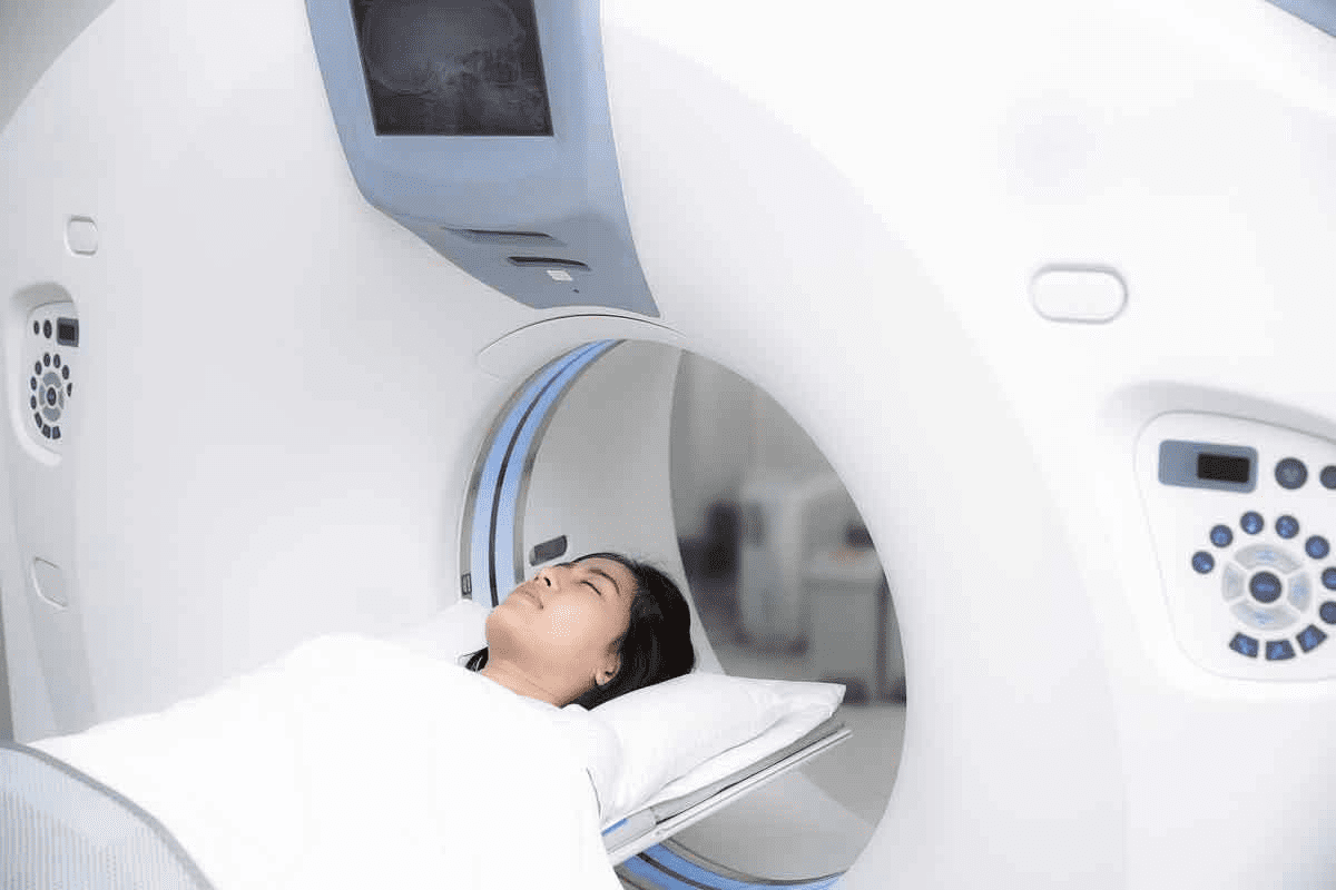
Patient Experience and Preparation
When patients prepare for a CT scan, knowing what to expect helps ease worries. A CT scan is a detailed medical imaging method that shows the body’s inside parts. We help patients understand the procedure, making sure they’re ready for it.
Before Your CT Scan
Getting ready for a CT scan is important. Patients should arrive at least 30 minutes early. This time is for paperwork, changing into a gown, and removing metal items or jewelry.
Specific Instructions:
- Patients might need to fast for a few hours before, if contrast is used.
- Telling the team about any allergies, like to iodine or contrast, is key.
- Informing about medical conditions, like diabetes or kidney disease, is also important.
Knowing these steps can make the process smoother and less stressful.
During and After the Procedure
During the scan, patients lie on a table that moves into a big machine. The scan is fast, lasting just a few minutes. If contrast is used, it’s given through an IV line.
After the scan, patients can usually go back to their normal day unless told not to. If contrast was used, watching for allergic reactions is important.
| Procedure Stage | What to Expect |
| Before the Scan | Arrive early, complete paperwork, change into a hospital gown, and remove metal objects. |
| During the Scan | Lie on a table that slides into the CT scanner; the scan is quick, typically a few minutes. |
| After the Scan | Resume normal activities unless instructed not to; watch for any bad reactions to contrast. |
Knowing about the CT scan process helps patients feel more at ease. Our medical team aims to support and inform, making sure patients are comfortable and confident.
“The more information patients have about their diagnostic procedures, the less anxiety they tend to experience.”
A medical professional’s insight
Conclusion: The Role of CT Scans in the Tumor Diagnosis Journey
CT scans are key in finding and understanding tumors. They help doctors decide what to do next. These scans show important details about tumors, like if they are cancerous.
At the start of finding a tumor, CT scans are very important. They give clear pictures of the tumor’s size, where it is, and what it looks like. This info helps doctors choose the best treatment.
Doctors use CT scans to make smart choices for patients. As we get better at imaging, CT scans will keep being a big help. They are essential in the fight against cancer.
FAQ
What is a CT scan, and how does it help in tumor diagnosis?
A CT scan uses X-rays and computer tech to create detailed images of the body. It helps doctors see tumors clearly. This is key for figuring out if a tumor is benign or malignant.
How do CT scans differentiate between benign and malignant tumors?
CT scans look at size, growth, density, and how the tumor looks. Benign tumors have smooth edges and uniform density. Malignant tumors have irregular edges and may invade nearby tissues.
What are the limitations of CT scans in tumor diagnosis?
CT scans are great for finding and describing tumors. But, they can’t always tell if a tumor is benign or malignant. Small tumors or those in hard-to-reach places can be tricky to spot.
How accurate are CT scans in classifying tumors?
CT scans are good at finding tumors but not always at telling if they’re benign or malignant. The accuracy depends on the tumor type, location, and image quality. They work best when used with other diagnostic tools.
What are some common benign tumors and their typical appearances on CT scans?
Benign tumors like lipomas, hemangiomas, adenomas, and fibromas have distinct looks on CT scans. Lipomas are low-density with clear edges. Hemangiomas show specific enhancement patterns. Adenomas have uniform density and smooth edges. Fibromas appear as well-defined masses with varied density.
How do CT scans evaluate tumors in different parts of the body?
CT scans give detailed images for each body area. In the brain, they spot hemorrhages or large tumors quickly. In the chest, they check lung nodules and masses. In the abdomen and pelvis, they look at liver, kidney, and other organ tumors.
What is the role of complementary imaging techniques like MRI and PET scans in tumor diagnosis?
MRI and PET scans add extra info to CT scans. MRI is better for soft tissue contrast, like brain tumors. PET scans show metabolic activity, helping spot active tumor tissues and cancer spread. They’re chosen based on the tumor’s type and location.
When is a biopsy necessary after CT findings?
A biopsy is needed when CT scans aren’t clear or suggest cancer. It gives a direct diagnosis by examining tissue samples. The decision to biopsy depends on the tumor’s appearance, clinical history, and patient risk factors.
How should I prepare for a CT scan?
Preparation for a CT scan includes removing metal objects and wearing comfy clothes. You might need to fast or avoid certain meds. Our team will give specific instructions based on your scan type and needs.
What can I expect during and after a CT scan?
During a CT scan, you’ll lie on a table that slides into the scanner. It’s quick and painless, but you might need to hold your breath. Afterward, you can usually go back to normal activities unless told not to. The images are reviewed by a radiologist, and the results are shared with your healthcare provider.
Reference
- National Cancer Institute. (2019). Advances in pancreatic cancer research. Cancer.gov. Retrieved from https://www.cancer.gov/types/pancreatic/research


