Last Updated on November 25, 2025 by
Urography is a key test that checks the urinary system. At our place, we make sure you’re comfortable and understand what’s happening.

Getting ready for tests can be scary. Urography lets us see the urinary system clearly, helping us find the right treatment. The process includes getting ready, the imaging, and sometimes a CT urogram for a full check.
We want to tell you about these steps. This way, you’ll know what to expect during your urogram procedure. We aim for a smooth experience for you.
Key Takeaways
- Urography is a diagnostic tool for evaluating the urinary system.
- The procedure involves several stages from preparation to imaging.
- A CT urogram is used for a detailed examination.
- Patient comfort and clarity are prioritized throughout the process.
- Understanding the stages helps in preparing for the procedure.
Understanding Urography: A Comprehensive Overview
Urography uses different imaging methods to check the urinary system’s health. It’s key to talk clearly about your health tests. Urography helps find and treat problems in the urinary tract.
Definition and Purpose of Urinary System Imaging
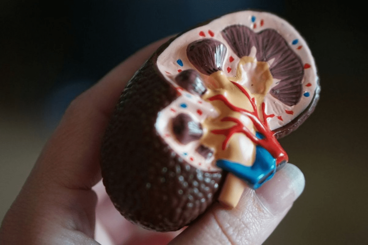
Imaging of the urinary system, like urography, shows the kidneys, ureters, bladder, and urethra. Its main goal is to spot and track issues like kidney stones, tumors, and infections. CT urography stands out for its detailed views.
Advanced imaging lets us see the urinary system’s shape and how it works. This info helps doctors create the best treatment plans for each patient.
Types of Urography: IVP vs CT Urography
There are many urography types, with Intravenous Pyelography (IVP) and CT Urography being top choices. IVP uses X-rays and a contrast agent to see the urinary tract. CT urography uses CT scans and contrast to show more details.
- IVP helps check the urinary tract’s shape and how it works.
- CT Urography gives a full view, spotting issues like tumors and stones.
Clinical Applications and Diagnostic Value
CT urography is key in finding and treating urinary tract problems. It gives clear images of the urinary system. This makes it very useful for doctors.
- It finds stones and blockages in the urinary tract.
- It spots tumors and cysts in the urinary tract.
- It checks for injuries and birth defects in the urinary tract.
Knowing about urography’s types and uses helps us see its value in healthcare.
Pre-Procedure Preparation Stages
Getting ready for a CT urogram is key to getting clear images. We guide our patients on what to do before the test. This includes eating and drinking rules and managing medications. Your help is crucial for a good diagnosis.

Patient Instructions: Fasting and Hydration Guidelines
Before your CT urogram, it’s crucial to follow our fasting and hydration guidelines. You should not eat or drink for a few hours before. But, the exact rules might change based on your needs and the contrast media used.
Bowel Preparation with Laxatives
In some cases, bowel preparation with laxatives may be necessary to clear the bowel. We’ll tell you if this is needed and give you clear instructions.
Medication Adjustments Before the Procedure
Some medications might need to be changed or stopped before the test. It’s essential to inform us about all medications you’re currently taking. This includes any supplements or over-the-counter drugs, so we can guide you properly.
Contrast Allergy Screening and Precautions
If you’re allergic to contrast media, it’s vital to inform your healthcare provider. We’ll take steps to avoid allergic reactions during the test. This might include giving you pre-medication or using different contrast agents.
By following these steps, we can make sure your CT urogram is safe and effective. This way, we get the best images for an accurate diagnosis.
Initial Imaging Stage: The Non-Contrast Scan
The first step in a CT urogram is a non-contrast scan. It’s key for setting a baseline. This stage helps us see the anatomy and find any issues before the diagnosis.
Purpose and Benefits of Baseline Imaging
The non-contrast scan’s main goal is to show the urinary system’s baseline. It helps spot calcifications or issues before we use contrast. The benefits include:
- Identifying existing conditions that may impact the diagnosis
- Providing a reference point for subsequent imaging stages
- Enhancing the accuracy of the CT urogram results
Starting with a non-contrast scan makes our diagnosis more accurate and complete.
Detection of Calcifications and Existing Abnormalities
The non-contrast scan is great for finding calcifications, like kidney stones. It also spots other issues, like cysts or tumors, in the urinary system.
Patient Experience During Non-Contrast Scanning
Some patients might feel anxious about a CT urogram. During the non-contrast scan, you’ll lie on a table that moves into the CT scanner. The scan is quick and doesn’t hurt, lasting just a few minutes. Our team will help you relax and make sure you’re comfortable.
We aim to give you top-notch care and support during the scan. Our goal is to make the experience easy and stress-free, while also getting you an accurate diagnosis.
CT Urogram Procedure and Its Three Critical Phases
The CT urogram is a detailed test that looks at the urinary tract in three main steps. We watch over you closely in these steps to get the right images for a correct diagnosis. Our team works hard to make this process easy and without pain.
Contrast Medium Administration Techniques
To start the CT urogram, we give you a special dye called a contrast medium. This dye is injected into a vein in your arm. It’s usually okay, but we take steps to make it as comfortable as possible. If you’re worried about the dye, like if you have allergies, tell us before we begin.
Corticomedullary Phase (25-40 seconds): Vascular Anatomy Visualization
The first step is the corticomedullary phase, which happens 25-40 seconds after the dye is given. At this time, the dye is in the blood vessels and the kidney’s outer layer. This lets us see the blood vessels of the kidneys and spot any problems.
Nephrographic Phase (80-120 seconds): Mass Detection with 95% Sensitivity
The next phase is the nephrographic phase, which starts 80-120 seconds after the dye is given. Now, the dye is in the kidney’s inner parts. This is the best time to find kidney masses. We can spot masses with about 95% accuracy during this phase.
Excretory Phase (5-15 minutes): Optimal Ureter and Bladder Assessment
The last phase is the excretory phase, which happens 5-15 minutes after the dye is given. By then, the dye is in the collecting system, ureters, and bladder. This lets us check these areas closely and find any issues, like blockages or tumors.
Patients often ask if the CT urogram hurts or about the “ct urogram CPT code.” The CT urogram is usually not painful, but you might feel a bit of discomfort from the dye injection. The CPT code for a CT urogram can change based on where you are and your insurance. So, it’s best to check with your doctor for the exact code.
Dynamic Imaging and Timed Interval Captures
Advanced CT urography techniques capture images at precise intervals. This lets us see how the kidneys and urinary tract work in real-time. It gives us important insights into their operation.
Standard Timepoints Protocol
A standard protocol for CT urography includes imaging at 0, 5, 10, and 20 minutes after contrast. This standardized protocol helps us track the contrast’s journey through the urinary tract. It helps us spot any problems or abnormalities.
- Early phase imaging (0-5 minutes) shows how the contrast is first taken up.
- Delayed phase imaging (10-20 minutes) shows how the contrast is excreted. This gives us insights into the urinary tract’s function.
Visualization of Kidney Filtration Function
Dynamic imaging at timed intervals lets us see how the kidneys filter. By taking images at specific times, we can see how well the kidneys filter the contrast. This is key for diagnosing kidney issues, as studies on PubMed Central show.
Assessment of Urinary Tract Excretion
Timed interval captures also let us see how the contrast is excreted through the urinary tract. By looking at the contrast’s flow at different times, we can spot any blockages or issues in excretion.
- The initial phase checks if the kidneys can take up the contrast.
- The later phases check how the contrast moves through the urinary tract.
Importance of Sequential Imaging for Comprehensive Diagnosis
Sequential imaging at timed intervals is key for a full diagnosis. It gives a detailed look at the urinary tract’s function. It helps us spot issues that might not show up on still images alone. Using cat scan urogram with timed captures, we can make a more accurate diagnosis and plan a better treatment.
Conclusion: Advanced Techniques and Future Directions
As we wrap up our look at the CT urogram procedure, it’s clear this tool is always getting better. We’re leading the way in using new tech like split-bolus protocols and artificial intelligence. This helps improve your care.
A CT urogram is a special CT scan for the urinary system. It gives us detailed images to diagnose and treat kidney, ureter, and bladder issues.
Using advanced methods like split-bolus protocols makes the utogram even better. This way, we can see the urinary tract and its functions more clearly. Our goal is to give you the most accurate diagnosis and treatment plan.
We’re always looking to use the latest technology and our knowledge to help your health. Our team stays up-to-date on CT urogram tech to ensure top-notch care for you.
FAQ
What is a CT urogram?
A CT urogram is a test that uses CT scans to see the urinary system. This includes the kidneys, ureters, and bladder.
What is the purpose of a CT urogram?
A CT urogram checks the urinary system for problems. This can include kidney stones, tumors, or blockages.
How do I prepare for a CT urogram?
To get ready for a CT urogram, you might need to fast for a few hours. You might also take laxatives and adjust your medications. Our team will give you all the details you need.
Is a CT urogram painful?
A CT urogram is usually not painful. But, you might feel some discomfort. This could be from lying still or getting an injection of contrast medium.
What is the difference between a CT urogram and an IVP?
A CT urogram uses CT scans for detailed images of the urinary system. An IVP, or intravenous pyelogram, uses X-rays to see the urinary tract.
What are the three critical phases of a CT urogram?
The three main phases of a CT urogram are the corticomedullary, nephrographic, and excretory phases. These phases help us see the urinary tract’s structures and how it works.
What is the CPT code for a CT urogram?
The CPT code for a CT urogram can change based on where you are and your insurance. Our team will figure out the right code and billing info for you.
How long does a CT urogram procedure take?
A CT urogram usually takes 30-60 minutes. This includes getting ready and doing the imaging.
What is the role of contrast medium in a CT urogram?
The contrast medium helps highlight the urinary tract’s structures. It makes it easier for us to spot any problems.
Are there any risks or side effects associated with a CT urogram?
Like any medical test, a CT urogram has some risks and side effects. These can include an allergic reaction to the contrast medium or radiation exposure. Our team will talk to you about these risks in more detail.
References
References:
- Dhaliwal, A. (2023). PDE5 Inhibitors. StatPearls Publishing.


