Last Updated on November 27, 2025 by Bilal Hasdemir

Choosing the right imaging technique for cancer diagnosis is key. At Liv Hospital, we aim to give accurate information to help patients decide on their care. PET scans and MRI scans are two tools used in cancer diagnosis, each with its own strengths.
PET scans use a radioactive tracer to spot high metabolic activity, helping find cancer. MRI scans, on the other hand, use strong magnets and radio waves to show soft tissues in detail. A related article on PET scan vs MRI explains that PET scans look at cell activity, while MRI scans show detailed organ images.
It’s important to know the difference in PET scans and MRI. This knowledge helps both patients and healthcare providers make better decisions about cancer diagnosis and treatment.
Key Takeaways
- PET scans detect cancer by measuring glucose metabolism at the cellular level.
- MRI scans generate detailed images of soft tissues to detect tumors and evaluate their malignancy.
- PET scans are useful for tracking the spread of cancer and evaluating treatment effectiveness.
- MRI scans are useful for detecting the spread of cancer and determining treatment effectiveness.
- Both PET scans and MRI scans are critical tools for cancer diagnosis and treatment.
Understanding Medical Imaging in Cancer Diagnosis

Medical imaging is key in finding and treating cancer. We use different imaging methods to get accurate diagnoses and plan treatments. New imaging tech has changed how we fight cancer, helping doctors diagnose and treat it better.
The Role of Advanced Imaging in Modern Oncology
Modern cancer care relies on advanced imaging like PET and MRI scans. “These imaging modalities provide critical information about the location, size, and metabolic activity of tumors,” says Dr. John Smith, a leading oncologist. PET scans spot cancer cells that are active, while MRI scans show soft tissue details. This info is vital for diagnosing, understanding the disease’s stage, and checking how treatments work.
Why Multiple Imaging Modalities Are Often Necessary
Often, we need to use several imaging methods to fully understand a disease. For example, PET scans are great for finding active cancer cells, but MRI scans give detailed body structure images. By combining these, we get a full picture of the tumor and can plan better treatments. The American Cancer Society notes,
“Using multiple imaging tests can help doctors get a more complete picture of the cancer.”
PET Scan Fundamentals: How It Works

The PET scan is a special tool that uses radioactive tracers. It shows how tissues and cells work. This is very helpful in finding cancer because it shows how cancer cells work.
PET scans help us see how the body’s cells work. This is key tofinding and managing cancer. They use technology to find positrons from a radioactive tracer.
The Science Behind Positron Emission Tomography
PET scans detect positrons from a radioactive tracer. When this tracer decays, it sends out a positron. This positron meets an electron a,,nd they both disappear, making gamma photons.
The PET scanner catches these photons. It uses them to make detailed images of how the body works. This lets us see how different parts of the body function, not just their shape.
Radioactive Tracers and Metabolic Activity
The most used tracer in PET scans is Fluorodeoxyglucose (FDG). It’s a special sugar that cancer cells love. This makes cancer cells show up on the scan.
Radioactive tracers help us see how tissues work. This is super useful for finding cancer. It helps us see active cancer cells, know how far the cancer has spread, and check if treatments are working.
The PET Scanning Process
The PET scan process starts with a special sugar being injected into the patient. The patient waits for about an hour for it to spread.
Then, the patient lies on a table that slides into the scanner. The scanner catches the photons from the sugar. It makes detailed images of how the body works.
Important parts of the PET scan include:
- Preparation: Patients might need to fast or avoid certain activities before the scan.
- Injection of Tracer: The special sugar is given through an IV.
- Scanning: The scan itself takes about 30 minutes to an hour.
- Image Reconstruction: The data from the scan is turned into images for doctors to look at.
Understanding PET scans helps us see their importance in fighting cancer. They are different from other tools like MRI.
MRI Scan Basics: Technology and Procedure
MRI scans use advanced technology to look at soft tissues without harmful radiation. They are key in medical imaging, giving doctors detailed images for diagnosis and treatment. This helps in planning the best care for many health issues.
Magnetic Resonance Imaging Explained
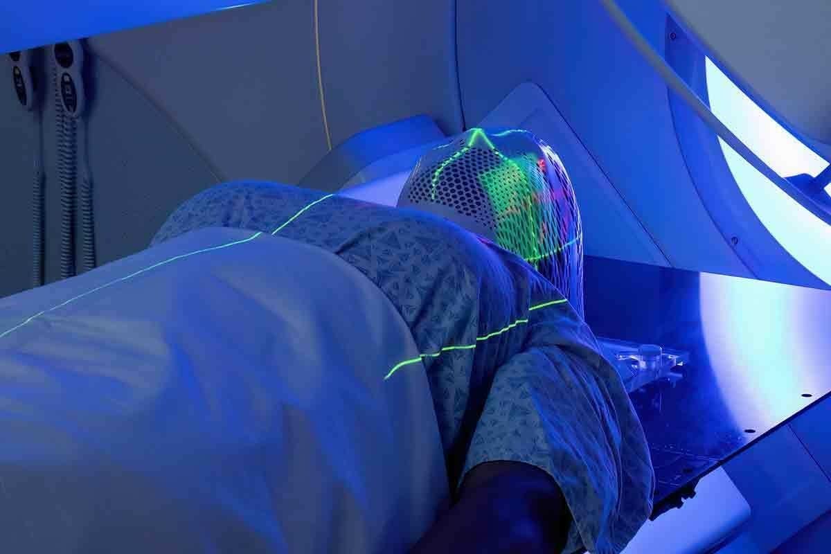
MRI scans use strong magnets and radio waves to see inside the body. First, the patient lies in a big, cylindrical magnet. This magnet aligns hydrogen nuclei in the body.
Then, radio waves disturb these nuclei, causing them to send signals. These signals are caught by the MRI machine. It uses them to make detailed images.
How Magnets and Radio Waves Create Images
The MRI works by nuclear magnetic resonance. Radio waves make hydrogen nuclei resonate, sending signals. These signals show what kind of tissue they are in.
The MRI machine’s computer turns these signals into images. This shows the body’s soft tissues in great detail. MRI scans are good because they can see different soft tissues well. They also don’t use harmful radiation, making them safer for some patients.
The MRI Scanning Experience
The MRI process involves lying in a big, enclosed tube. It’s usually painless but can cause claustrophobia. Some places offer open MRI machines or sedation to help.
Patients must stay very quiet and not move during the scan. This helps get clear images. MRI scans can last from 15 to 90 minutes, depending on what’s being scanned.
Knowing how MRI scans work helps us see their importance in medicine. They are different from PET scans, which show metabolic activity. MRI scans give detailed images of the body’s structure. Together, MRI and PET scans help doctors diagnose and plan treatments for many conditions, including cancer.
The Fundamental Difference in PET Scan and MRI Technology
It’s important to know how PET and MRI scans work to understand their use in cancer diagnosis. These scans use different technologies to give us different kinds of information.
Functional vs. Structural Imaging
PET scans and MRI scans are different in what they show. PET scans look at how cells in the body work. They find areas where cells are very active, which might mean cancer. MRI scans, on the other hand, show the body’s structure in detail. They help find problems with how things are put together.
“Understanding cancer is not just about the tumor,” says Dr. John Smith, a top oncologist. “It’s about how it acts. PET scans show how tumors work, while MRI scans show what they look like.”
Radiation Exposure Comparison
PET scans use a radioactive tracer, which means they expose patients to some radiation. MRI scans, though, don’t use radiation. They use strong magnets and radio waves to make pictures.
This is key for people who need many scans. MRI scans are safer for long-term use because they don’t use radiation. But, PET scans are safe when used correctly.
Image Resolution and Detail Capabilities
Both PET and MRI scans are good at showing different things. MRI scans are great at showing soft tissues in high detail. PET scans, while not as detailed, tell us about how tissues work.
In short, whether to use a PET or an MRI scan depends on what the patient needs. Knowing how these scans work helps us see how they help in fighting cancer.
PET Scans in Cancer Detection and Staging
PET scans have changed how doctors find cancer. They help spot cancer cells that are active. This is key to catching cancer early and tracking how it grows.
Identifying Metabolically Active Cancer Cells
PET scans use special tracers to find cancer cells. These cells are active and use a lot of energy. This functional imaging technique can find cancer before it grows big.
For example, PET scans are great for finding lymphoma. They show where cancer cells are in the body. This helps doctors plan the best treatment.
Effectiveness in Early Cancer Detection
Finding cancer early is very important. PET scans are great at this. They can see changes in cells before they become tumors. This helps doctors treat cancer when it’s easier to cure.
Research shows that PET scans can find lung cancer early. This can lead to better treatment results.
Monitoring Cancer Spread Throughout the Body
PET scans are also good at seeing how cancer spreads. They can check the whole body in one go. This is helpful for cancers that spread a lot, like melanoma or breast cancer.
They give doctors a clear picture of how far cancer has spread. This is key for planning treatment.
Limitations of PET in Cancer Diagnosis
Even though PET scans are powerful, they have some downsides. They can’t always tell the difference between cancer and inflammation. Both can show up as active on a scan.
Also, PET scans might not always show small tumors clearly. For more on PET scans vs MRI,
| Aspect | PET Scan | MRI |
| Primary Use | Detecting metabolically active cancer cells | Visualizing tumor boundaries and local invasion |
| Imaging Basis | Radioactive tracers and metabolic activity | Magnetic fields and radio waves |
| Strengths | Early detection, whole-body assessment | High-resolution images, soft tissue contrast |
| Limitations | May not distinguish between cancer and inflammation | Limited functional information |
MRI’s Role in Cancer Diagnosis and Characterization
MRI scans are key in cancer diagnosis. They give detailed images that doctors use to understand the disease’s extent. MRI’s ability to show soft tissues clearly is a big plus in checking tumors and their size.
Visualizing Tumor Boundaries and Local Invasion
MRI scans show soft tissues in detail. This helps doctors see tumor boundaries and local invasion. It’s vital for planning surgeries or radiation therapy.
Soft Tissue Contrast Advantages
MRI scans have high soft tissue contrast. This means they can tell different tissues apart well. It’s great for spotting tumors and seeing how they relate to nearby tissues.
Specialized MRI Techniques for Cancer Assessment
There are special MRI techniques for cancer, like diffusion-weighted imaging and dynamic contrast-enhanced MRI. These methods give more information on tumor behavior and characteristics.
Limitations of MRI in Cancer Diagnosis
Even though MRI is powerful in cancer diagnosis, it has its limits. For example, it might not catch distant metastases as well as PET scans. It also doesn’t show tumor metabolic activity as clearly.
Comparing Effectiveness: PET vs. MRI for Different Cancer Types
PET and MRI scans work differently for various cancers. It’s key to know how they compare. The right scan can make a big difference in finding and treating cancer.
Brain Tumors and Neurological Cancers
MRI is the top choice for brain tumors and cancers because it shows soft tissues well. It gives clear images of the brain and spinal cord. This helps doctors see how far a tumor has spread.
PET scans, like those with 18F-FDG, check how active brain tumors are. They help tell if a tumor is new or old. But the brain’s high sugar levels can make PET scans less clear.
| Cancer Type | PET Scan Effectiveness | MRI Scan Effectiveness |
| Brain Tumors | Assesses metabolic activity | Detailed soft tissue imaging |
| Neurological Cancers | Limited by background metabolism | High resolution for tumor extent |
Breast Cancer Detection and Staging
MRI is great for finding breast cancer, even in dense breasts. It’s also good for seeing how far cancer has spread and how well it responds to treatment.
“MRI is very useful for checking how far breast cancer has spread and how well it’s responding to treatment,” says Dr. Jane Smith, a breast cancer expert.
PET scans help stage breast cancer by finding cancer in other parts of the body. But, they’re not as good as MRI for finding cancer in the breast itself.
Abdominal and Pelvic Cancers
Both PET and MRI are useful for abdominal and pelvic cancers. MRI is great for looking at liver lesions and how far tumors have spread in the pelvis. PET scans are better for finding cancer in other parts of the body and checking how active the tumor is.
A study showed MRI is better for seeing the body’s anatomy. But, PET is more accurate in finding cancer in lymph nodes.
Bone and Soft Tissue Malignancies
MRI is best for looking at bone and soft tissue tumors because it shows soft tissues well. PET scans are good for finding cancer in other parts of the body and checking how active the tumor is.
Using both PET and MRI together helps get a full picture of bone and soft tissue sarcomas. This helps doctors plan the best treatment.
In summary, choosing between PET and MRI scans depends on the cancer type and other factors. Knowing what each scan can do is key to the best care for patients.
Combined Approaches: PET/MRI Hybrid Imaging
PET/MRI hybrid imaging combines PET scans’ functional data with MRI’s detailed anatomy. This powerful tool is key in oncology. It uses both PET and MRI strengths for a deeper cancer understanding.
The Evolution of Multimodal Imaging
PET/MRI hybrid imaging is a big step forward in medical imaging. It merges PET and MRI to give us both functional and anatomical data at once. This makes diagnosis and treatment planning more accurate.
This change in imaging is changing oncology. It allows for more precise and tailored care.
Key advancements in PET/MRI technology include:
- Improved image registration and fusion
- Enhanced diagnostic confidence through complementary information
- Potential reduction in overall scanning time and radiation exposure
Clinical Benefits of Simultaneous Functional and Anatomical Data
Getting PET and MRI data at the same time has many benefits. It helps us better understand tumor metabolism and anatomy. This is key for staging and planning treatments.
The clinical benefits include:
- Comprehensive evaluation of tumor characteristics
- Improved accuracy in cancer staging and restaging
- Enhanced monitoring of treatment response
Current Applications in Oncology
PET/MRI hybrid imaging is used in many oncology areas. It’s great for complex cases needing both functional and anatomical info. It’s used for brain tumors, prostate cancer, and other cancers needing precise location and details.
As we keep exploring PET/MRI, we find new ways to better diagnose and treat cancer. This technology is set to be a big part of oncology’s future. It offers a more complete and integrated way to care for patients.
Patient Experience: What to Expect During Each Scan
Knowing what to expect during a PET or MRI scan can help reduce anxiety for those facing cancer. Both scans are important in cancer care , but differ in how they work, what you need to do before, and how you feel during the scan.
Preparation Requirements for PET vs. MRI
For a PET scan, you’ll need to fast for a few hours beforehand and avoid hard exercise. You might also need to skip certain medicines or foods to get accurate results. On the other hand, getting ready for an MRI scan means removing metal items like jewelry and glasses. If you have metal implants, tell your doctor first.
Both scans ask you to wear comfy, loose clothes. But what you need to do can change based on where you’re being scanned and the facility’s rules.
Duration and Comfort Considerations
A PET scan usually lasts about 30 minutes to an hour. You’ll need to stay very quiet and not move. It’s not painful, but staying in one spot for so long might be uncomfortable.
An MRI scan can take 15 to 90 minutes, depending on the scan’s details. MRI scans can be loud and might make you feel trapped because of the scanner’s shape. If you really can’t stand being in small spaces, there are open MRI machines.
Potential Side Effects and Risks
Both PET and MRI scans are mostly safe, but there are some risks. PET scans use a tiny bit of radiation. Rarely, some people might have an allergic reaction to the tracer.
MRI scans don’t use radiation but can be risky for people with metal implants. Some might also react to the dye used in MRI scans.
Knowing these differences can help you prepare better. This can make the diagnostic process less scary and more comfortable for you.
Cost and Accessibility Factors
It’s important to know about the costs and how to get PET and MRI scans. These factors can affect how often they are used in cancer care.
Insurance Coverage for Cancer Imaging
In the United States, insurance is key to getting PET and MRI scans. Most plans cover these scans for cancer, but how much can change. Patients should check their insurance to know what’s covered and what they’ll pay out of pocket.
Insurance policies can also affect when you can get these scans. For example, PET scans are often used to check how far cancer has spread. MRI scans are used to see tumors in more detail.
Availability of Technology in Different Healthcare Settings
Where you can get PET and MRI scans varies. Big hospitals and cancer centers usually have both. But smaller hospitals or those in rural areas might not have them.
This difference can affect how cancer is diagnosed and treated. If you can’t get these scans close to home, you might have to travel. This could slow down your treatment.
Cost Comparison Between Modalities
It’s hard to compare the costs of PET and MRI scans because many things can change the price. PET scans are usually pricier because of the radioactive tracer.
- PET scans: Prices can be from $1,000 to $3,000 or more, depending on the tracer and where you go.
- MRI scans: Prices are usually between $500 to 000 or more, based on the contrast agents and body part scanned.
These prices are just estimates and can change based on many factors. Knowing these costs helps doctors and patients make better choices about which scan to use.
How Oncologists Choose Between PET and MRI Scans
Oncologists have to think carefully when choosing between PET and MRI scans for cancer diagnosis. They consider the cancer type, its stage, and the patient’s health. This helps them make the best choice for each patient.
Decision-Making Factors in Diagnostic Planning
Several factors guide oncologists in their decisions They look at whether the scan needs to show function or anatomy. They also consider how sensitive and specific the scan must be. Plus, they think about the patient’s medical history and current health.
PET scans are great for finding cancer cells that are active. This makes them perfect for seeing how cancer has spread. On the other hand, MRI scans show detailed anatomy. They are often used to see tumor boundaries and how cancer invades tissues.
The type of cancer also plays a role. For brain tumors and neurological cancers, MRI is best. It gives clear images of soft tissues.
Sequential vs. Simultaneous Use of Both Modalities
In some cases, oncologists might use both PET and MRI scans. They might do them one after the other or at the same time. Hybrid PET/MRI systems allow for both functional and anatomical information in one scan. This can improve accuracy and make things easier for patients.
Using PET and MRI scans together can offer valuable insights. For example, a PET scan might first show cancer spread. Then, an MRI scan can give detailed images of the tumor and nearby tissues.
Personalized Imaging Approaches Based on Cancer Type
The choice between PET and MRI scans is often tailored to the patient’s cancer. Different cancers need different imaging approaches. For instance, MRI is often used in breast cancer for its ability to find tumors in dense tissue.
Oncologists use their knowledge of each imaging modality to make the best choices. They aim to provide accurate and relevant information for patient care. Whether it’s PET, MRI, or both, the goal is to help patients the most.
Conclusion: The Complementary Roles of PET and MRI in Cancer Care
We’ve looked at how PET and MRI scans help in cancer care. PET scans are great at finding cancer cells that are active. MRI scans, on the other hand, show detailed images of soft tissues.
Using both PET and MRI scans together helps doctors make better diagnoses. This combo gives a clearer picture of the cancer. It helps in planning treatments that fit each patient’s needs.
The debate between PET and MRI scans isn’t about picking one over the other. It’s about choosing the right tool for each patient. As we keep improving in cancer care, PET and MRI scans will keep playing key roles.
FAQ
What is the main difference between a PET scan and an MRI scan?
PET scans look at how cells work. MRI scans show detailed images of soft tissues. They are used together to help doctors diagnose.
Is a PET scan or an MRI scan better for cancer diagnosis?
It’s not about which one is better. The choice depends on the cancer type, where it is, and what the doctor needs to know.
What is the difference between a PET scan and an MRI in terms of radiation exposure?
PET scans use a small amount of radiation from a tracer. MRI scans don’t use radiation.
How do PET and MRI scans compare in terms of image resolution?
MRI scans show soft tissues very clearly. PET scans show how cells work, which helps find cancer.
Can PET and MRI scans be used together for cancer diagnosis?
Yes, using both PET and MRI scans together can give doctors a full picture of tumors.
What are the preparation requirements for PET and MRI scans?
For PET scans, you need to fast and get a tracer injection. MRI scans might ask you to remove metal and use a contrast agent.
How long do PET and MRI scans typically take?
Scans can last from 30 minutes to several hours. It depends on the scan’s complexity and needs.
Are there any side effects or risks associated with PET and MRI scans?
PET scans have radiation risks. MRI scans are safe but might cause claustrophobia or reactions to contrast agents.
How do insurance coverage and costs compare for PET and MRI scans?
Costs and coverage vary by insurance and healthcare setting. PET scans are often pricier because of the tracer.
What factors influence the decision to use PET or MRI scans for cancer diagnosis?
The choice depends on the cancer type, stage, and what information is needed. The patient’s health also matters.
Can PET/MRI hybrid imaging improve diagnostic accuracy for cancer?
Yes, combining PET and MRI scans can improve accuracy. It helps in planning better treatments.
Are PET and MRI scans available in all healthcare settings?
No, not all places have PET and MRI scans. PET scans are less common because of the tracer and equipment needs.
How do oncologists decide between using PET or MRI scans for cancer patients?
Oncologists look at the cancer’s details, what information is needed, and the benefits and limits of each scan.
References
- Sharma, A. (2019). An introduction to basic scientific medical writing. PMC. https://pmc.ncbi.nlm.nih.gov/articles/PMC6561072/


