Last Updated on November 27, 2025 by Bilal Hasdemir

When a PET scan shows activity, people often worry it’s cancer. At Liv Hospital, we use global standards and advanced imaging. We help patients understand what PET scan results mean for benign and malignant tumors.Learn how do benign tumors light up on PET scan and what it really means for diagnosis and cancer detection accuracy.
A PET scan finds areas where cells are very active. Cancer cells often show this activity. But infections, inflammation, and regenerating tissues can also exhibit heightened activity.
Benign tumors, though not cancerous, can sometimes show up on PET scans. This is because of different reasons. It causes concern and needs more investigation.
Key Takeaways
- PET scans identify highly active cells in the body.
- Benign tumors can show increased uptake on PET scans.
- Various factors can cause benign tumors to “light up.”
- Further investigation is often necessary after a PET scan.
- Liv Hospital provides advanced imaging and ethical care.
Understanding PET Scan Technology

PET imaging combines radiotracers and metabolic processes in a complex way. It’s a key tool in medical diagnostics, mainly in oncology.
How PET Scans Work
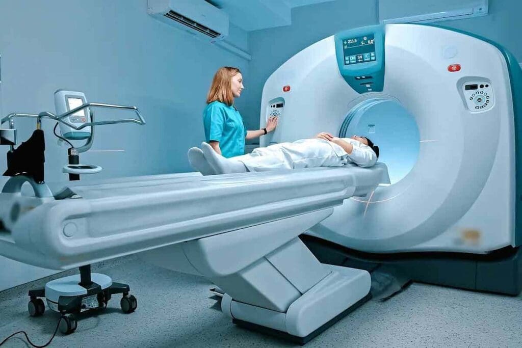
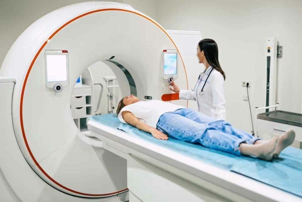
PET scans use a mildly radioactive liquid called a radiotracer. This liquid shows where cells are more active than usual. We inject it into the body, and the PET scanner detects the radiation.
The tracer is usually Fluorodeoxyglucose (FDG), a glucose molecule with a radioactive atom. Cells with high metabolic rates absorb more FDG, making them visible on the scan.
Radiotracer Uptake and Metabolic Activity
The amount of radiotracer a cell takes up shows its metabolic activity. Cells in growing tumors take up more, making them show up on scans. This helps us find abnormal cell activity, which can mean cancer.
But increased radiotracer uptake doesn’t always mean cancer. Inflammatory processes and certain benign conditions can also cause it, leading to false positives.
The Role of FDG in PET Imaging
FDG is the main radiotracer used in PET imaging. It acts like glucose, helping us see how cells use glucose. This is key because many tumors use more glucose than normal tissues.
FDG’s ability to find metabolically active cells makes it very useful. But one must remember its limitations. Some non-cancerous conditions can also show high glucose use.
Do Benign Tumors Light Up on PET Scan?
Benign tumors can show up on PET scans, making them look like cancer. This can cause false alarms, leading to worry and making diagnosis harder.
We must understand why this happens to correctly read PET scan results.
Prevalence of Radiotracer Uptake in Benign Lesions
More than 25 percent of benign lesions show up on PET scans. This shows how important it is to think about benign conditions when looking at PET scans.
Benign lesions can take up radiotracers because of inflammation, infection, or metabolic activity.
Mechanisms Behind False Positive Results
False positives on PET scans can happen for several reasons. Inflammatory processes are a big one, as they increase glucose metabolism and radiotracer uptake.
Other reasons include granulomatous diseases, infectious processes, and post-surgical changes. Knowing these reasons helps us understand PET scan results better.
Common Benign Conditions That Show PET Activity
Many benign conditions can show up on PET scans, which can lead to wrong diagnoses. These include:
- Fibroids: Uterine fibroids can show a lot of radiotracer uptake.
- Adenomas: Certain adenomas, like those in the thyroid or adrenal glands, can be PET-positive.
- Cysts: Some cysts, with inflammatory parts, may show more radiotracer uptake.
- Inflammatory lesions: Conditions like sarcoidosis or other granulomatous diseases can cause PET activity.
Knowing about these benign conditions helps avoid wrong diagnoses and ensures the right care for patients.
What Makes Tumors “Light Up” on PET Scans
Tumors “light up” on PET scans because of their high metabolic activity and glucose use. Both benign and malignant tumors have more activity than normal tissues.
Metabolic Activity and Glucose Consumption
Cancer cells grow fast and need lots of energy. They use more glucose than regular cells. This is why PET scans use Fluorodeoxyglucose (FDG) to spot them.
FDG is a glucose analog that cells take up based on their glucose use. Cancer cells take up FDG, making them glow on PET scans because of their high metabolic rate.
Inflammatory Processes and Radiotracer Uptake
Not just cancer cells can show up on PET scans. Inflammation can also cause increased radiotracer uptake. Inflammation is a natural response to injury or infection, with increased blood flow and metabolic activity, leading to more FDG uptake.
This is why some non-cancerous conditions, like infections or inflammatory diseases, can look like cancer on PET scans.
Why Both Benign and Malignant Lesions Can Glow
Both benign and malignant lesions can show up on PET scans because of their metabolic activity or inflammation. The key factor is not just the presence of a tumor, but its metabolic characteristics and the context in which it appears. Understanding these factors is key to accurately reading PET scan results and telling benign from malignant conditions.
By looking at metabolic activity, glucose use, and inflammation, we can understand why some tumors show up on PET scans. This knowledge helps healthcare providers make better decisions for patient care and treatment plans.
What Cancer Looks Like on PET Scans
Understanding cancer on PET scans is key to accurate diagnosis and treatment. PET scans show how tumors work, helping doctors make better decisions for patients.
Typical Appearance of Malignant Tumors
Malignant tumors show up as bright spots on PET scans. This is because cancer cells use more glucose than normal cells. The scan uses a glucose-like substance to spot these active areas.
The brightness of these spots can show how aggressive the cancer is. For example, fast-growing tumors show up brighter.
Variations by Cancer Type and Location
Cancer looks different on PET scans based on its type and where it is. Some cancers may not show up well because they don’t use much glucose. This makes them harder to see.
Where the tumor is also matters. Tumors in busy areas like the brain can be harder to spot because of the background activity.
Standardized Uptake Values (SUV) in Cancer Diagnosis
Standardized Uptake Values (SUV) are important for reading PET scans. SUV shows how much glucose a tumor uses compared to the body. It helps doctors see how active the tumor is and if treatment is working.
A higher SUV means the tumor might be more aggressive. But it’s important to look at all the information together. SUV values help doctors compare tumors over time, guiding treatment plans.
Differentiating Between Benign and Malignant PET Findings
It’s key for doctors to tell apart benign and malignant PET scan results. This helps them make the best choices for their patients. Accurate PET scan readings are vital for spotting and treating cancer right.
Intensity Patterns and Distribution of Activity
Doctors look at how active the radiotracer is and where it goes in the body. Malignant tumors usually take up more radiotracer because they’re more active than benign ones.
- Intensity: Higher SUV values often mean it’s cancer, but not always.
- Distribution: The way the radiotracer spreads can give clues. For example, even spread might be benign, while uneven spread could be cancer.
Morphological Characteristics of Integrated CT
Using PET/CT scans together gives doctors a big advantage. They get metabolic info from PET and body details from CT. This helps them see the difference between benign and malignant lesions better.
For instance, some benign lesions look different on CT scans. They might have calcifications or special patterns of enhancement. These signs can help doctors figure out what they are.
- Size and Shape: Big, irregular shapes often mean cancer.
- Border Characteristics: Clear borders might mean it’s benign. But if the borders are messy or spread out, it could be cancer.
- Associated Findings: Seeing lymph nodes or metastases on CT can point to cancer.
Temporal Changes in Follow-up Scans
Follow-up PET/CT scans are very important. They show how lesions change over time. Benign lesions usually don’t grow or change much.
- Stability: If a lesion stays the same or gets smaller, it’s likely benign.
- Growth: If it gets bigger or more active, it might be cancer.
By looking at intensity, shape, and how things change over time, doctors can better tell benign from malignant PET findings. This helps improve patient care.
Limitations of PET Scan in Cancer Detection
PET scans are a key tool in finding and tracking cancer. Yet, they have some limits that doctors must keep in mind. These scans have changed how we diagnose and treat cancer. But, like any tool, they have their own set of challenges.
Types of Cancers Poorly Visualized on PET
Not every cancer shows up well on PET scans. Low-grade tumors or those with low metabolic activity might not be seen clearly. For example, some prostate cancers and certain neuroendocrine tumors are hard to spot because they don’t use much glucose.
Factors Affecting Scan Sensitivity
Many things can change how well a PET scan works. The type and size of the tumor, the patient’s blood glucose levels, and the specific radiotracer used all play a role. High blood sugar, for instance, can make it harder for the scan to find cancer cells.
Why Some Cancers Don’t “Light Up”
Some cancers don’t show up on PET scans because of their nature. They might be small, low metabolic rate, or be in areas with a lot of background activity. Knowing these reasons helps doctors understand PET scan results better and make the right treatment plans.
By understanding the limits of PET scans, doctors can use them more wisely. They can pair PET scans with other tests and patient information for better care.
Integrated PET/CT Imaging: Improving Diagnostic Accuracy
PET and CT imaging together have changed oncology. They give both metabolic and anatomical details. This helps doctors make better choices for patient care.
Benefits of Combined Anatomical and Metabolic Imaging
Using PET/CT together has many advantages. It mixes metabolic activity from PET with CT’s detailed anatomy. This helps spot and understand abnormalities better, even in hard-to-see areas.
Key benefits include:
- Improved lesion localization and characterization
- Enhanced detection of metabolically active tumors
- Better differentiation between benign and malignant lesions
- More accurate staging of cancers
How CT Helps Characterize PET-Positive Lesions
CT is key in figuring out PET-positive lesions. PET shows metabolic activity, while CT shows anatomy. Together, they help know what a lesion is.
For example, a PET-positive lesion might look benign on CT. But if it’s fatty, it’s likely not cancer. On the other hand, a PET-positive lesion with odd CT features might need more tests or a biopsy.
| Lesion Characteristics | PET Findings | CT Findings | Interpretation |
| Metabolically Active | High FDG uptake | Suspicious morphology | Potential malignancy |
| Metabolically Inactive | Low FDG uptake | Benign features | Likely benign |
| Metabolically Active | High FDG uptake | Benign features | Further evaluation needed |
Case Examples of PET/CT Correlation
Here are some examples of how PET/CT helps:
Case 1: A lung cancer patient has a PET-positive adrenal gland lesion. The CT shows it’s a benign adenoma. So, the PET/CT says it’s probably not cancer.
Case 2: A lymphoma patient has a PET-positive lymph node in the neck. The CT shows it’s big and looks odd. The PET/CT confirms it’s an active lymphoma.
These examples show how PET/CT improves diagnosis. It combines metabolic and anatomical info for better patient care.
Growth Patterns: Do Benign Tumors Grow on Follow-up Imaging?
It’s key to know how benign tumors grow to manage patient care. We watch how these tumors change over time on imaging studies.
Growth Characteristics of Benign vs. Malignant Lesions
Benign tumors grow more slowly than cancerous ones. Their growth patterns tell us a lot about the tumor.
A study showed that benign tumors grow slowly over time. But cancerous tumors grow fast.
| Tumor Type | Typical Growth Rate | Characteristics |
| Benign | Slow | Gradual increase in size, often stable |
| Malignant | Rapid | Quick growth, potentially invasive |
Stability as a Predictor of Benignity
Stable or slow growth on follow-up scans often means the tumor is benign. Tumors that stay the same size for a long time are likely benign.
“The stability of a tumor on serial imaging is a reassuring sign of its benign nature.”
A medical expert, an Oncologist
When Growth Doesn’t Equal Malignancy
Growth alone doesn’t mean a tumor is cancerous. Some benign tumors grow due to hormones or inflammation.
So, when we see growth on scans, we must look at the whole picture. This includes the patient’s history and other test results.
Conclusion: Understanding Your PET Scan Results
Understanding PET scan results is key to making good health choices. We’ve looked at how PET scans work and the role of radiotracers. We’ve also seen how different conditions show up on scans.
When you get your PET scan results, it’s important to think about what they mean. This includes knowing if a PET scan will show cancer. It’s also vital to know how to tell if the results are normal or not.
PET scan results can be tricky, but knowing what to look for helps. We’ve talked about how some benign tumors can show up on scans, leading to false positives. Malignant tumors usually show up differently. Understanding these implications is important for your health.
By understanding your PET scan results, you can make better choices about your health. We’re here to help you understand your results. This way, you can make informed decisions about your health.
FAQ
Do benign tumors light up on PET scans?
Yes, benign tumors can sometimes show up as active on PET scans. This can cause concern and may need more investigation.
What does it mean when a PET scan lights up?
When a PET scan “lights up,” it means there’s high activity in certain areas. This can be due to cancer, inflammation, or infection.
How does cancer appear on a PET scan?
Cancer shows up as areas with more radiotracer uptake on PET scans. The intensity and pattern vary based on the cancer type and location.
What is the role of FDG in PET imaging?
FDG is a radiotracer used in PET scans. It helps show areas with high glucose consumption, often seen in cancer and other active processes.
Can PET scans detect all types of cancer?
No, PET scans don’t work equally well for all cancers. Some cancers may not show up or are poorly visualized on PET scans.
How do doctors differentiate between benign and malignant lesions on PET scans?
Doctors look at intensity patterns, morphological characteristics on CT, and changes over time. These help tell if a lesion is benign or malignant.
What are the limitations of PET scans in cancer detection?
PET scans have limitations. They can have false positives and negatives, and sensitivity can vary. This can affect their accuracy in finding cancer.
How does integrated PET/CT imaging improve diagnostic accuracy?
Integrated PET/CT imaging combines metabolic information from PET with anatomical information from CT. This gives a better understanding of lesions, improving diagnostic accuracy.
Do benign tumors grow on follow-up imaging?
Yes, benign tumors can grow on follow-up imaging. But their growth pattern and characteristics can help tell them apart from malignant lesions.
What does a normal PET scan result mean?
A normal PET scan result means no significant abnormal metabolic activity is detected. But it doesn’t rule out cancer or other conditions entirely.
What does a PET scan show for cancer?
A PET scan can show areas of increased metabolic activity that might indicate cancer. But further evaluation is usually needed to confirm the diagnosis.
References
- Cook, G. J. R., & Fogelman, I. (2011). Positron emission tomography (PET) for benign and malignant breast lesions. Seminars in Nuclear Medicine, 41(1), 18-30. https://pmc.ncbi.nlm.nih.gov/articles/PMC3021752/


