Last Updated on November 27, 2025 by Bilal Hasdemir

When we diagnose lymphoma, we need to know how to spot cancerous lymph nodes. We use PET scans to see if swollen lymph nodes have lymphoma or to find small cancer spots.
PET scans are key in finding areas with high activity, which is a sign of cancerous cells. Cancerous lymph nodes light up on a lymphoma PET scan. This means they use more glucose than healthy cells. Do cancerous lymph nodes show up on pet scan? Learn detection in lymphoma patients.
Key Takeaways
- PET scans help identify cancerous lymph nodes by detecting high metabolic activity.
- Cancerous lymph nodes show increased uptake of the tracer on a PET scan.
- PET scans aid in diagnosis and staging of lymphoma.
- The level of tracer uptake can indicate the aggressiveness of lymphoma.
- PET scans are useful for monitoring response to treatment.
Understanding Lymphoma and the Role of Lymph Nodes

Lymphoma is a cancer that starts in the lymphocytes, a type of white blood cell. These cells are key to our immune system. To understand lymphoma, we need to know how the lymphatic system works and how cancer affects it.
Types of Lymphoma: Hodgkin’s vs. Non-Hodgkin’s
There are two main types of lymphoma: Hodgkin’s and non-Hodgkin’s. The main difference is in the type of lymphocytes affected. Hodgkin’s lymphoma has Reed-Sternberg cells, which are not found in non-Hodgkin’s lymphoma.
| Characteristics | Hodgkin’s Lymphoma | Non-Hodgkin’s Lymphoma |
| Presence of Reed-Sternberg Cells | Present | Absent |
| Lymphocyte Type Affected | B lymphocytes | B or T lymphocytes |
| Pattern of Spread | Predictable, contiguous spread | Variable, can be widespread |
| Age of Onset | Bimodal peaks (20s and 50s+) | More common in older adults |
Function of Healthy Lymph Nodes
Healthy lymph nodes are vital for our immune system. They filter lymph fluid, trap pathogens, and house lymphocytes. These cells help fight infections.
How Lymphoma Affects the Lymphatic System
Lymphoma disrupts the lymphatic system by causing lymphocytes to grow uncontrollably. This leads to swollen lymph nodes, fever, and other symptoms. Understanding this is key to seeing why PET scans are important in detecting lymphoma.
Knowing the basics of lymphoma and its effects on the lymphatic system is important. It helps us understand the role of accurate diagnosis and treatment. PET scans are key in identifying lymphoma and checking if treatment is working.
The Basics of PET Scan Technology

PET scans are a key tool in medicine. They use radioactive tracers to see how the body works. This is very important for finding and treating lymphoma.
What is a PET Scan?
A PET scan is a test that uses a special sugar molecule to find diseases. It spots cancer because cancer cells use more sugar than normal cells. This makes PET scans great for finding active areas in the body.
How PET Scans Differ from CT and MRI
PET scans show how tissues work, not just what they look like. CT scans use X-rays to show body parts. MRI scans use magnetic fields to show soft tissues. PET scans find areas that use a lot of sugar, which can mean cancer.
When PET scans are used with CT or MRI, they give even more information. This helps doctors make better diagnoses.
The Role of FDG (Fluorodeoxyglucose) Tracers
FDG is the main tracer used in PET scans for cancer. It’s a sugar molecule with a radioactive atom. When it’s injected, it goes to areas that use a lot of sugar, like cancer. The PET scan then shows where this sugar is.
| Imaging Modality | Primary Information Provided | Key Features |
| PET Scan | Metabolic Activity | Detects cancerous tissues based on glucose uptake |
| CT Scan | Anatomical Details | Provides detailed X-ray images of internal structures |
| MRI Scan | Soft Tissue Details | Uses magnetic fields and radio waves for imaging |
Knowing how PET scans work is key to understanding their role in fighting lymphoma. They help doctors make better choices for patient care by showing how the body works.
Do Cancerous Lymph Nodes Show Up on PET Scan?
PET scans are key in finding cancerous lymph nodes. They use a special tracer to see how active cells are in the body. This helps doctors spot cancer in lymph nodes.
Cancerous lymph nodes soak up more of this tracer than healthy cells. This makes PET scans great for finding cancer in these nodes.
The “Lighting Up” Phenomenon Explained
The term “lighting up” means cancerous lymph nodes show up bright on a PET scan. This happens because they take in more of the tracer. It’s a big help in finding out how far cancer has spread.
The brightness of these areas can tell doctors how fast the cancer is growing. Areas that light up a lot are usually more aggressive.
SUV (Standardized Uptake Value) Measurements
SUV measurements show how much tracer is taken in by lymph nodes. A higher number means more tracer, which often means cancer. Doctors use these numbers to see how serious the cancer is and if treatment is working.
By looking at SUV numbers before and after treatment, doctors can see if the treatment is effective. This helps them decide what to do next.
Visual Characteristics of Malignant Lymph Nodes
Malignant lymph nodes look different on a PET scan. They are much brighter than the rest of the tissue. Their size and shape can also hint at their nature, with bigger ones being more concerning.
Even though PET scans are very good, they’re not 100% accurate. Some infections or inflammation can also make lymph nodes light up. So, doctors need to understand the whole picture before making conclusions.
PET/CT Combination: Enhanced Diagnostic Capabilities
The mix of PET and CT scans has changed how we diagnose diseases, like lymphoma. It gives doctors a clearer picture of the disease.
Benefits of Integrated Imaging
PET/CT scans are better at finding and staging diseases. They combine CT’s body images with PET’s metabolic data. This makes diagnosing tumors more accurate.
Key advantages of PET/CT combination include:
- Enhanced diagnostic accuracy
- Improved staging capabilities
- Better assessment of treatment response
- More precise detection of residual disease
Digital PET/CT Technology Advancements
New digital PET/CT scanners are even better. They can spot smaller tumors and track metabolic changes more accurately.
A study in Frontiers in Nuclear Medicine shows how digital PET/CT has improved lymphoma diagnosis and staging.
Anatomical and Metabolic Information Integration
PET and CT scans together give a full view of lymphoma. They show both the body’s structure and metabolic activity. This helps doctors plan better treatments.
| Modality | Anatomical Information | Metabolic Information | Diagnostic Capability |
| CT Scan | Detailed structural information | Limited | Good for assessing tumor size and location |
| PET Scan | Limited | Detailed metabolic activity information | Excellent for assessing tumor activity and spread |
| PET/CT Combination | Detailed structural information | Detailed metabolic activity information | Excellent for complete lymphoma staging and assessment |
Sensitivity and Specificity of PET Scans for Lymphoma Detection
Knowing how well PET scans work is key for lymphoma patients. These scans are a big help in finding cancer in lymph nodes. They are good at spotting the disease.
Understanding Diagnostic Accuracy Metrics
Accuracy in diagnosis comes from sensitivity and specificity. Sensitivity is about finding people with the disease. Specificity is about finding those without it.
PET scans are very good at finding cancer. They help see how far the disease has spread. This is important for planning treatment.
Current Sensitivity Rates
Research shows PET scans are very good at finding lymphoma. They correctly identify about 96% of patients with the disease. Newer PET scanners are even better, helping doctors more.
Specificity Considerations and Limitations
PET scans are very sensitive but not always specific. Sometimes, they can show cancer when there isn’t any. This can happen if there’s inflammation or infection.
For patients with lymphoma stage 1 or lymphoma stage 4 cancer, PET scans are very useful. They help see how far the disease has spread. For those with non-Hodgkin’s lymphoma, they help track how the disease is changing.
Understanding PET scans helps doctors make better choices for patients. This ensures those with lymphoma get the right treatment.
The PET Scan Procedure for Lymphoma Patients
The PET scan is key in finding lymphoma. It shows how active lymph nodes are. Knowing the steps helps make the process smoother and more successful.
Patient Preparation Guidelines
Before your PET scan, there are steps to take. Fasting is needed for a while before the scan. This ensures the results are accurate. You might also need to stop certain medications or activities that could mess with the scan.
- Avoid eating or drinking anything except water for the specified fasting period.
- Inform your doctor about any medications you are currently taking.
- Remove any metal objects, such as jewelry or glasses, before the scan.
Stay hydrated and comfortable while preparing. Wear loose, comfortable clothing. Avoid caffeine and sugary drinks to keep your blood sugar steady.
What to Expect During the Scan
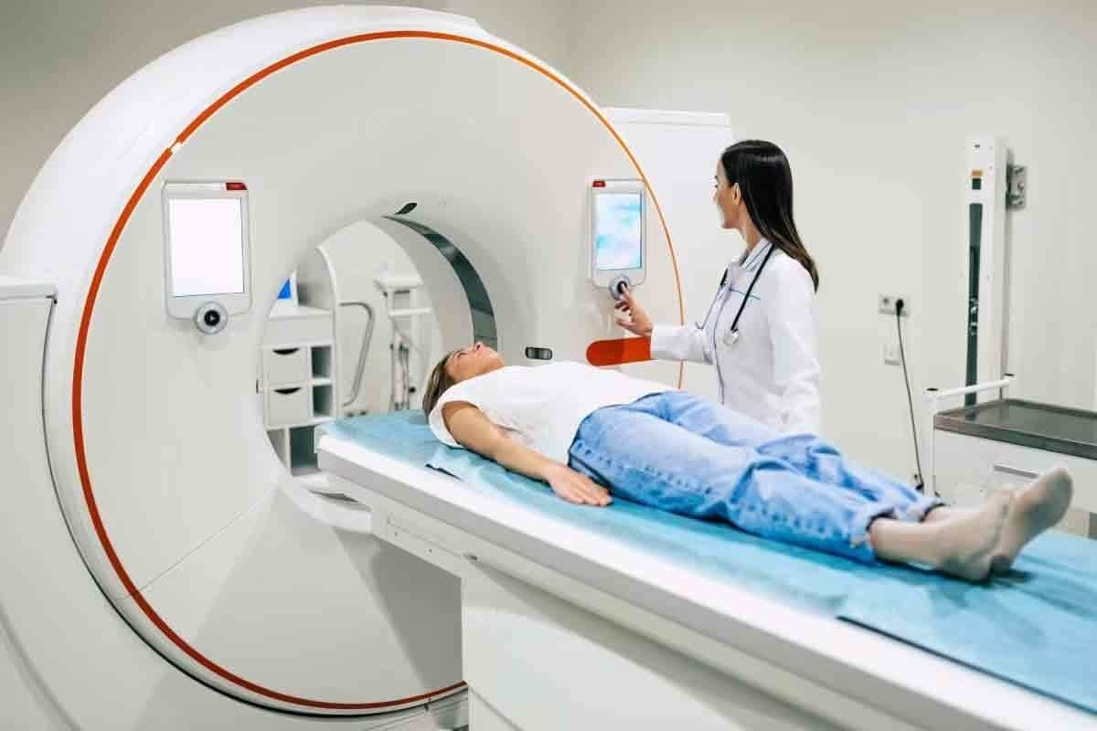
During the PET scan, you’ll get a small amount of FDG (Fluorodeoxyglucose). This tracer shows where cancerous lymph nodes are. The scan is done in a big, doughnut-shaped machine. It’s painless and non-invasive.
- You will lie on a comfortable table that slides into the PET scanner.
- The scan will capture images of your body, focusing on the areas where lymphoma is suspected.
- The entire process typically takes about 30 to 60 minutes, depending on the complexity of the scan.
Feeling a bit anxious is normal. Our medical team is there to support you. We know undergoing a PET scan is a big step in your diagnosis and treatment. We’re here to make sure you’re comfortable and safe.
Post-Scan Protocols and Safety
After the PET scan, you can usually go back to your normal activities. But, it’s important to follow any instructions from your doctor. This ensures your safety and the accuracy of the scan results.
- Drink plenty of water to help flush out the FDG tracer from your body.
- Follow any specific dietary recommendations provided by your healthcare team.
- Attend any scheduled follow-up appointments to discuss your scan results.
PET scans are generally safe, but there can be mild side effects. Our team is ready to monitor your health and address any concerns.
Knowing what to expect from the PET scan helps you feel more prepared. We’re here to support you every step of the way.
Interpreting Lymphoma PET Scan Results
Understanding PET scan results for lymphoma patients is key. PET scans are vital in finding cancer. They show how active cancer cells are.
Reading the Scan Images
PET scan images show how active lymph nodes and tissues are. Bright spots mean cancer cells are active. The Standardized Uptake Value (SUV) measures this activity.
Key aspects to consider when reading PET scan images include:
- The size and location of lymph nodes
- The intensity of FDG uptake
- The presence of any other areas of abnormal uptake
Deauville Criteria for Lymphoma Assessment
The Deauville criteria help assess lymphoma treatment response. It scores tumor uptake against liver and mediastinum uptake. Scores range from 1 to 5.
The Deauville criteria are as follows:
- No uptake
- Uptake ≤ mediastinum
- Uptake > mediastinum but ≤ liver
- Uptake moderately higher than liver
- Uptake markedly higher than liver and/or new lesions
How Oncologists Use PET Scan Information for Staging
Oncologists use PET scans to stage lymphoma. This is important for treatment planning. PET scans show how far the disease has spread.
The staging process involves:
- Assessing the number and location of involved lymph nodes
- Evaluating the presence of extranodal disease
- Determining the overall metabolic activity of the tumor
By combining PET scan results with other data, oncologists create a treatment plan. This plan is tailored to each patient’s needs.
When Lymph Nodes Light Up on PET Scan: Cancer vs. Other Causes
When we look at PET scan results, it’s key to know that lymph nodes can show up for reasons other than cancer. PET scans help see how active tissues are, like lymph nodes. They’re great for finding cancer, but other things can also make lymph nodes look active. Knowing these reasons helps doctors make the right diagnosis and treatment plan.
Inflammatory Conditions That Mimic Cancer
Inflammatory conditions can make lymph nodes look active on PET scans, just like cancer. For example, sarcoidosis can cause this. Autoimmune diseases and other inflammation can also make lymph nodes light up, making diagnosis tricky.
A study on PubMed Central shows that knowing how FDG uptake patterns work can help tell if it’s cancer or not.
Infection-Related Uptake Patterns
Infections can also make lymph nodes look active on PET scans. This can be due to bacterial, viral, or fungal infections. The way the uptake looks can hint at the cause. For example, a spread-out uptake might mean a body-wide infection, while a focused uptake could point to a specific area.
When Biopsy Is Necessary for Confirmation
PET scans give useful info on lymph node activity, but they’re not enough to be sure. Often, a biopsy is needed to confirm if active lymph nodes are cancerous. A biopsy lets doctors look at the tissue directly, giving a clear diagnosis.
Deciding to do a biopsy depends on many things. This includes the patient’s overall health, how the lymph nodes look on other scans, and the PET scan’s SUV measurements. A high SUV value might suggest cancer, but it’s not a sure sign.
| Cause | PET Scan Appearance | Diagnostic Considerations |
| Cancer | High FDG uptake, often focal | Biopsy usually necessary for confirmation |
| Inflammatory Conditions | Variable uptake, can be diffuse | Clinical correlation, possibly biopsy |
| Infections | Moderate to high uptake, variable pattern | Clinical context, potentially biopsy or follow-up scans |
In summary, PET scans are great for finding active lymph nodes, but there are many reasons why they might show up. It’s important to use all the information we have, including clinical judgment, imaging, and sometimes biopsy, to make an accurate diagnosis and plan the right treatment.
PET Scans in Non-Hodgkin’s Lymphoma Diagnosis and Staging
PET scans are key in diagnosing and staging Non-Hodgkin’s Lymphoma (NHL). They give detailed insights into how far and severe the disease is. This info is vital for making effective treatment plans.
Specific Features of NHL on PET Scans
Non-Hodgkin’s Lymphoma shows unique features on PET scans. It has high metabolic activity, which makes it stand out. This activity helps doctors tell NHL apart from other diseases.
We use SUV (Standardized Uptake Value) to measure how much FDG tracer is taken up. Higher SUV values mean more aggressive disease. This helps doctors understand how severe NHL is and decide on treatment.
Aggressive vs. Indolent NHL Appearance
PET scans show different things for aggressive and indolent NHL. Aggressive NHL has higher FDG uptake and spreads more. Indolent NHL has lower activity and spreads less.
Knowing these differences is key for accurate diagnosis and treatment. Aggressive NHL usually needs more intense treatment.
Extranodal Disease Detection
PET scans are great for finding NHL in places outside the lymph nodes. This includes the spleen, liver, and bone marrow. Finding these sites is important for accurate staging and treatment planning.
| Feature | Aggressive NHL | Indolent NHL |
| FDG Uptake | High | Variable, often lower |
| Disease Spread | More extensive | Less widespread |
| Treatment Approach | Intensive therapy | Watchful waiting or less intensive |
PET scans give detailed info on NHL’s extent and activity. This helps doctors create treatment plans that fit each patient’s needs.
PET Scans for Treatment Response Assessment
PET scans are key in checking how well lymphoma treatment works. They show detailed images of the body’s metabolic activity. This helps doctors see if the treatment is cutting down cancer cells.
Interim PET Scanning During Therapy
Interim PET scans are done during treatment to see how it’s going. They help find out who’s responding well and who might need a different plan.
These scans are very important for:
- Deciding if treatment needs to change
- Guessing how a patient will do
- Figuring out if to keep, change, or stop the current treatment
End-of-Treatment Evaluation
At the end of treatment, a PET scan checks the final response. It’s key to see if the treatment got rid of the lymphoma or if more is needed.
A negative PET scan means a good outlook. But a positive scan might mean more tests or treatment are needed.
Differentiating Residual Cancer from Scar Tissue
It’s hard to tell if it’s cancer left behind or scar tissue. PET scans help by showing areas with high activity, which are likely cancer.
In summary, PET scans are essential in lymphoma treatment assessment. They give vital info at different treatment stages. This helps doctors give patients more tailored and effective care.
Conclusion: Advances and Future Directions in Lymphoma Imaging
PET/CT technology has made a big leap in finding and staging lymphoma. It gives vital info for planning treatments. Lymphoma PET scan images are key in diagnosing and managing the disease. They help make more accurate assessments of lymphoma stage 4 cancer PET scan results.
PET/CT scans have improved how we diagnose diseases. They help doctors understand how far the disease has spread. As we keep working on PET scan and lymphoma diagnosis, we see better patient results. This is because of more precise staging and treatment response checks.
The future of lymphoma imaging looks bright. We’re working to make PET scans even better. With ongoing research and new tech, we expect to see even more effective treatments for lymphoma patients.
FAQ
How do PET scans help identify cancerous lymph nodes in lymphoma patients?
PET scans spot high metabolic activity, which cancer cells show. This helps doctors diagnose and tailor treatments.
What is the difference between Hodgkin’s and non-Hodgkin’s lymphoma?
Hodgkin’s lymphoma has Reed-Sternberg cells. Non-Hodgkin’s lymphoma doesn’t. They are different types of lymphomas.
How do PET scans work?
PET scans use a radioactive tracer, like FDG, to find active areas. These areas might be cancerous cells.
What is the role of FDG tracers in PET scans?
FDG tracers show where cells are very active, like cancer cells. This helps doctors diagnose.
What does it mean when lymph nodes “light up” on a PET scan?
“Light up” means the nodes are active. This could mean cancer is present.
How do SUV measurements help in diagnosing lymphoma?
SUV measurements show how active lymph nodes are. This helps doctors tell healthy from diseased tissue.
What are the benefits of PET/CT combination in lymphoma diagnosis?
PET/CT gives both metabolic and anatomical info. This improves diagnosis and staging.
How sensitive are PET scans in detecting lymphoma?
PET scans are very sensitive, around 96%, in finding lymphoma. They are a reliable tool.
How should I prepare for a PET scan?
Follow dietary and exercise guidelines. This ensures a smooth and accurate scan.
How are PET scan images interpreted?
Images are read using the Deauville criteria. This helps doctors assess activity and stage lymphoma.
Can PET scans detect non-Hodgkin’s lymphoma?
Yes, PET scans can find non-Hodgkin’s lymphoma. They’re great for spotting disease outside lymph nodes and checking treatment.
What is the role of interim PET scans during treatment?
Interim PET scans check how treatment is working. They help adjust therapy for better results.
Can PET scans differentiate between residual cancer and scar tissue?
Yes, PET scans can tell the difference. This is key for knowing if treatment is working.
Are there other reasons why lymph nodes may show increased activity on PET scans?
Yes, inflammation and infections can also show up. A biopsy might be needed to confirm the cause.
Reference
- Kameyama, K., et al. (2022). New PET/CT criterion for predicting lymph node metastasis in head and neck cancer patients. Cancer Medicine, 11(2), 345-355. https://www.ncbi.nlm.nih.gov/pmc/articles/PMC8888156/


