
Reaching the 8-week mark in your pregnancy is a big deal. An 8 week ultrasound is key to making sure your pregnancy is healthy.Find out if do you get an ultrasound at 8 weeks is standard practice in your region for early confirmation. Know do you get an ultrasound at 8 weeks.
This ultrasound gives important info on how your baby is growing. It checks if the embryo is in the right place in the uterus and if it has a heartbeat.
Medical Expert, “Eight weeks is a great time for confirming dating and viability.” At 8 weeks, your baby looks like a small bean, about 16mm long.
We know how important this time is to you. That’s why we offer caring, expert ultrasound services with the latest tech.
Key Takeaways
- An 8 week ultrasound confirms the viability of your pregnancy.
- It detects the fetal heartbeat and ensures correct implantation in the uterus.
- The fetus measures approximately 16mm long at 8 weeks.
- This stage is key for spotting problems early.
- Expert-led ultrasound services offer accurate and caring care.
The Significance of Early Pregnancy Scans

First-trimester ultrasounds are more than just a peek at the baby; they’re a critical tool for monitoring fetal development and identifying any issues. Early pregnancy scans have become a key part of prenatal care. They give expectant parents a lot of information about their baby’s health and development.
Medical Importance of First-Trimester Ultrasounds
The American Institute of Ultrasound Medicine highlights the importance of first-trimester ultrasounds. These scans can confirm if the pregnancy is viable, spot multiple gestations, and find any issues with the embryo’s development. Detailed images of fetal development during the first trimester are vital for medical professionals to check the health of the pregnancy.
Key medical benefits of early pregnancy scans include:
- Confirming the pregnancy is viable and the embryo is developing as expected
- Identifying multiple gestations (twins, triplets, etc.)
- Detecting any issues with the embryo’s development or the placenta
- Providing a baseline for future ultrasounds to compare against
Medical Benefit | Description |
Viability Confirmation | Confirms the pregnancy is viable and the embryo is developing |
Multiple Gestations | Identifies if there are multiple embryos |
Potential Issues Detection | Detects any issues with the embryo or placenta |
Emotional Impact for Expectant Parents
The emotional impact of early pregnancy scans is significant. For many, the first sonogram at 8 weeks is a momentous occasion. It marks the start of a deeper connection with their unborn child. Seeing the embryo’s heartbeat and its tiny movements can be very emotional. It reassures parents that their baby is growing and developing as expected.
The first ultrasound is often a highlight of the pregnancy journey, creating lasting memories for families.
Setting Realistic Expectations
Early pregnancy scans also help set realistic expectations for the pregnancy. They provide a clear picture of the embryo’s development. This helps manage expectations and reduce anxiety. Expectant parents can better understand what to expect in the coming weeks and months. This allows them to prepare emotionally and practically for their baby’s arrival.
As we continue through the pregnancy journey, the information from early pregnancy scans is invaluable. It helps monitor the baby’s growth and development. Understanding the significance of these scans helps expectant parents appreciate the importance of prenatal care. It ensures a healthy pregnancy.
Do You Get an Ultrasound at 8 Weeks?

Expectant parents often wonder when they’ll have their first ultrasound. The answer varies based on several factors. Some get their first ultrasound at 8 weeks, while others wait longer. This depends on medical guidelines, health circumstances, and insurance.
Standard Timing for First Ultrasound in the US
In the United States, the first ultrasound’s timing isn’t fixed at 8 weeks. Medical Expert 12 weeks. But, some doctors might do an earlier scan if there are health concerns.
Medical Reasons for Scheduling an 8-Week Scan
There are medical reasons for an 8-week ultrasound. These include:
- History of miscarriage or previous pregnancy complications
- Vaginal bleeding or spotting during the current pregnancy
- Severe morning sickness or other concerning symptoms
- Multiple pregnancy (twins, triplets, etc.)
- Previous ectopic pregnancy or other uterine conditions
An early ultrasound can give vital information about the pregnancy’s health and viability.
Insurance Coverage Considerations
Insurance coverage affects when the first ultrasound is done. Most plans cover at least one ultrasound during pregnancy. But, the timing can vary. Some plans might cover an early ultrasound if it’s medically necessary, while others might only cover a later scan.
It’s key for expectant parents to check their insurance coverage. They should also talk to their healthcare provider about any concerns. Knowing what influences the timing of the first ultrasound helps prepare for this important pregnancy milestone.
What to Expect During Your 8-Week Ultrasound Appointment
Knowing what to expect at your 8-week ultrasound can ease your worries. This check-up is key to confirming your pregnancy and checking on your embryo’s health.
How to Prepare for Your Scan
Before your 8-week ultrasound, find out if it’s a transvaginal or transabdominal scan. Transvaginal ultrasounds are more common at this stage because they give clearer images. You might be asked to drink water to fill your bladder for better transabdominal images.
For a transvaginal ultrasound, you’ll need to undress from the waist down and wear a gown. Choose comfortable clothes that are easy to remove.
Duration and Comfort Considerations
An 8-week ultrasound usually lasts 15 to 30 minutes. Most women find it relatively comfortable, though some might feel a bit uncomfortable during a transvaginal scan. The sonographer will make sure you’re comfortable and explain the process.
Who Performs the Ultrasound
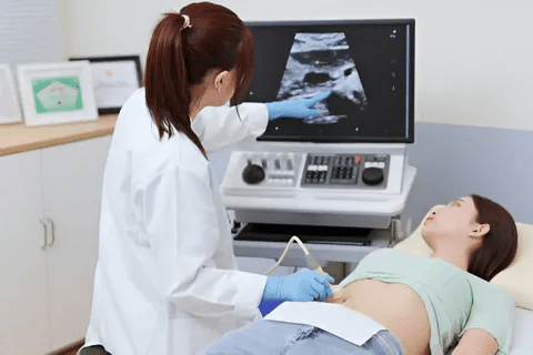
Ultrasounds are done by trained sonographers or ultrasound technicians. They have the skills to get the right images and share their first thoughts. But, a healthcare provider will give the final say.
What Happens During the Procedure
The sonographer will apply gel to your abdomen for a transabdominal scan or use a special probe for a transvaginal scan. The gel helps get better images by reducing air bubbles. They will take measurements and pictures of your embryo, like the gestational sac and embryonic pole.
A medical expert says, “The 8-week ultrasound is a critical milestone in pregnancy, providing valuable insights into fetal development and health.”
“The first ultrasound is always a special moment for expectant parents, giving them their first look at their baby.”
— Medical Expert, OB-GYN
Types of Ultrasound Techniques Used at 8 Weeks
Ultrasound technology is key in tracking fetal growth. At 8 weeks, two main methods are used. Knowing about these can make expectant parents feel more at ease and informed.
Transvaginal Ultrasound: Procedure and Benefits
A transvaginal ultrasound uses a special probe in the vagina to see the uterus and embryo. It’s often used early in pregnancy because it gives clear, detailed images.
The benefits of transvaginal ultrasounds at 8 weeks include:
- Closer to the uterus for clearer images
- Can spot the fetal heartbeat and milestones early
- Shows the embryo and its surroundings well
Healthcare experts say, “Transvaginal ultrasound is great in early pregnancy for its detailed images.”
“This method lets us see the embryo and placenta up close in the first trimester.”
Transabdominal Ultrasound: When It’s Used
Transabdominal ultrasounds use gel on the belly and a transducer to see through the wall. It’s more common later in pregnancy but can be used earlier if needed.
Transabdominal ultrasounds are used when:
- The uterus is big enough to see through the belly
- There’s a medical reason to avoid the vaginal method
- The patient prefers it or is uncomfortable with the vaginal method
Comparing Image Quality Between Methods
Image quality varies between transvaginal and transabdominal ultrasounds at 8 weeks. Transvaginal ultrasounds usually give clearer images because they’re closer to the uterus.
Ultrasound Method | Image Quality at 8 Weeks | Primary Use |
Transvaginal | High-resolution images | Early pregnancy, detailed examination |
Transabdominal | Variable, often less clear than transvaginal | Later pregnancy stages, overall fetal assessment |
Safety of Both Ultrasound Types
Both transvaginal and transabdominal ultrasounds are safe when done by skilled healthcare professionals. The key is to use the least amount of ultrasound waves needed.
Ultrasound safety is maintained by:
- Following the ALARA principle for ultrasound exposure
- Ensuring the operator is well-trained and skilled
- Adhering to guidelines for ultrasound use in pregnancy
Fetal Development at 8 Weeks
At 8 weeks, the embryo’s growth is amazing and complex. It has grown to about 16mm in length. Its development is happening fast.
Size and Appearance of the Embryo
The embryo is about 16mm long at 8 weeks. It’s the size of a small bean or raspberry. Its shape is like a bean, which is normal at this stage.
The Bean-Like Shape Explained
The embryo’s shape is due to its fast growth and the start of body parts forming. Its head and tail fold towards its chest. This creates the bean-like shape.
Developmental Milestones Reached by 8 Weeks
By 8 weeks, many important milestones are reached. The embryo’s major organs like the heart, lungs, and liver start forming. But they’re not working fully yet.
The neural tube, which will become the brain and spinal cord, closes. Limb buds, which will turn into arms and legs, are also there.
Internal Structures Forming but Not Yet Visible
Even though the embryo’s outside shape is clear, many inside parts are forming unseen. These include the start of the digestive system, pancreas, and gallbladder. These parts are key for the embryo’s growth.
Knowing what happens by 8 weeks helps parents see how fast and complex their baby’s growth is. As the pregnancy goes on, more will be seen during ultrasounds.
What Your Doctor Can See on an 8-Week Ultrasound
Your doctor can see important parts of your baby’s growth at 8 weeks with an ultrasound. This scan gives valuable info about your pregnancy’s health and how it’s going.
Gestational Sac Visualization and Measurements
The gestational sac is one of the first things seen on an ultrasound. It’s a fluid-filled area around the embryo, helping it grow safely. By 8 weeks, it’s about 2-3 cm big. Measuring it helps check if the pregnancy is on track.
Yolk Sac Identification and Function
The yolk sac is seen inside the gestational sac by 5-6 weeks and at 8 weeks too. It’s key for early growth, giving nutrients before the placenta does. It looks like a small, round shape. Seeing it and its size means the pregnancy is likely healthy.
Amniotic Sac Development
The amniotic sac surrounds the embryo, filled with fluid that protects it. By 8 weeks, it’s visible on the ultrasound, around the embryo. Its growth is important for the baby’s movement and growth.
Embryonic Pole and Fetal Pole
At 8 weeks, the embryonic pole is seen, showing early fetal development. As it grows, it’s called the fetal pole. Measuring it helps date the pregnancy accurately. Seeing these signs means the baby is developing as it should.
Key features seen at an 8-week ultrasound include:
- Gestational sac size and shape
- Presence and size of the yolk sac
- Amniotic sac development
- Embryonic or fetal pole length
These details give a full view of early pregnancy, helping doctors check the pregnancy’s health and chances of success.
Detecting the Heartbeat at 8 Weeks
The 8-week ultrasound is a key moment. It’s when we can first see the baby’s heartbeat. This gives parents their first real sign of their baby’s life.
When Cardiac Activity Becomes Visible
By 6 weeks, we can start to see the heartbeat. By 8 weeks, it’s clear during an ultrasound. Seeing the heartbeat is a big sign that the baby is doing well.
Normal Fetal Heart Rate Ranges
At 8 weeks, a baby’s heart beats between 160 to 180 times per minute. This can change a bit for each baby, but it usually stays in this range.
How the Heartbeat is Measured
At the ultrasound, a sonographer uses Doppler ultrasound. This technology sends sound waves to show the baby’s heartbeat and activity.
What it Means if No Heartbeat is Detected
If we don’t see a heartbeat at 8 weeks, it might mean there’s a problem. It could be because the due date was wrong. Or it might mean we need to do more tests.
Knowing when we can hear the baby’s heartbeat is important. It helps us keep an eye on how the baby is growing. Here’s a quick summary of what we know about hearing the heartbeat at 8 weeks:
Aspect | Details | Significance |
Timing of Detection | Typically visible by 8 weeks | Indicator of fetal viability |
Normal Heart Rate | 160-180 bpm | Reassuring sign of fetal health |
Measurement Method | Doppler Ultrasound | Accurate measurement of heart activity |
No Heartbeat Detected | Potential for miscalculated gestational age or need for further testing | May require additional monitoring |
Dating Your Pregnancy with Crown-Rump Length
The crown-rump length is a key part of early ultrasounds. It helps doctors figure out how far along you are and when you might give birth. This measurement goes from the top of the embryo’s head to its bottom.
How crown-rump length is measured
An 8-week ultrasound measures the crown-rump length to find out the gestational age. This is done with either a transvaginal or transabdominal ultrasound. The length is a good way to tell how far along you are in the first trimester.
Key aspects of crown-rump length measurement include:
- The embryo is measured from the crown (top of the head) to the rump (bottom of the buttocks).
- This measurement is most accurate between 7 and 14 weeks of gestation.
- The crown-rump length is used to estimate the gestational age and, subsequentl, the due date.
Calculating estimated due dates
After measuring the crown-rump length, doctors can guess when you’ll give birth. They use the average growth of embryos in the first trimester. By comparing the length to growth charts, they can figure out the gestational age and due date.
It’s important to remember that growth can vary from person to person.
Accuracy of early dating scans
Scans between 8 and 12 weeks are very accurate for figuring out gestational age. The crown-rump length is reliable during this time because embryos grow at a steady rate in the first trimester.
Factors contributing to the accuracy of early dating scans include:
- The consistent growth rate of embryos during the first trimester.
- The use of standardized growth charts to compare measurements.
- The skill and experience of the sonographer performing the ultrasound.
When measurements might be adjusted later
Even though the crown-rump length is usually right, sometimes it needs to be changed later. This can happen if there are differences in growth or if there are discrepancies between ultrasounds. These changes can affect the estimated due date.
In conclusion, the crown-rump length is a key measurement for early pregnancy dating. It shows how important early ultrasounds are for tracking fetal growth and predicting when you’ll give birth.
Understanding Your Ultrasound Images
Understanding your ultrasound images is key to your pregnancy journey. These images, though black and white, hold vital info about your baby’s growth. We’ll help you interpret these images and share them with your loved ones.
Interpreting Black and White Images
Ultrasound images are in black and white, which can be tricky to understand. Different shades show different tissues and structures. For example, fluid looks black, and bones are white or light gray.
Key to Interpretation: The contrast between black and white helps spot various structures. Soft tissues show up in shades of gray, giving a detailed view of your baby’s body.
Common Features in Ultrasound Images
At 8 weeks, several important features can be seen. The gestational sac, yolk sac, and embryonic pole are among them. The gestational sac is the first sign, looking like a fluid-filled area. The yolk sac feeds the embryo before the placenta forms, and the embryonic pole is the embryo’s first sign.
Understanding the Limitations
Ultrasound images are very informative, but there are limits, even at 8 weeks. Some details, like the baby’s sex, might not be clear yet. But, seeing a heartbeat and the embryo’s growth are signs of a healthy pregnancy.
Sharing Your First Baby Pictures
Sharing your ultrasound images is a great way to share your pregnancy news. You can make a digital or physical album to keep this moment. These images can also be used for personalized announcements or gifts.
Tips for Sharing: When you share your ultrasound pictures, include the scan date and any important measurements or notes from your doctor. This adds context and makes the moment even more special.
Understanding your ultrasound images is more than just seeing your baby for the first time. It’s about learning about their development and health. By knowing what you see, you can better enjoy your pregnancy journey.
Conclusion: What Your 8-Week Ultrasound Tells You
An 8-week ultrasound is a key moment in pregnancy. It gives us a peek into how the baby is growing and if they are healthy. We can see the gestational sac, yolk sac, and the start of the embryo.
This ultrasound tells us a lot about the pregnancy. It shows if the baby has a heartbeat, which is a big sign of health. It also helps us check if there are any problems early on.
Seeing the baby at 8 weeks is more than just a tiny embryo. It’s a sign that the pregnancy is moving forward. This first check-up helps us plan the best care for the mom and baby.
FAQ
What can you see on an 8-week ultrasound?
At 8 weeks, an ultrasound can show the gestational sac, yolk sac, and amniotic sac. It also shows the embryonic pole. The embryo looks like a small bean, and its heartbeat can be seen.
Is an 8-week ultrasound necessary?
Sometimes, an 8-week ultrasound is needed. It helps confirm if the pregnancy is viable and can spot early problems.
What is the difference between a transvaginal and transabdominal ultrasound?
A transvaginal ultrasound uses a probe in the vagina for a closer look. A transabdominal ultrasound is done on the belly. Both are safe and chosen based on the pregnancy stage and medical needs.
Can you hear the baby’s heartbeat at 8 weeks?
Yes, by 8 weeks, the heartbeat is visible and can be heard during an ultrasound. A normal heart rate is between 100 to 160 beats per minute.
How is the crown-rump length measured during an 8-week ultrasound?
The crown-rump length is measured from the top of the embryo’s head to its buttocks. This helps determine the gestational age and estimate the due date.
What if no heartbeat is detected during the 8-week ultrasound?
If no heartbeat is detected, it may mean there are issues. More tests or ultrasounds might be needed to check the pregnancy’s viability.
How do I prepare for my 8-week ultrasound appointment?
To prepare for an 8-week ultrasound, understand the procedure and what to expect. Knowing who will perform the ultrasound and what happens can help reduce anxiety.
What do ultrasound images show at 8 weeks?
At 8 weeks, ultrasound images show the embryo as a small bean. The gestational sac, yolk sac, and amniotic sac are also visible. These images can be hard to understand because they are black and white.
Can I get an ultrasound at 8 weeks if it’s not medically necessary?
Insurance coverage affects when the first ultrasound is done. Some plans cover an early scan if medically necessary, while others may not.
How accurate is the estimated due date from an 8-week ultrasound?
The crown-rump length measurement at 8 weeks is usually accurate for dating the pregnancy. But, later ultrasounds or other factors might adjust the due date.
References
National Center for Biotechnology Information. 8-Week Ultrasound: Assessing Early Pregnancy and Embryonic Development. Retrieved from https://pubmed.ncbi.nlm.nih.gov/30579966/


