Last Updated on November 27, 2025 by Bilal Hasdemir
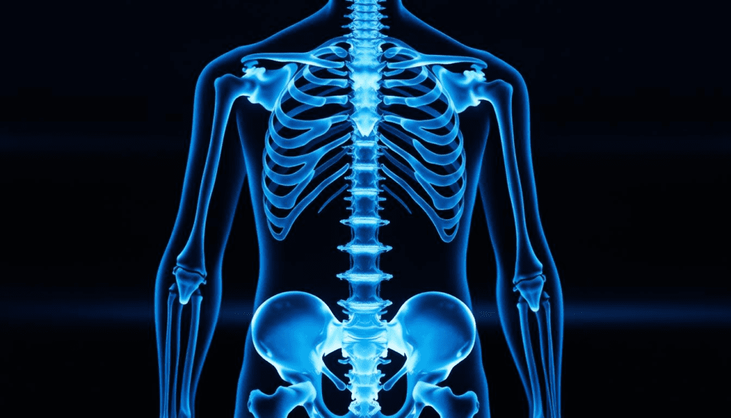
At Liv Hospital, we know how key accurate diagnosis is for bone health. A bone scan, or bone scintigraphy, is a special radiology test. It looks at the skeleton for different conditions.
This tool uses a radioactive isotope to find where bone activity is high. This can show signs of inflammation, infection, fractures, or even cancer. A radionuclide bone scan helps spot areas of concern. It gives insights into arthritis or bone cancer.
Knowing what a bone scan shows is important for both patients and doctors. It helps make better decisions about diagnosis and treatment. We aim to give the latest information and care that puts patients first.
Key Takeaways
- A bone scan is a diagnostic tool that examines the skeleton for various bone-related conditions.
- It uses a radioactive isotope to detect increased bone activity.
- Bone scans can indicate the presence of arthritis or bone cancer.
- Accurate diagnosis is key to managing bone health well.
- Liv Hospital is dedicated to providing patient-centered care and the latest insights.
Understanding Bone Scans: The Basics
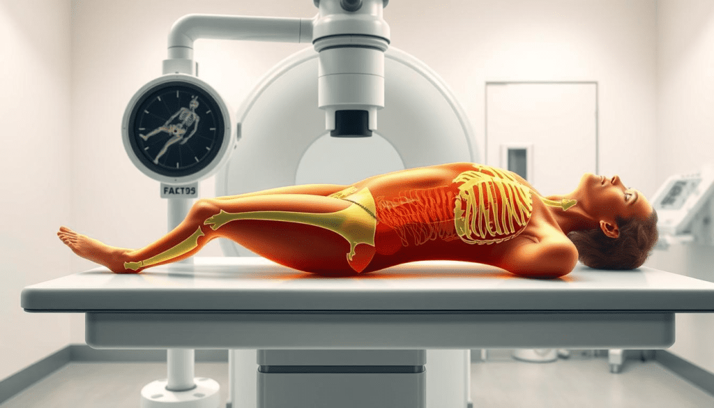
To understand bone scans, we need to know how they work. A bone scan is a test that shows doctors where bones are active. This helps find bone problems like arthritis and cancer.
What Is a Bone Scan (Bone Scintigraphy)?
Bone scintigraphy, or a bone scan, is a detailed imaging test. It uses a tiny bit of radioactive material to find bone issues.
This method is great for spotting areas of bone that are more active. This can mean different things, like arthritis or cancer.
How Bone Scans Work
First, a radioactive isotope is injected into a vein. Then, it goes to bones that are changing, like because by disease or injury.
The radionuclide sends out gamma radiation. A scanner picks up this radiation, making clear images of the bones.
The Radioactive Isotope Process
The radioactive substance, or tracer, builds up in bone areas that are different. This shows where the bone is active.
The Bone Scan Procedure: What to Expect
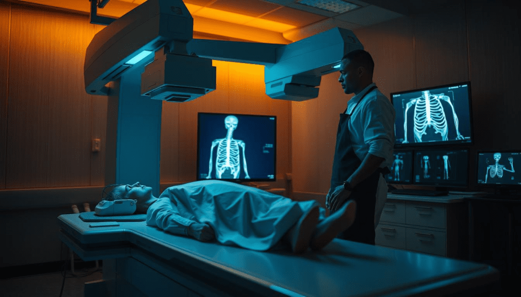
Learning about the bone scan procedure can ease worries and get you ready. A bone scan, or bone scintigraphy, is a test that finds and watches bone problems.
Before the Scan
You usually don’t need to do anything special before a bone scan. You won’t need to fast or get sedated. But, tell your doctor about any medicines you take and if you’re expecting or breastfeeding.
Drinking water is important: You’ll be asked to drink lots of water before and after. This helps get rid of the radioactive tracer from your body.
During the Procedure
You’ll get a small amount of radioactive tracer injected into a vein. This tracer goes to your bones, showing where there’s unusual activity. Then, you’ll wait a few hours before the scan starts.
The scan itself takes 30 to 60 minutes. You’ll lie on a table while a camera moves around you, taking pictures of your bones. You might need to stay very quiet and hold your breath sometimes for clear pictures.
After the Scan
After the scan, you can go back to your usual activities unless your doctor says not to. The tracer will leave your body through urine and feces. Drinking lots of water helps this process.
- Drink plenty of fluids to help eliminate the tracer.
- Avoid close contact with pregnant women and young children for a short period, as advised by your doctor.
- Follow any specific instructions provided by your healthcare team.
Radiation Exposure Considerations
One worry about bone scans is radiation. Though the amount is small, it’s good to know about the risks and benefits. The tracer used in bone scans decays fast, reducing exposure time.
It’s worth noting that the benefits of a bone scan are often greater than the risks. This is true, even for serious conditions. Your doctor will talk about radiation risks and answer your questions.
Knowing what to expect from a bone scan helps patients prepare. If you have any worries or questions, always talk to your healthcare provider.
When Doctors Order Bone Scans
Doctors often suggest bone scans when patients show certain symptoms. These scans help diagnose bone issues like cancer, arthritis, and infections. They are a key tool for doctors.
Common Clinical Indications
Doctors order bone scans for many reasons. They check for bone diseases or track known conditions. Symptoms like bone pain, fractures, and cancer are common reasons.
Symptoms That Warrant Investigation
Some symptoms lead doctors to suggest bone scans. These include ongoing bone pain, unexpected fractures, and bone infections. A scan can uncover the cause and guide treatment.
Role in Disease Monitoring
Bone scans are vital for tracking bone diseases and treatment success. For cancer patients, they spot metastases and check treatment response.
Bone scans help diagnose and monitor many conditions. They’re great for finding metastatic cancer, checking bone trauma, and infections. Knowing when a bone scan is needed helps patients understand their value.
Does a Bone Scan Show Arthritis? Detecting Joint Inflammation
Bone scans are key in finding bone-related issues, like arthritis. They help see how much damage and inflammation there is in joints. This info is vital for figuring out what’s wrong and how to treat it.
Identifying Osteoarthritis
Osteoarthritis is a disease where cartilage wears down and bones change shape. A bone scan shows up as more activity in the affected joints. This is because the body is trying to fix the damage.
Key features of osteoarthritis on a bone scan include:
- Focal or multifocal increased uptake in weight-bearing joints
- Uptake often corresponds to areas of joint space narrowing and osteophyte formation
- Typically involves joints such as the hips, knees, and spine
Detecting Inflammatory Arthritis
Inflammatory arthritis, like rheumatoid arthritis, causes inflammation and damage in joints. Bone scans can spot this by showing more activity in many joints. This is because of the inflammation and bone changes.
Characteristics of inflammatory arthritis on a bone scan:
- Symmetrical increased uptake in multiple joints
- Involvement of small joints in the hands and feet
- Uptake is often more diffuse compared to osteoarthritis
Characteristic Patterns in Arthritic Joints
The way activity shows up on a bone scan can tell us about different types of arthritis. Osteoarthritis usually has more focused activity. Inflammatory arthritis, on the other hand, shows up more evenly across joints.
Knowing these patterns is key to making the right diagnosis and treatment plan. Bone scans are good at showing bone activity, but they’re not perfect for arthritis. They’re best used with other tests and what the doctor sees in the patient.
Limitations of Bone Scans in Arthritis Diagnosis
Using bone scans to diagnose arthritis has its challenges. They are very good at showing changes in bone activity. But they can’t always tell why these changes happen.
Specificity Issues
Bone scans have a big problem: they’re not specific. They can spot areas where bone activity is up, but this can mean many things. For example, infections, fractures, and tumors can also show up on a scan.
Specificity issues mean bone scans can’t tell what’s causing the activity. So, even if a scan shows signs of arthritis, it’s not a sure thing. More tests or a doctor’s opinion are needed to confirm.
False Positives and Negatives
Bone scans can sometimes say you have arthritis when you don’t. This can happen if other things, like recent injuries, cause bone activity. On the flip side, they might miss early signs of arthritis or not catch small changes.
This shows why it’s important to look at bone scan results with the whole picture in mind. It’s not just about the scan itself.
When Other Imaging Tests Are Preferred
Often, other tests are better for diagnosing arthritis. X-rays show bone and joint details well. MRIs are great for soft tissues like cartilage and tendons. These tests give more specific information about joints and tissues.
When it’s hard to tell what’s going on, doctors might choose these tests over bone scans. They help get a clearer picture of what’s happening in the body.
Bone Scans in Cancer Detection: An Overview
Bone scans are key in finding cancer, mainly when it spreads to the bones. They help see if cancer has moved to the bones, which happens in many cancers.
Primary vs. Metastatic Bone Cancer
It’s important to tell the difference between bone cancer that starts in bones and cancer that spreads to bones. Bone scans are great at finding cancer that has spread to the bones, which is more common.
Most often, cancer that spreads to the bones comes from breast, prostate, or lung cancers. These cancers like to go to the bones, causing bone metastases. But, bone cancers that start in bones, like osteosarcoma and Ewing’s sarcoma, are rarer but just as serious.
What Cancers Can a Bone Scan Detect?
Bone scans are good at finding many cancers that have spread to the bones. They’re very helpful for spotting metastases from cancers like:
- Breast cancer
- Prostate cancer
- Lung cancer
- Kidney cancer
- Thyroid cancer
Sensitivity for Different Cancer Types
The effectiveness of bone scans can change with the cancer type. For example, they work well for finding osteoblastic metastases, common in prostate cancer. But, they might not catch osteolytic metastases as well, which are more common in cancers like multiple myeloma.
Knowing how well bone scans work for different cancers is key to understanding the results. Our doctors look at many things, like the cancer type and the bone metastases, to give a full diagnosis.
Identifying Bone Metastases from Common Cancers
Cancer spreading to the bone is a big worry for people with breast, prostate, and lung cancers. Bone metastases happen when cancer cells move from the main tumor to the bone. This can cause a lot of damage and make the disease harder to manage.
Breast Cancer Metastases
Breast cancer often spreads to the bone. It usually goes to the spine, pelvis, and ribs. Bone scans are great for finding these metastases early, helping to start treatment sooner.
The signs of breast cancer metastases on bone scans include:
- Multiple focal areas of increased uptake
- Involvement of the axial skeleton
- Presence of both osteolytic and osteoblastic lesions
Prostate Cancer Spread to Bones
Prostate cancer also spreads to bone, often causing osteoblastic lesions. These show up as areas of increased uptake on bone scans. The spine, pelvis, and proximal femur are the most affected bones.
Key features of prostate cancer bone metastases on bone scans:
- Intense uptake in the affected bones
- Predominantly osteoblastic pattern
- Often multifocal involvement
Lung Cancer Bone Involvement
Lung cancer can also spread to bone, but less often than breast or prostate cancer. It usually causes osteolytic lesions. Bone scans can spot these metastases, but they might be harder to see than osteoblastic lesions.
Important points about lung cancer bone metastases:
- Often present with osteolytic lesions
- May be associated with hypercalcemia
- Can cause significant bone pain and pathological fractures
Other Common Sources of Bone Metastases
Other cancers, like the kidney, thyroid, and melanoma, can also spread to the bone. The way these cancers affect bones can be different.
Knowing how different cancers spread to bone is key to accurate diagnosis and treatment. Bone scans are a big help, giving doctors the info they need to make treatment plans.
Primary Bone Cancer Detection Through Scintigraphy
Bone scans are key in finding primary bone cancers, like several aggressive types. These cancers are rare but serious, so finding them early is vital. We use bone scintigraphy to spot and track these cancers, helping plan treatments.
Osteosarcoma
Osteosarcoma is the most common primary bone cancer, hitting kids and young adults. It starts in the long bones’ growth areas, like the femur or tibia. On a bone scan, it shows up bright because of its active bone growth.
Ewing Sarcoma
Ewing sarcoma is another aggressive cancer, mainly in kids and young adults. It can pop up in any bone, but often is in the pelvis, chest, and long bones. The scan shows it as a bright spot, helping see how big it is and if treatments are working.
Chondrosarcoma
Chondrosarcoma starts from cartilage cells and affects older adults. It can show up in many bones, like the pelvis, femur, and humerus. On scans, it might look different, sometimes appearing dark if it’s mostly cartilage.
Multiple Myeloma Considerations
Multiple myeloma is a cancer of plasma cells in the bone marrow. It’s not always seen as a primary bone cancer, but it affects bones a lot. Scans might not catch it as well as other tests, like PET scans, because it doesn’t always show up bright on scans.
Bone scans are very helpful in finding and managing primary bone cancers. Knowing how different cancers look on scans helps doctors make better diagnoses and treatment plans.
Bone Scan Patterns: Differentiating Arthritis from Cancer
To tell arthritis from cancer on bone scans, we need to understand the patterns each shows. We’ll look at how the scan’s uptake can hint at the cause.
Typical Arthritis Distribution Patterns
Arthritis usually shows symmetrical uptake on bone scans, mainly in joints. This symmetry is key to telling it apart from other conditions.
Osteoarthritis often shows up in weight-bearing joints, and those with trauma or overuse. The uptake is usually focused on the affected joint.
Characteristic Cancer Patterns
Cancer can show different patterns on bone scans, depending on its type and stage. Metastatic disease often has scattered, asymmetric uptake across the skeleton.
Primary bone cancers like osteosarcoma show intense, localized uptake. Sometimes, there’s a “cold” area due to bone destruction.
When Patterns Overlap
Arthritis and cancer patterns can sometimes look alike, making diagnosis hard. For example, someone with arthritis might also have metastatic bone disease.
In these cases, looking at the patient’s history, other images, and sometimes biopsies is key for a correct diagnosis.
The Role of the Radiologist
Radiologists are essential in reading bone scan patterns. They use their knowledge to tell if a process is benign or malignant.
By studying the uptake’s distribution and intensity, and correlating it with the patient’s history, radiologists offer insights that help in treatment planning.
| Condition | Typical Bone Scan Pattern | Characteristics |
| Osteoarthritis | Focal or localized uptake | Symmetrical, weight-bearing joints |
| Metastatic Cancer | Multiple, scattered areas of uptake | Asymmetric, variable intensity |
| Primary Bone Cancer (e.g., Osteosarcoma) | Localized, intense uptake | May have “cold” areas with destruction |
Complementary Diagnostic Tests
Bone scans give us important info, but we often need more tests to fully understand a patient’s health. These tests help doctors diagnose and treat bone issues better.
X-rays and Bone Scans
X-rays are key in diagnosing bone problems. X-rays show us the bones’ details, like fractures or diseases. Together, X-rays and bone scans give a clearer view of bone health.
For example, a bone scan can spot active bone areas. An X-ray then shows the bone’s structure in those spots.
CT Scans and MRIs
CT scans and MRIs give us detailed images of the body. CT scans are great for complex bones and finding bone cancer. MRIs are best for soft tissues, like tumors in the bone marrow.
PET Scans
PET scans help find cancer cells by showing where they’re active. PET scans spot high activity, helping in cancer diagnosis and treatment tracking. With CT scans, they give even more info.
Biopsy: The Definitive Test
Even with new imaging, biopsy is the best way to diagnose many bone issues, like cancer. A biopsy takes bone tissue for a detailed look. This gives a clear diagnosis when images aren’t enough.
Conclusion: The Value of Bone Scans in Diagnosis
Bone scans are key in finding many bone issues, like arthritis and cancer. We’ve looked into how they work, what they’re used for, and their limits. They help spot problems in bones, like arthritis and cancer, and track how they change.
These scans show how bones are working, helping doctors find and track bone issues. This info is vital for diagnosing and treating bone problems. It helps both patients and doctors make better care plans.
In short, bone scans are very important for diagnosing and treating bone issues. They can spot many bone problems, making them a must-have in medicine. As medical tech gets better, bone scans will keep helping patients get better care.
FAQ
What is a bone scan, and how does it work?
A bone scan uses a tiny amount of radioactive material to highlight bone activity. It involves injecting this material into a vein. Then, a scanner picks up the gamma radiation, creating detailed bone images.
Does a bone scan show arthritis?
Yes, a bone scan can spot arthritis by showing where bone activity is high. But it’s not always clear-cut. Doctors usually look at other signs and tests too.
What cancers can a bone scan detect?
Bone scans can find many cancers, like osteosarcoma and breast cancer. They’re good at spotting cancers that have spread to the bones.
How does a bone scan differentiate between arthritis and cancer?
Bone scans show different signs for arthritis and cancer. Arthritis looks like a pattern in joints, while cancer shows up as spots. A doctor looks at these signs to tell them apart.
What are the limitations of bone scans in arthritis diagnosis?
Bone scans aren’t always clear about arthritis. They can give false results. Other tests, like X-rays or MRI,s might be better for a sure diagnosis.
Can a bone scan detect bone cancer?
Yes, bone scans can find bone cancer. They’re great at showing where bone activity is high, which might mean cancer.
What is the role of radionuclide bone scanning in disease monitoring?
Radionuclide bone scanning is key to tracking disease and treatment. It shows how well the treatment is working and if the disease is changing.
How should I prepare for a bone scan?
To prepare for a bone scan, drink lots of water. This helps get rid of the radioactive tracer. Your doctor will give you specific instructions.
What are the radiation exposure considerations for a bone scan?
The radiation from a bone scan is usually safe. But follow your doctor’s advice to stay hydrated. This helps keep exposure low.
Are there complementary diagnostic tests to bone scans?
Yes, tests like X-rays and MRIs work well with bone scans. They give more details for diagnosis and planning treatment.
What is the definitive diagnostic test for bone conditions?
For many bone conditions, like cancer, a biopsy is the best test. It takes a bone sample for detailed examination.
References
- Kim, J. H., et al. (2012). Effectiveness of bone scans in the diagnosis of temporomandibular joint osteoarthritis. Oral Surgery, Oral Medicine, Oral Pathology and Oral Radiology, 114(4), 477-483. https://www.ncbi.nlm.nih.gov/pmc/articles/PMC3520285/


