Last Updated on November 27, 2025 by Bilal Hasdemir

Understanding the difference between a bone scan and a bone density scan is key. A bone scan is a nuclear medicine test. It finds abnormal activity in bones, helping spot bone cancers and metastases from other cancers.
A bone density scan checks bone mineral content and strength. It’s mainly for diagnosing osteoporosis. At Liv Hospital, we focus on patient care and accurate diagnosis. Find out: does a bone scan show cancer in organs? Get crucial answers on the limitations and what it truly detects in the skeletal system.
Key Takeaways
- Understand the difference between bone scans and bone density scans.
- Bone scans detect abnormal bone activity indicative of cancer metastasis.
- Bone density scans measure bone mineral content and strength.
- Bone scans are used to detect cancer spread to the bones.
- Bone density scans are typically used to diagnose osteoporosis.
Understanding Bone Scans: The Basics

Bone scans are key in finding bone problems, like cancer. We’ll look at what bone scans are, how they work, and when they’re used.
What is a Bone Scan?
A bone scan is a test that uses tiny amounts of radioactive material. It helps find and track bone issues, like cancer, arthritis, and fractures. It shows how our bones are doing.
How Bone Scans Work
First, a radioactive tracer is injected into your blood. It goes to your bones. Then, a camera picks up the radiation from the tracer. This makes images of your bones, showing any odd activity.
When Doctors Recommend Bone Scans
Doctors suggest bone scans for many reasons. They help find bone cancer, check if cancer has spread to bones, and see if treatments are working. They also help figure out why you might be in pain or to see how far a bone disease has spread.
Radioactive Tracers and Their Function
The tracers in bone scans target bone tissue. They build up in areas with lots of bone activity, like cancer spots. The camera picks up this radiation, making clear images of your bones.
| Condition | Bone Scan Findings | Clinical Significance |
| Bone Cancer | Increased tracer uptake in the tumor area | Helps in diagnosing primary bone cancer |
| Metastatic Cancer | Multiple areas of increased tracer uptake | Indicates cancer spread to multiple bone sites |
| Osteoarthritis | Increased tracer uptake in joint areas | Reflects active bone remodeling |
Knowing how bone scans work and what radioactive tracers do helps patients. It shows how this test is important for checking bone health and finding conditions like cancer.
Does a Bone Scan Show Cancer in Organs?
Bone scans are key in diagnosing health issues, but they have their limits. They mainly check bone health and find problems like fractures or bone cancer. They don’t directly spot cancer in organs.
The Limitations of Bone Scans for Organ Cancer Detection
A bone scan uses a radioactive tracer that goes through the blood and sticks to bones. It’s great for showing bone activity, but it can’t find cancer in organs. For example, it won’t find cancer in the lungs, liver, or prostate.
How Bone Scans Detect Metastatic Cancer from Organs
But bone scans are good at finding cancer that has moved from the organs to the bones. When cancer reaches the bones, it changes bone activity. A bone scan can spot these changes, like in prostate cancer spreading to the bones.
Doctors use bone scans on patients over 40 to see if cancer has reached the bones. This helps figure out the cancer’s stage and plan treatment.
Complementary Tests for Complete Cancer Detection
Bone scans are helpful for finding cancer in bones, but not a full replacement for organ checks. Doctors often suggest CT scans, MRI scans, or PET scans for a detailed look at organs. These tests can spot cancer in organs directly.
In short, bone scans don’t show cancer in organs directly, but are key for finding cancer in bones. By using bone scans with other tests, we get a full picture of cancer. This helps us create a good treatment plan.
Types of Cancer a Bone Scan Can Detect
Bone scans are great at finding cancer in bones. They are most useful for cancers that start elsewhere but spread to the bones. These scans are key in helping doctors understand how far cancer has spread. This information helps them plan the best treatment.
Metastatic Cancers to Bone
Metastatic cancer means cancer has moved from its original place to another part of the body. Bones are a common place for this to happen, often from breast, prostate, or lung cancers. Bone scans are very good at finding these metastases, which means doctors can act quickly.
When cancer reaches the bones, it can change the bone in ways a scan can spot. For example, a scan might show “hot spots” where cancer has landed. This means the bone is more active than usual.
Common Cancers That Spread to the Bones
Some cancers are more likely to spread to the bones. These include:
- Breast Cancer: Breast cancer often goes to the bones, and scans help track this.
- Prostate Cancer: Prostate cancer often spreads to the bones, and scans help doctors see how far it has gone.
- Lung Cancer: Lung cancer can also spread to the bones, and scans are used to find these spots.
Knowing which cancers tend to spread to the bones helps doctors use bone scans better. This leads to quicker diagnosis and treatment. It also helps improve life quality for those with cancer.
Bone Scan vs. Bone Density Scan: Key Differences

It’s important to know the differences between bone scans and bone density scans. Both tests check bone health but in different ways. They use different technologies for different reasons.
Purpose and Technology Differences
A bone scan looks for changes in bone activity. It might find cancer, infection, or fractures. It uses a small radioactive tracer that shows up on a gamma camera.
A bone density scan, or DEXA scan, checks bone mineral density. It helps find osteoporosis and fracture risks. DEXA scans use X-rays to see your bones.
Key differences in technology and purpose:
| Characteristics | Bone Scan | Bone Density Scan (DEXA) |
| Purpose | Detects bone activity, often for cancer or infection | Measures bone mineral density, typically for osteoporosis |
| Technology | Uses radioactive tracers and gamma camera | Employs low doses of X-rays |
Radiation Exposure Comparison
Both tests use radiation, but in different amounts. Bone scans use a small amount of radioactive tracer. DEXA scans use low X-ray doses.
Bone scans can expose patients to 2 to 6 millisieverts (mSv) of radiation. DEXA scans expose patients to much less, about 0.001 to 0.01 mSv.
Patient Experience During Each Procedure
Bone scans involve a radioactive tracer injection a few hours before. Patients lie on a table while images are taken. The whole process can take 30 minutes to hours.
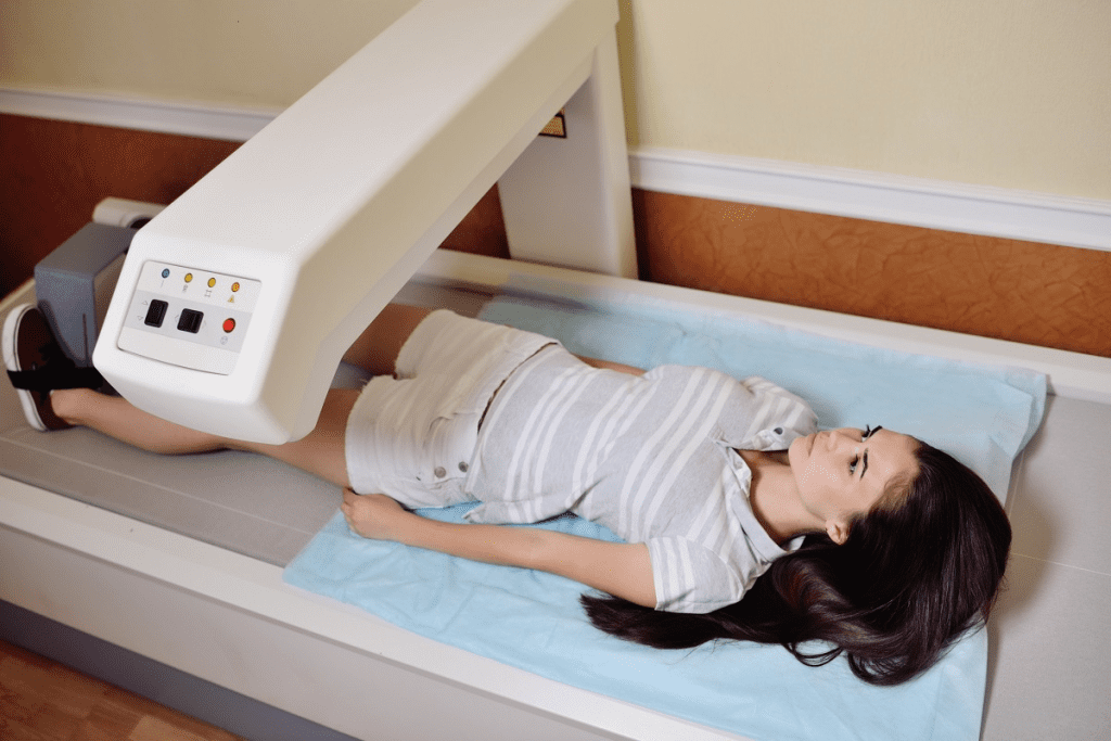
DEXA scans are quicker. Patients lie on a table while the scanner measures their bones. It usually takes less than 10 minutes.
Knowing these differences helps patients and doctors choose the right test. It’s all about understanding your needs.
Understanding Bone Density Scans (DEXA)
A DEXA scan is a non-invasive test that measures bone mineral density. It helps doctors check the risk of osteoporosis. This test is key for spotting those at risk of fractures and osteoporosis problems.
Purpose of DEXA Scans
The main goal of a DEXA scan is to measure bone density. This is vital for diagnosing osteoporosis and assessing fracture risk. DEXA scans are often suggested for:
- Postmenopausal women
- Older adults
- Individuals with a history of fractures
- Those taking medications that may affect bone density
Healthcare providers use DEXA scans to spot risks. They then suggest preventive steps or treatments.
How DEXA Technology Works
DEXA technology uses low-level X-rays to measure bone density. It compares your bone density to a healthy young adult. This gives a T-score that shows your bone health.
Key aspects of DEXA technology include:
- Low radiation exposure
- Quick and painless procedure
- High accuracy in measuring bone density
Interpreting Bone Density Results
Bone density results are based on the T-score. It compares your bone density to a healthy young adult’s. A T-score of -2.5 or lower means you have osteoporosis. A score between -1 and -2.5 means you have osteopenia.
Understanding your T-score is key:
- A T-score above -1 means your bone density is normal.
- A T-score between -1 and -2.5 means you have low bone mass, or osteopenia.
- A T-score of -2.5 or lower means you have osteoporosis.
Knowing your bone density and T-score helps you and your doctor make better decisions about your bone health.
Will a Bone Density Scan Show Cancer?
Many patients ask if a bone density scan can find cancer. But it’s important to know what each test is for. A bone density scan, or DEXA scan, mainly checks bone health and spots osteoporosis.
Why DEXA Scans Cannot Identify Tumors
DEXA scans are not made to find cancer. They use low-level X-rays to see how dense bones are. This helps understand bone health, not find tumors or cancer cells.
When to Choose a Bone Scan Over a DEXA Scan
If you think you might have cancer, a bone scan is better. Bone scans look for where bones are more active. This might show cancer.
| Characteristics | Bone Scan | DEXA Scan |
| Purpose | Detects areas of bone activity, often used for cancer detection | Measures bone density, used for osteoporosis diagnosis |
| Technology | Uses a radioactive tracer to highlight areas of bone activity | Low-level X-rays to measure bone mineral density |
| Cancer Detection | Can detect metastatic cancer to the bone | Not designed for cancer detection |
It’s key to know the differences between these tests for the right diagnosis and care. DEXA scans are great for bone health, but bone scans are better for finding cancer.
Bone Scan Arthritis vs. Cancer: Differential Diagnosis
Bone scans help tell arthritis from cancer, two bone health issues. They show if bone problems are from inflammation, like arthritis or cancer.
Inflammation on Bone Scans
Arthritis can cause inflammation seen on bone scans. This inflammation makes the affected areas show up because of the tracer used.
Key Features of Arthritis on Bone Scans:
- Increased tracer uptake in joint areas
- Symmetrical involvement
- Correlation with clinical symptoms like pain and stiffness
Distinguishing Cancer from Arthritis
Cancer, like metastatic cancer, can also show up on bone scans. But, there are key differences that help tell it apart from arthritis.
| Characteristics | Arthritis | Cancer |
| Tracer Uptake Pattern | Typically, around joints, symmetrical | Often focal, can be anywhere |
| Clinical Correlation | Correlates with joint pain and stiffness | May not directly correlate with pain |
As noted by a medical expert,
“The ability to distinguish between arthritis and cancer on a bone scan is key for patient care. It helps decide on further tests and treatment.”
Additional Tests for Confirmation
While bone scans are helpful, more tests are needed to confirm a diagnosis. These might include:
- X-rays or CT scans to check bone structure
- MRI for detailed soft tissue look
- Blood tests for inflammation or tumor markers
In summary, bone scans are vital in telling arthritis from cancer. By looking at tracer uptake patterns and clinical signs, doctors can make accurate diagnoses. This leads to better treatment plans.
The Procedure: What to Expect During a Bone Scan
Getting ready for a bone scan involves a few important steps. These steps help make sure the test goes well and gives accurate results. We know that getting a diagnostic test can be scary. But knowing what to expect can make you feel better.
Preparation Requirements
Before your bone scan, there are a few things you need to do. First, tell your doctor about any medicines you’re taking. Some medicines can affect the scan. Also, take off any jewelry or metal items that might get in the way of the scan.
On the day of the scan, a small amount of radioactive tracer will be injected into you. This is done in a nuclear medicine department. The tracer goes into your bones, showing where there might be problems.
- Arrive at least 30 minutes before your scheduled appointment time.
- Be prepared to change into a hospital gown.
- Remove any metal objects, including jewelry and glasses.
The Scanning Process
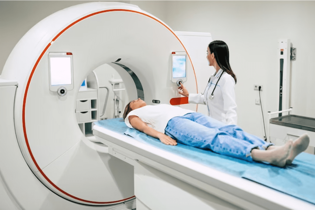
The scanning process is easy. After the tracer is absorbed, you’ll lie on a table. A gamma camera will then move over your body, taking pictures of your bones. The whole scan usually takes about 30 to 60 minutes.
You’ll need to stay very quiet and not move during the scan. This helps get clear pictures of your bones. The gamma camera picks up the radiation from the tracer, showing detailed images of your bones. These images can help find problems like cancer or bone disease.
| Procedure Step | Description | Time Required |
| Tracer Injection | Injection of radioactive tracer | 5-10 minutes |
| Tracer Absorption | Waiting period for tracer absorption | 30-60 minutes |
| Scanning | Gamma camera imaging | 30-60 minutes |
After the scan, you can go back to your usual activities. The radioactive tracer will slowly lose its strength over a few hours. It will be removed from your body through your urine and feces.
The Procedure: What to Expect During a Bone Density Scan
Knowing what to expect during a bone density scan can ease your worries. This scan, also known as a DEXA scan, is a non-invasive test. It measures bone mineral density to diagnose conditions like osteoporosis.
Preparation for DEXA Scan
Before the scan, you need to prepare. You’ll be asked to remove metal items like jewelry and glasses. Also, wear loose, comfortable clothing.
An expert clinic says, “You may be asked to remove some clothing, depending on the area being scanned. You may be given a gown to wear during the procedure.”
The Scanning Process
The scan is quick and painless. You’ll lie on a table, and a scanner will pass over the tested areas, like the spine or hip. The scanner uses two X-ray beams to measure bone density.
| Step | Description |
| 1 | Lie on the scanning table |
| 2 | The scanner passes over the designated areas |
| 3 | X-ray beams measure bone density |
Post-Scan Information
After the scan, you’ll get a report with your bone density measurements. These are compared to a young adult’s average bone density, known as the T-score. A T-score of -1.0 or above is normal. Scores between -1.0 and -2.5 show low bone mass (osteopenia). Scores of -2.5 or lower indicate osteoporosis.
“The DEXA scan is a key tool for checking bone health and fracture risk,” says an osteoporosis expert. “Knowing your bone density helps you and your doctor plan your treatment.”
Understanding the DEXA scan process, from start to finish, can make you feel more at ease. If you have questions, always talk to your healthcare provider.
Interpreting Bone Scan Results
Understanding bone scan results is key for both patients and doctors. Bone scans help in diagnosing cancer. But, it’s important to know what the images really mean.
Understanding “Hot Spots” and “Cold Spots”
Bone scans use a special tracer that shows where bones are active. “Hot spots” show where the tracer builds up, which might mean cancer, fractures, or other issues. On the other hand, “cold spots” show where the tracer doesn’t build up, possibly indicating dead bone or certain bone lesions.
Common Benign Findings
Not every “hot spot” means cancer. Conditions like osteoarthritis, fractures, or infections can also show up. Knowing the patient’s health history and symptoms helps in understanding these findings.
Concerning Patterns That Suggest Cancer
Some patterns on a bone scan might suggest cancer. Seeing many “hot spots” in places like the spine, pelvis, or ribs could mean cancer has spread. The way the tracer builds up and the pattern of uptake, along with other information, helps tell if it’s cancer or not.
To understand the difference between benign and malignant findings, see the table below:
| Characteristics | Benign Findings | Malignant Findings |
| Tracer Uptake Pattern | Focal or localized, often related to known conditions like arthritis | Multiple or diffuse, potentially indicating metastatic disease |
| Intensity of Uptake | Variable, often less intense | Typically more intense |
| Clinical Correlation | Often related to degenerative changes or trauma | May be associated with systemic symptoms or a known primary cancer |
Conclusion: Choosing the Right Bone Imaging Test
Understanding the difference between a bone scan and a bone density scan is key. At Liv Hospital, we aim to provide top-notch healthcare. We also offer full support and guidance to international patients.
Choosing the right test is not just about picking one. It’s about getting the right diagnosis. A bone scan finds cancer in bones. On the other hand, a bone density scan, or DEXA scan, checks for osteoporosis risk.
The main difference between these tests is their purpose and technology. While they might seem similar, they meet different diagnostic needs. Knowing what each test does helps patients make better health choices.
Deciding between a bone scan and a bone density scan depends on your health needs. We suggest talking to a healthcare expert to pick the best test. This way, patients get accurate diagnoses and effective treatments.
FAQ
What is the difference between a bone scan and a bone density scan?
A bone scan looks for abnormal bone activity, like cancer. A bone density scan checks bone strength and mineral content. It’s used to find osteoporosis.
Does a bone scan show cancer in organs?
A bone scan doesn’t find cancer in organs. But it can spot cancer that has spread to bthe ones from other places.
What types of cancer can a bone scan detect?
Bone scans can find cancer that has spread to the bones. This includes cancers from the breast, prostate, lung, and more.
Will a bone density scan show cancer?
No, a bone density scan, or DEXA scan, doesn’t find cancer. It checks bone density to see if you might get osteoporosis or fractures.
How does a bone scan differ from a bone density scan in terms of technology and purpose?
Bone scans use a radioactive tracer to show bone activity. Bone density scans use X-rays to measure bone mineral content. Bone scans look for cancer, while bone density scans check bone health.
Can a bone scan detect arthritis?
Yes, a bone scan can spot arthritis by showing inflammation. But it’s hard to tell if it’s arthritis or cancer. More tests might be needed.
What should I expect during a bone scan?
During a bone scan, you’ll get a small amount of radioactive tracer. Then, you’ll have a scan of your bones. It’s painless and takes about 30 minutes to an hour.
How do I prepare for a DEXA scan?
For a DEXA scan, you just need to remove any clothes or jewelry that might get in the way. You might also be told not to take calcium supplements or certain medications before the scan.
What do “hot spots” and “cold spots” mean on a bone scan?
“Hot spots” on a bone scan mean areas with more activity, like cancer or infection. “Cold spots” mean areas with less activity, which could be cancer or bone damage.
Can a DEXA scan diagnose osteoporosis?
Yes, a DEXA scan can diagnose osteoporosis by measuring bone density. It helps figure out your risk of fractures and guides treatment.
When should I choose a bone scan over a DEXA scan?
Choose a bone scan if your doctor thinks you might have cancer or other bone issues. Use a DEXA scan to check bone density and diagnose osteoporosis.
References
- Agrawal, A., Kharn, M., Varde, V., & Nijhawan, R. (2016). Metastatic mimics on bone scan: “All that glitters is not metastatic. Seminars in Nuclear Medicine, 46(2), 146“159. https://pmc.ncbi.nlm.nih.gov/articles/PMC4918480/


