Last Updated on December 1, 2025 by Bilal Hasdemir
About 1 million people in the United States have Parkinson’s disease. It affects how they move, balance, and coordinate. Finding out if someone has Parkinson’s can be hard because its signs are similar to other brain disorders.
New neuroimaging techniques have changed how we look at the brain. They let doctors see the brain’s structure and function in great detail. This article will talk about how brain scan technology helps find Parkinson’s. We’ll look at the different scans used and what they can show.
Key Takeaways
- Parkinson’s disease diagnosis is challenging due to its similar symptoms with other neurological disorders.
- Neuroimaging techniques play a critical role in diagnosing Parkinson’s.
- Different types of brain scans are used for Parkinson’s detection.
- Brain scan technology has advanced significantly, enabling detailed visualization of brain structure and function.
- Accurate diagnosis is essential for effective treatment and management of Parkinson’s.
The Neurobiology of Parkinson’s Disease

Exploring Parkinson’s Disease shows us how it progresses. It’s a complex disorder that affects millions, causing motor and non-motor symptoms.
Dopamine Depletion and Substantia Nigra Degeneration
Parkinson’s Disease is marked by a lack of dopamine. This neurotransmitter is key for movement. The loss of dopamine-producing neurons in the substantia nigra disrupts the brain’s motor control.
This disruption leads to symptoms like tremors, rigidity, and slow movement. Researchers say the loss of these neurons is a key sign of Parkinson’s. Lewy bodies, abnormal protein clumps, also form in nerve cells.
Pathological Changes in the Basal Ganglia
The basal ganglia help control movement. In Parkinson’s, dopamine loss messes with their function. This causes an imbalance in the brain’s movement pathways.
This imbalance is what leads to Parkinson’s symptoms. The brain’s motor control is severely affected.
Clinical Diagnosis vs. Neuroimaging in Parkinson’s Disease
Diagnosing Parkinson’s Disease (PD) is tough because it looks like other diseases. Doctors mainly use clinical evaluation. But, neuroimaging helps add more details to the diagnosis.
The UKPDS Brain Bank Criteria
The UK Parkinson’s Disease Society Brain Bank (UKPDSBB) criteria are key for diagnosing PD. They focus on bradykinesia (slow movement), rigidity, and tremor. These symptoms, along with ruling out other diseases, help doctors make a diagnosis.
The UKPDSBB criteria also look at how well a patient responds to dopamine therapy. They check for signs that might point to another disease. But, diagnosing PD early can be tricky.
The Complementary Role of Brain Imaging
Brain scans like MRI and PET scans help in diagnosing PD. They’re not the only tool used, but they’re very helpful. These scans can rule out other diseases that might look like PD.
Advanced scans are being studied to find signs of PD early. Using both clinical checks and brain scans could lead to better care for patients.
Conventional Structural Imaging Techniques
Imaging techniques like MRI and CT scans are key in diagnosing Parkinson’s disease. They help doctors see changes in the brain. This is important for understanding and treating the disease.
Magnetic Resonance Imaging (MRI) Findings
MRI gives detailed images of the brain. It can show changes in Parkinson’s patients, but these are not always clear signs. MRI findings in Parkinson’s patients may include:
- Narrowing of the substantia nigra
- Signal changes in the brainstem
- Atrophy in various brain regions
Advanced MRI methods can show more details. They help understand tissue health and iron levels in the brain. This is important for Parkinson’s research.
| MRI Technique | Findings in Parkinson’s |
| Diffusion-weighted imaging | Changes in water diffusion in brain tissues |
| Susceptibility-weighted imaging | Increased iron deposition in substantia nigra |
Computed Tomography (CT) Limitations
CT scans are fast but have big drawbacks for Parkinson’s. CT scans are not sensitive to the early changes associated with Parkinson’s. They are mainly used to check for other health issues.
Why Standard Imaging Often Misses Early Parkinson’s
Standard imaging like MRI and CT scans can’t catch early Parkinson’s. The changes are small and hard to see. The diagnosis of early Parkinson’s remains mainly clinical, based on symptoms and a doctor’s exam.
Creating better imaging tools is key for spotting Parkinson’s early. This would help track the disease’s progress.
Functional Brain Imaging Approaches
Functional brain imaging has led to new ways to study Parkinson’s disease. It looks at brain activity and how different parts of the brain connect. This helps doctors understand the disease better and find new ways to treat it.
Brain Activity Patterns and Functional MRI (fMRI)
Functional MRI (fMRI) is a key tool for studying Parkinson’s. It shows how brain activity changes by looking at blood flow and oxygen levels. Studies have found that Parkinson’s patients have unique brain activity patterns, mainly in motor areas.
Key findings from fMRI studies include:
- Altered activity in the basal ganglia and motor cortex
- Changes in connectivity between different brain regions
- Compensatory mechanisms in early-stage Parkinson’s
Arterial Spin Labeling (ASL) for Perfusion Analysis
Arterial Spin Labeling (ASL) is a non-invasive method that looks at brain blood flow. It helps spot areas where blood flow changes in Parkinson’s. This can show how the disease progresses.
Recent studies have highlighted the ASL’s role in:
- Assessing changes in cerebral blood flow
- Monitoring disease progression
- Evaluating the effectiveness of therapeutic interventions
Resting-State Functional Connectivity
Resting-state functional connectivity MRI looks at brain connections when it’s not actively working. It has given insights into Parkinson’s network changes.
| Network | Changes in Parkinson’s |
| Default Mode Network | Altered connectivity |
| Motor Network | Disrupted connectivity |
| Cognitive Control Network | Compensatory changes |
As research goes on, brain imaging is key to understanding Parkinson’s. It helps doctors see how the disease affects the brain.
“The integration of functional brain imaging into clinical practice has the potential to revolutionize the diagnosis and management of Parkinson’s disease.”
Parkinson’s Brain Scan Detection Using Nuclear Medicine
Nuclear medicine is key in finding Parkinson’s disease early. It uses tiny amounts of radioactive tracers. These help diagnose and treat many diseases, like cancers and heart issues.
Positron Emission Tomography (PET) and Single-Photon Emission Computed Tomography (SPECT) are important for Parkinson’s. They show how the brain works and its structure. This helps doctors spot dopamine loss and other Parkinson’s signs.
Positron Emission Tomography (PET) Techniques
PET scans use radioactive tracers to see brain function. For Parkinson’s, they measure dopamine and find brain changes. Fluorodopa PET checks dopamine production, showing how well the brain makes dopamine.
“PET imaging has revolutionized the field of neurology, opening a new window into the brain’s function.”
Single-Photon Emission Computed Tomography (SPECT)
SPECT imaging looks at brain function too. It uses a radioactive tracer that binds to brain targets, like dopamine transporters. DaTSCAN is a SPECT scan that checks dopamine transporters in the striatum, a key area in Parkinson’s.
PET and SPECT have big benefits for Parkinson’s diagnosis and treatment. They give detailed brain function and dopamine loss info. This helps doctors make better treatment plans.
DaTscan: The Gold Standard for Dopaminergic Imaging
Dopaminergic imaging with DaTscan is now the top choice for diagnosing Parkinson’s disease. This method has changed neurology by giving accurate and dependable diagnostic info.
Technical Aspects of DaTscan Acquisition
DaTscan uses a special radiopharmaceutical, Ioflupane (I-123), to bind to dopamine transporters in the brain. It starts with Ioflupane given through a vein, followed by a Single-Photon Emission Computed Tomography (SPECT) scan.
The details of DaTscan acquisition are key for accurate diagnosis. Things like Ioflupane dosage, SPECT scan timing, and image reconstruction algorithms affect image quality and accuracy.
Characteristic Patterns in Parkinson’s Disease
In Parkinson’s disease, DaTscan shows specific patterns of dopaminergic degeneration. It shows less Ioflupane uptake in the striatum, mainly in the putamen. It also shows unevenness between the brain’s two hemispheres.
Quantitative Analysis Methods
Quantitative analysis of DaTscan images measures Ioflupane binding ratio in brain regions. It compares striatum uptake to a reference area, like the occipital cortex.
| Region | Specific Binding Ratio |
| Striatum | 2.5 ± 0.5 |
| Putamen | 1.8 ± 0.4 |
| Caudate | 3.2 ± 0.6 |
Clinical Utility and Limitations
DaTscan is very useful in clinical practice. It helps tell Parkinson’s disease apart from other parkinsonian syndromes and tremors. But, it has its limits. Things like medication effects, image issues, and needing skilled interpretation can affect its use.
Despite these challenges, DaTscan is a valuable tool in Parkinson’s disease diagnosis and management. It gives doctors a better way to check dopaminergic function.
Brain Scan Findings in Different Stages of Parkinson’s
As Parkinson’s disease moves through its stages, brain scans show different patterns. These patterns help doctors diagnose and plan treatments. Knowing these changes is key for good care and new treatments.
Prodromal and Early-Stage Imaging Biomarkers
In the early stages of Parkinson’s, certain signs can be seen on scans. These signs include:
- Dopamine transporter imaging: DaTscan shows less dopamine in the brain, which is a sign of disease.
- Functional MRI (fMRI): Scans can spot changes in brain activity, like in the motor areas.
- Arterial Spin Labeling (ASL): This method measures blood flow in the brain, which changes early in Parkinson’s.
These signs are important for catching the disease early and starting treatment sooner.
Mid-Stage Disease Progression Markers
In the mid-stage of Parkinson’s, more changes are seen on scans:
- Progression of nigrostriatal degeneration: Dopamine levels keep dropping.
- Changes in brain connectivity: fMRI shows how brain networks change.
- Structural changes: Some brains show shrinkage in certain areas, but it varies.
These signs help doctors track the disease and adjust treatments.
Advanced Parkinson’s Structural Changes
In advanced Parkinson’s, big changes are seen:
- Widespread brain atrophy: Not just the substantia nigra, but other areas too.
- White matter changes: Imaging shows changes in brain tracts.
- Iron accumulation: Some studies find more iron in certain brain spots.
Correlation Between Imaging and Clinical Symptoms
There’s a complex link between what scans show and symptoms in Parkinson’s:
- Dopaminergic imaging correlates: How much dopamine is lost matches how bad symptoms are.
- Functional connectivity changes: Changes in brain networks link to symptoms.
- Structural changes and clinical progression: How much the brain shrinks and changes links to how long the disease lasts and symptoms get worse.
Understanding these links is key to making treatments better and helping patients more.
Emerging Biomarkers in Parkinson’s Neuroimaging
New biomarkers are changing how we see Parkinson’s disease. They help us find the disease early and track it over time. These markers are key to understanding Parkinson’s and finding better treatments.
Alpha-Synuclein Imaging Development
Alpha-synuclein is a protein linked to Parkinson’s. New imaging tools can spot this protein in the brain. This could mean catching the disease sooner and tracking how it changes.
Current Challenges: Making these tools work well is a big hurdle.
Neuroinflammation Markers
Neuroinflammation is a big part of Parkinson’s. New markers are being made to see this inflammation. PET scans with special tracers might help measure it.
Example: Tracers for the translocator protein (TSPO) show how active microglia are in the brain.
Mitochondrial Dysfunction Assessment
Mitochondrial problems are common in Parkinson’s. New markers can check how well mitochondria work in the brain. They look at dopamine and mitochondrial complex I activity.
| Biomarker | Target | Imaging Modality |
| Alpha-synuclein probes | Alpha-synuclein aggregation | PET |
| TSPO tracers | Microglial activation | PET |
| Dopamine metabolism markers | Mitochondrial function | PET, SPECT |
Multimodal Biomarker Integration
Using many biomarkers together is exciting. It combines different imaging methods like MRI, PET, and SPECT. This gives a fuller picture of Parkinson’s.
Together, these biomarkers help doctors diagnose better and track the disease. They also help in making treatments that really work.
Future Directions in Parkinson’s Neuroimaging
Parkinson’s disease research is on the verge of a new era. This is thanks to advanced neuroimaging technologies. These tools are helping us understand the disease better and find better treatments.
Artificial Intelligence and Machine Learning Applications
Artificial intelligence (AI) and machine learning (ML) are changing neuroimaging. AI can spot patterns in data that humans might miss. This is key in Parkinson’s, where catching the disease early is vital.
Machine learning can tell Parkinson’s apart from other diseases based on images. This means doctors can give more accurate diagnoses and treatments.
Novel PET and SPECT Tracers
New PET and SPECT tracers are opening up new ways to see Parkinson’s disease. These tracers can focus on specific parts of the disease, like alpha-synuclein clumps or inflammation. This gives us a deeper look into what’s happening in the brain.
For example, tracers for alpha-synuclein could help find the disease early. Tracers for inflammation could show how well treatments are working.
Ultra-High Field MRI Developments
Ultra-high field MRI is making big strides. It lets us see the brain in even more detail. This means we can spot tiny changes and understand Parkinson’s better.
It also helps find important signs of Parkinson’s, like iron buildup in the brain.
Portable Imaging Technologies
Portable imaging technologies are changing the game. They make it easier to get brain scans in different places. This could include clinics or even homes.
This could mean we can check on the brain more often. This could help catch problems sooner and treat them faster.
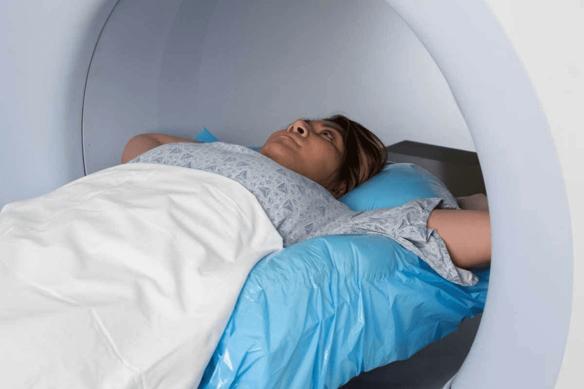
Patient Experience: Preparing for Brain Imaging
Getting ready for your brain imaging can ease your worries. Brain scans are key in understanding Parkinson’s disease. Knowing what to expect makes the process easier.
What to Expect During Different Scan Types
There are various brain scans for Parkinson’s disease. Knowing the differences can make you feel more at ease.
- MRI (Magnetic Resonance Imaging): MRI scans use a strong magnetic field and radio waves to create detailed images. You’ll lie on a table that slides into a large tube-like machine.
- CT (Computed Tomography) Scan: CT scans use X-rays to make images of the brain. You’ll lie on a table that moves through a doughnut-shaped machine.
- PET (Positron Emission Tomography) Scan: PET scans involve injecting a small amount of radioactive material into your bloodstream. This helps visualize brain activity.
Medication Considerations Before Imaging
Tell your doctor about any medications you’re taking before a brain scan. Some medications might need to be adjusted or stopped for accurate results.
| Medication Type | Considerations |
| Dopamine agonists | May need to be stopped or adjusted before a DaTscan |
| Antidepressants | Inform your doctor, as some may interfere with scan results |
Managing Anxiety and Claustrophobia
Feeling anxious or claustrophobic during a scan is common. Let your healthcare provider know about your concerns. They can offer support or make adjustments.
“I was nervous about the MRI, but the technician explained everything and made sure I was comfortable. It made a big difference.” – A patient
Questions to Ask Before and After Your Scan
Being informed is key for a smooth experience. Here are some questions to ask your healthcare provider:
- What type of scan will I be having and what does it entail?
- Are there any specific preparations I need to make before the scan?
- How long will the scan take, and what can I expect during the procedure?
- When will I receive the results, and who will explain them to me?
Being prepared and informed helps you make the most of your brain imaging experience. It also helps you actively manage your Parkinson’s disease.
Conclusion: The Role of Brain Imaging in Parkinson’s Management
Brain imaging is key in managing Parkinson’s disease. It helps from the start of diagnosis to tracking how the disease grows. Techniques like MRI, PET, and DaTscan are very helpful in spotting Parkinson’s and seeing how it changes.
Using brain imaging has made diagnosing Parkinson’s more accurate. It also lets doctors keep a closer eye on how the disease is moving. This helps them create better treatment plans for patients, leading to better results.
As research gets better, brain imaging will play an even bigger part in managing Parkinson’s. New biomarkers and imaging methods are on the horizon. These could make diagnosing and tracking Parkinson’s even more precise. By keeping up with these advances, doctors can give the best care to those with Parkinson’s.
FAQ
What is Parkinson’s brain scan detection?
Parkinson’s brain scan detection uses imaging to diagnose and monitor Parkinson’s disease. Techniques like MRI, CT, PET, and SPECT scans show changes in brain structure and function. These changes are linked to the disease.
Can a brain scan diagnose Parkinson’s disease?
A brain scan alone can’t confirm Parkinson’s disease. But, it offers valuable insights. It can rule out other conditions and spot changes typical of Parkinson’s.
What is DaTscan, and how is it used in Parkinson’s diagnosis?
DaTscan is a dopaminergic imaging method. It uses a radioactive tracer to see the dopamine system in the brain. It helps diagnose Parkinson’s by finding abnormalities in dopamine transporter density, common in the condition.
How does MRI help in diagnosing Parkinson’s disease?
MRI detects changes in brain structure and function. It can spot atrophy in certain areas and rule out other conditions. This helps in diagnosing Parkinson’s disease.
What is the role of PET scans in Parkinson’s disease diagnosis?
PET scans detect changes in brain metabolism and dopamine function. They help diagnose Parkinson’s and monitor its progression. They also assess treatment effectiveness.
Can brain imaging detect Parkinson’s disease in its early stages?
New imaging biomarkers, like alpha-synuclein imaging, show promise for early detection. But, more research is needed to fully develop these techniques.
How do I prepare for a brain scan for Parkinson’s diagnosis?
Follow your healthcare provider’s instructions to prepare for a brain scan. This may include removing metal objects, avoiding certain medications, and managing anxiety or claustrophobia.
What are the limitations of current brain imaging techniques for Parkinson’s diagnosis?
Current techniques have limitations. They require specialized equipment, involve radiation, and can have false positives or negatives. Some are experimental.
How will future advancements in neuroimaging impact Parkinson’s disease diagnosis and treatment?
Future advancements in neuroimaging will improve diagnosis and monitoring. New PET and SPECT tracers, ultra-high field MRI, and portable technologies are expected to enhance accuracy and accessibility.
Can artificial intelligence and machine learning improve Parkinson’s disease diagnosis using brain imaging?
Yes, artificial intelligence and machine learning can improve diagnosis. They analyze brain imaging data to find patterns that humans might miss.

