Last Updated on November 25, 2025 by Ugurkan Demir

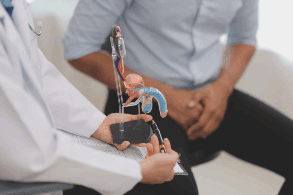
See 10 real enlarged prostate gland images and photos. A visual guide to help you understand the appearance of BPH.
As men get older, the chance of having an enlarged prostate grows. It’s important to know about this condition to take care of our urinary health. At Liv Hospital, we use the latest imaging methods to show clear pictures. These help us diagnose and treat enlarged prostate issues.
Benign Prostatic Hyperplasia (BPH) is a common issue for men as they age. It makes the prostate gland grow bigger. This can cause problems with urination.
The exact reason for BPH isn’t known. But, it’s thought to be linked to hormonal changes in older men. These changes can make the prostate gland grow.
Men with BPH might find it hard to start urinating or have a weak flow. They might also need to go to the bathroom more often. Doctors use a digital rectal examination (DRE) and other tests to diagnose BPH.

Medical imaging is key in finding out if a prostate is enlarged. Tools like ultrasound and MRI help see how big the prostate is. They also help decide the best treatment.
Getting the right diagnosis is very important. It helps doctors choose the best treatment for an enlarged prostate. Ultrasound lets doctors see the prostate and check its size.
With medical imaging, doctors can make treatment plans that fit each patient’s needs.
Diagnosing an enlarged prostate gland uses different imaging technologies. MRI Technology is key for checking the prostate’s size and shape. Ultrasound imaging helps evaluate the prostate gland too.
CT scans are used to diagnose prostate enlargement. They give detailed images of the prostate gland.

Magnetic Resonance Imaging (MRI) is key in spotting an enlarged prostate. It shows the prostate gland’s details and how it affects the urethra. This helps doctors figure out the best treatment.
Using MRI for prostate problems is now more common. It’s non-invasive and gives clear images.
TRUS pictures give us important insights into how an enlarged prostate affects the urethra. These images are key for diagnosing and treating Benign Prostatic Hyperplasia (BPH). We use TRUS to see the prostate gland and its effects on nearby areas.
TRUS shows how an enlarged prostate presses on the urethra. This pressure can cause symptoms like a weak flow and needing to urinate often. Doctors can see how much the urethra is compressed by looking at TRUS images.
For example, a study might use TRUS to measure how much the urethra narrows due to the prostate. This information helps decide the best treatment.
TRUS is also good for measuring prostate volume. Knowing the exact volume is key for diagnosing prostate enlargement and checking if treatments are working.
| Prostate Volume (cc) | Typical Interpretation |
| < 30 | Normal size |
| 30-60 | Mild enlargement |
| > 60 | Significant enlargement |
TRUS can also find structural problems in the prostate. It can spot cysts, calcifications, or other irregularities that might affect the patient’s health.
“The use of TRUS has significantly improved our ability to diagnose and manage prostate-related issues. It’s a valuable tool in our diagnostic arsenal.” – Medical Expert, Urologist
By using TRUS images, we can understand a patient’s condition better. This leads to more effective treatments.

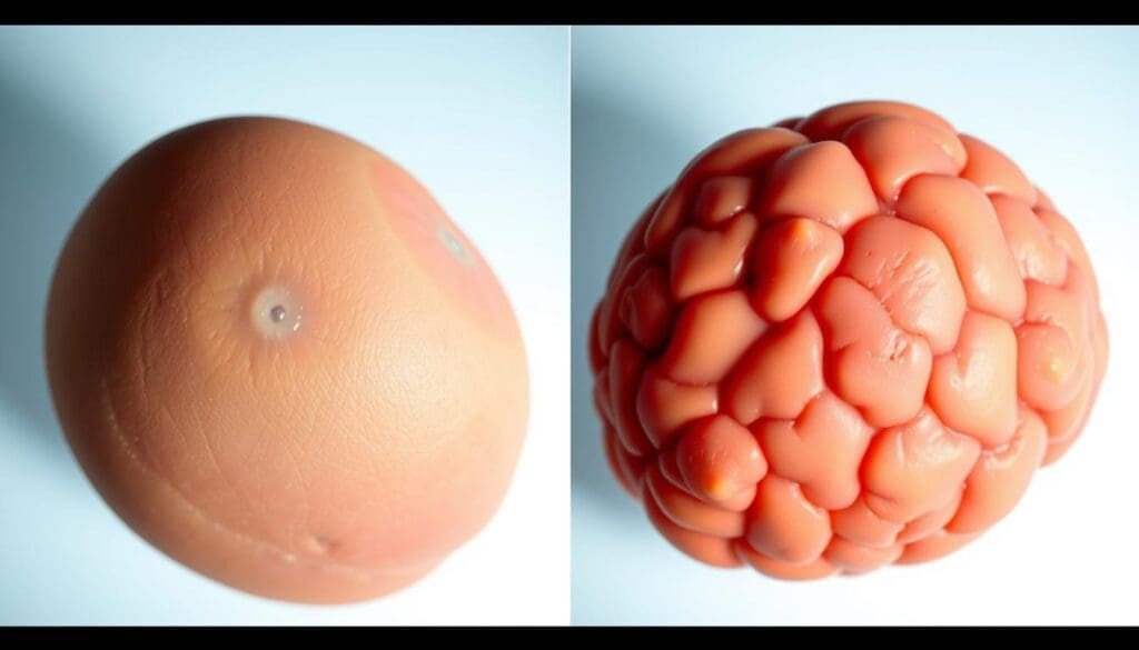
Many men face enlarged prostate, also known as benign prostatic hyperplasia (BPH), as they get older. It’s important to know the difference between a normal and an enlarged prostate. This knowledge helps in diagnosing and treating the condition.
The images below show the main differences between a normal and an enlarged prostate.
These pictures highlight the big differences between a healthy prostate and one that’s enlarged.
It’s key for patients to understand these differences. This helps them grasp their condition and the best treatment options.
We can now see the prostate in amazing detail thanks to new 3D images from MRI. This technology helps us diagnose and treat Benign Prostatic Hyperplasia (BPH) better.
Multiparametric MRI is key for making 3D images of the prostate. These images give us a full view of the prostate’s shape and size. They help us see how much it’s enlarged and how it affects nearby areas. The multiparametric MRI also helps us understand the prostate’s tissue better, which is important for making accurate diagnoses.
Advanced 3D images are very important for surgical planning for BPH. They show the prostate and nearby areas in detail. This helps surgeons plan the best treatment, like choosing the right surgery or minimally invasive treatment options.
| Surgical Planning Aspect | Benefit of 3D Reconstruction Images |
| Pre-operative assessment | Detailed visualization of prostate anatomy |
| Surgical approach planning | Identification of optimal surgical pathways |
| Treatment outcome prediction | Enhanced accuracy in predicting patient recovery |
These advanced images are great for more than just initial diagnosis and planning. They also help monitor how well treatments work. By comparing before and after treatment images, doctors can see if the treatment is effective. This is key for giving personalized care and getting the best results for BPH patients.
In conclusion, advanced 3D images from MRI are changing how we manage enlarged prostate issues. They provide detailed views, help with planning surgeries, and track treatment success. This leads to better care and outcomes for patients with BPH.
At Liv Hospital, we aim to give top-notch care to our patients. Our facilities and technology are the latest, helping us find and treat prostate problems well.
Our skilled team uses the newest in medical imaging. This way, we make sure our patients get the best care possible.
Getting a correct diagnosis is key to treating an enlarged prostate well. New imaging methods help us understand this condition better.
A: Imaging is key in spotting an enlarged prostate. It lets doctors see the prostate gland’s size and shape.
A: MRI gives clear pictures of the prostate gland. Doctors can then check its size, shape, and any oddities. This is vital for diagnosing and treating prostate growth.
A: A normal prostate is smaller and has a regular shape. An enlarged prostate, though, is bigger and might be irregular. This can press on the urethra, leading to urinary issues.
A: Doctors use tools like ultrasound, MRI, and CT scans to see the prostate gland. These help spot BPH by checking the prostate’s size and shape, and any oddities.
A: 3D reconstruction makes detailed, three-dimensional images of the prostate gland. This gives doctors a better view of its size, shape, and structure.
A: Liv Hospital uses the latest tech, like advanced imaging, to diagnose and treat prostate growth. They offer care and plans that fit each patient’s needs.
National Center for Biotechnology Information. (2025). 10 Enlarged Prostate Gland Images Real Pictures and. Retrieved from https://www.ncbi.nlm.nih.gov/books/NBK558920/
Subscribe to our e-newsletter to stay informed about the latest innovations in the world of health and exclusive offers!