At Liv Hospital, we use electrophysiological testing to find and fix heart rhythm problems. An EP study is a small procedure that checks the heart’s electrical system. It helps us find out why arrhythmias happen.
In an electrophysiology evaluation, we use catheters through a vein or artery. These tools record the heart’s electrical signals. This lets our experts find where the heart rhythm problems start and plan the best treatment.
Looking at the electrophysiology of the heart gives us important information. It helps us see how the heart works and find any problems. Our team works hard to give patients the best care. We make sure they get the right treatment for their heart issues.
Key Takeaways
- EP studies are minimally invasive procedures used to evaluate the heart’s electrical system.
- Electrophysiological testing helps diagnose arrhythmias and understand heart rhythm disorders.
- Liv Hospital’s specialists use advanced technologies for accurate diagnosis and personalized care.
- Catheters are used to record the heart’s electrical activity during an electrophysiology evaluation.
- Understanding the electrophysiology of the heart is key for making good treatment plans.
The Fundamentals of Cardiac Electrical Activity

Knowing how the heart’s electrical system works is key to fixing heart rhythm problems. The heart’s electrical system is complex. It makes sure the heart beats in sync and pumps blood well.
Normal Cardiac Conduction Pathways
The heart’s electrical journey starts with the SA node in the right atrium. This node is the heart’s natural clock. The signal then goes to the AV node, then the Bundle of His, and ends in the ventricles.
This path makes sure the heart muscle contracts together. This is how the heart pumps blood well.
The cardiac conduction system is key for a regular heartbeat. Any problem here can cause irregular heartbeats.
How Electrical Signals Control Heart Contractions
Electrical signals are vital for heart muscle contractions. When an electrical signal hits the heart muscle cells, it starts a chain of chemical reactions. This is how the heart pumps blood.
The electrical activity of the heart is complex. It adjusts to what the body needs. When we exercise or are stressed, the heart beats faster. This is because the SA node sends more electrical signals.
Understanding how electrical signals control heart contractions is important. It helps us see why electrophysiologic studies (EP studies) are used to find and fix heart rhythm issues. EP studies use catheters to map and record the heart’s electrical activity. They help find where abnormal rhythms start.
What is an EP Test and Why is it Performed?

An EP test, or electrophysiological study, checks the heart’s electrical system. It’s key for finding and fixing heart rhythm problems, or arrhythmias.
EP tests help us understand the heart’s electrical activity. This lets us find the cause of arrhythmias and choose the best treatment.
Definition and Purpose of Electrophysiological Studies
An electrophysiological study, or EP study, is a test that looks at the heart’s electrical activity. It checks the heart’s electrical system to find any problems that might cause arrhythmias.
To do an EP study, we insert catheters through a vein in the leg. These catheters record the heart’s electrical signals. This helps us diagnose and sometimes treat arrhythmias.
Indications for EP Testing
The main reason for an EPS is to check for cardiac arrhythmias. EP testing is great for finding arrhythmias like atrial fibrillation. It helps us decide if medication or ablation is the best treatment.
Some reasons for EP testing include:
- Recurrent or persistent arrhythmias
- Symptoms like palpitations, dizziness, or fainting
- Family history of sudden cardiac death
- Abnormal electrocardiogram (ECG) or Holter monitor results
By studying the heart’s electrical activity with EP studies, we can create specific treatment plans. This helps manage arrhythmias well.
Types of Arrhythmias Diagnosed Through Electrophysiological Studies
Electrophysiology evaluation helps doctors spot different arrhythmias and predict future heart rhythm problems. Studies like EPS give important details for diagnosing and planning treatment. They help with issues like sinus node dysfunction, atrioventricular conduction problems, and various tachyarrhythmias.
Supraventricular Arrhythmias
Supraventricular arrhythmias start above the ventricles. They include atrial fibrillation, atrial flutter, and SVT. These arrhythmias cause irregular heartbeats due to abnormal electrical signals in the atria. EPS helps find the exact type and plan the best treatment.
Ventricular Arrhythmias
Ventricular arrhythmias, like VT and VF, are serious and start in the ventricles. EPS is key for diagnosing and figuring out the risk. It helps decide the best treatment, like medication, ablation, or an ICD.
Bradyarrhythmias and Conduction Disorders
Bradyarrhythmias and conduction disorders are diagnosed with EPS. They include slow heart rates and problems with electrical signals. Sick sinus syndrome and AV block are examples. EPS helps see how severe they are and if a pacemaker is needed.
EPS gives deep insights into arrhythmias, helping manage heart rhythm disorders. We use EPS findings to create treatment plans tailored to each patient’s needs.
The Electrophysiology Evaluation Process
Understanding the electrophysiology evaluation process is key. It involves several steps to diagnose and treat heart rhythm disorders accurately.
Initial Assessment and Diagnostic Tests
The process starts with a detailed initial assessment. This includes a medical history and physical exam. Diagnostic tests are then used to find the cause of arrhythmias.
- Electrocardiogram (ECG) to record the heart’s electrical activity
- Holter monitoring for continuous ECG recording over 24-48 hours
- Event monitoring for longer-term ECG recording
- Echocardiogram to assess heart structure and function
These tests help understand the heart’s electrical system. They guide further evaluation and treatment.
Risk Stratification Before EP Studies
Before electrophysiologic testing, risk stratification is vital. It helps identify risks and ensure patient safety. This involves looking at the patient’s health, medical history, and other factors.
Important factors include:
- Withholding antiarrhythmic therapy for at least 4 half-lives before the procedure
- Ensuring appropriate anticoagulation management to minimize stroke risk
- Evaluating kidney function to avoid contrast-induced nephropathy
This careful risk assessment makes the EP study safer and more effective.
The Electrophysiology Team
A successful electrophysiology evaluation needs a team. This team includes:
- Electrophysiologists with specialized training in heart rhythm disorders
- Cardiac nurses and technicians skilled in EP procedures
- Anesthesiologists and support staff for patient comfort and safety
This team works together. They ensure the EP study is precise and safe. This leads to accurate diagnoses and effective treatments.
Advances in electrophysiological tests and heart mapping have improved diagnosis and treatment. Knowing the electrophysiology evaluation process helps patients understand their care better. It allows them to make informed decisions about their treatment.
Preparing for an Electrophysiological Test
Getting ready for your EP study is key to its success and your safety. We know medical tests can make you nervous. So, we’re here to help you through every step.
Pre-Procedure Instructions
To get ready for your EP study, you’ll need to:
- Not eat or drink for up to 12 hours before. This is called fasting.
- Have someone drive you home. You might take medicine that makes driving hard.
- Wear loose, comfy clothes to the hospital.
Following these steps is very important for a safe and effective test.
Medication Adjustments
Your doctor might ask you to change your meds before the test. This could mean:
- Stopping some meds that might mess up the test.
- Changing how much of your current meds you take.
Talking to your doctor about your meds is a must to know what changes you need.
What to Bring to the Hospital
When you go to the hospital for your EP study, remember to bring:
- A list of your meds and how much you take.
- Any important medical records or test results.
- Comfortable clothes and any personal items you might need.
By following these tips, you can make sure your EP study goes well. And you’ll be comfortable the whole time. If you have any questions or worries, just ask your healthcare team.
The EP Study Procedure Step by Step
During an EP study, catheters are carefully guided to the heart to record its electrical activity. This procedure is key for diagnosing and understanding various heart rhythm disorders.
Catheter Insertion and Navigation
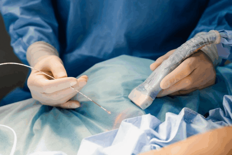
The first step in an EP study is the insertion of catheters through a vein. This is usually done in the groin or neck area. The catheters are then guided to the heart under fluoroscopic guidance.
Once in place, the catheters record the heart’s electrical signals from different locations. This data is vital for understanding the heart’s electrical system and spotting any issues.
Electrophysiologic Mapping of the Heart
Electrophysiologic mapping is a key part of the EP study. It creates a detailed map of the heart’s electrical activity. By analyzing signals from various catheter locations, the electrophysiologist can pinpoint the source of arrhythmias and understand the heart’s electrical pathways.
This mapping helps diagnose the cause of a patient’s arrhythmia. It’s essential for planning the right treatment.
Provocative Testing to Induce Arrhythmias
To better diagnose and understand a patient’s arrhythmia, provocative testing may be done. This involves using electrical stimulation or medications to induce arrhythmias under controlled conditions.
By inducing arrhythmias, the electrophysiologist can observe the heart’s response. This gives valuable insights into the arrhythmia’s mechanism. This information is key for choosing the best treatment strategy.
The EP study procedure, including catheter insertion, electrophysiologic mapping, and provocative testing, offers deep insights into the heart’s electrical activity. This information is vital for diagnosing arrhythmias and guiding treatment decisions.
Advanced Mapping Technologies in EP Tests
Advanced mapping technologies have changed electrophysiology, making EP studies more precise. These tools help us better diagnose and treat complex heart rhythm problems.
3D Electroanatomic Mapping Systems
Three-dimensional electroanatomic mapping systems make detailed maps of the heart’s electrical activity. They let us see the heart’s anatomy and electrical paths in real-time. This helps us find the sources of arrhythmias more accurately.
Key benefits of 3D electroanatomic mapping include:
- Enhanced accuracy in identifying arrhythmia mechanisms
- Improved catheter navigation and positioning
- Reduced procedural times and fluoroscopy exposure
Intracardiac Echocardiography
Intracardiac echocardiography (ICE) gives us real-time ultrasound images from inside the heart. It helps us see cardiac structures clearly. This makes it safer and more effective to place catheters.
“The integration of ICE with 3D mapping systems has significantly enhanced our ability to perform complex ablation procedures.” – Dr. John Smith, Electrophysiologist
High-Density Mapping Techniques
High-density mapping uses special catheters to record many electrical signals from the heart. These signals help create detailed maps of the heart’s electrical activity. This allows us to pinpoint arrhythmia mechanisms with great precision.
The advantages of high-density mapping include:
- Improved resolution of complex arrhythmias
- Enhanced understanding of arrhythmia mechanisms
- Better guidance for ablation therapy
By using these advanced mapping technologies, we can make EP studies more effective. This leads to more accurate diagnoses and better treatment plans for patients with complex arrhythmias.
Interpreting Results from Electrophysiological Tests
Understanding electrophysiological (EP) study results is key to diagnosing and treating cardiac arrhythmias. It involves looking at the shape and timing of electrical signals compared to the surface ECG.
It’s vital for doctors to grasp the details of EP study results. This knowledge helps them make the best decisions for their patients. We’ll look at how to tell normal from abnormal conduction, find out what causes arrhythmias, and how these insights shape treatment plans.
Normal vs. Abnormal Conduction Parameters
EP studies check on how well electrical signals move through the heart. Normal results mean everything is working right. But, if the results are off, it could point to problems or diseases.
A long AH interval might show a trouble spot in the AV node. An odd HV interval could mean a problem with the His-Purkinje system. Doctors use these clues to plan the best treatment.
Identifying the Mechanism of Arrhythmia
EP studies help doctors figure out why arrhythmias happen. They look at the heart’s electrical activity. This helps them see if it’s due to a loop, automatic firing, or something else.
Knowing the cause helps doctors pick the right treatment. For example, if it’s a loop causing the problem, removing it might be the best option.
How Findings Guide Treatment Planning
EP study results are very important for planning treatment. Doctors use them to decide on the best course of action. This could be medication, a procedure, or a device to help the heart.
If the study shows a specific arrhythmia, like SVT, doctors might suggest a procedure to fix it. Sometimes, the results point to the need for medication or other treatments.
In summary, understanding EP study results is complex but essential. By looking at conduction, identifying arrhythmia causes, and planning treatments, doctors can give patients the best care for their heart issues.
Treatment Options Following EP Studies
EP studies help doctors understand the heart’s electrical activity. This knowledge guides them in choosing the best treatment. The treatment depends on the arrhythmia type and how it affects the heart. We’ll discuss the available options, like medication, catheter ablation, and implantable devices.
Antiarrhythmic Medication Therapy
Antiarrhythmic medications are often the first choice for arrhythmias. They help control the heart’s rhythm and prevent irregular beats. Beta-blockers and anti-arrhythmic drugs are commonly used to manage symptoms and improve life quality.
The right medication depends on the arrhythmia type, patient health, and possible side effects. It’s important to monitor the treatment closely to adjust it as needed.
Catheter Ablation Procedures
Catheter ablation is a minimally invasive procedure. It uses energy to destroy abnormal heart pathways causing arrhythmias. This method is very effective for supraventricular tachycardia (SVT) and some ventricular arrhythmias.
The procedure involves inserting catheters through a leg vein and guiding them to the heart. Advanced technologies help pinpoint the exact location of the problem, allowing for precise treatment.
Implantable Cardiac Devices
Implantable cardiac devices, like pacemakers and implantable cardioverter-defibrillators (ICDs), manage arrhythmias and prevent sudden death. These devices send electrical impulses or shocks to correct the heart rhythm.
Pacemakers treat bradyarrhythmias, while ICDs protect against dangerous ventricular arrhythmias. The choice of device depends on the patient’s condition and risk level.
Recovery and Follow-Up After Electrophysiological Studies
Recovery and follow-up after an electrophysiological study are key parts of treatment. It’s important to make sure patients get the right care during this time. This helps them stay well and get the best results from the procedure.
Immediate Post-Procedure Care
Right after the EP study, patients stay in a recovery area for a few hours. Medical staff watch for any problems, like bleeding or heart rhythm issues. Close monitoring means they can act fast if something goes wrong.
Patients might feel some pain or bruising where the catheter was put in. This usually goes away on its own and can be treated with over-the-counter pain meds. We also teach them how to take care of the site to avoid infection.
Activity Restrictions and Wound Care
Patients are told to avoid heavy lifting and strenuous activities for a while. Gentle care of the catheter sites is also important to prevent problems.
- Avoid strenuous activities for 24-48 hours
- Keep the catheter sites clean and dry
- Monitor for signs of infection, such as redness or swelling
Long-Term Monitoring and Follow-Up Testing
Long-term follow-ups are important to see how well the EP study worked. This might include visits to a cardiologist, more tests, or changes to medication. Regular monitoring helps manage the patient’s condition and adjust their treatment as needed.
We also teach patients about the importance of sticking to their follow-up schedule. Knowing when to seek immediate medical help is also key. This way, we can help our patients get the best results.
Conclusion: The Evolving Role of EP Studies in Cardiac Care
EP studies are key in finding and treating heart rhythm problems. Their role is growing as electrophysiology advances.
The field of electrophysiology is changing fast. New tech and methods make EP studies better and more accurate. This leads to better care for patients with heart issues.
As we explore new possibilities in electrophysiology, EP studies will keep leading in heart care. They give us deep insights into arrhythmias. This helps doctors make better treatment plans and find new ways to help patients.
The future of heart care depends on EP studies getting even better. We’re excited to see how they will help us treat heart rhythm problems more effectively.
FAQ:
What is an EP study, and how does it evaluate heart rhythms?
An EP study, or electrophysiological study, checks the heart’s electrical activity. It helps find and understand arrhythmias. Doctors use catheters to record the heart’s signals and create arrhythmias safely.
What are the indications for undergoing an EP test?
EP testing is used to check for arrhythmias and the heart’s electrical system. It helps find the best treatment for issues like irregular heartbeats and conduction problems.
How do I prepare for an electrophysiological test?
To get ready for an EP test, follow the doctor’s instructions. This might include changing your medications, fasting, and getting a ride home. Tell your doctor about any allergies or health conditions.
What happens during the EP study procedure?
During the test, catheters are put through a vein and reach the heart. The team records the heart’s electrical signals and maps the heart. They might also try to create arrhythmias to study them.
What are the risks associated with EP studies?
EP studies are mostly safe but can have risks like bleeding and infection. Your team will talk about these risks and try to avoid them.
How are the results of an EP study interpreted?
The team looks at the heart’s electrical activity to understand arrhythmias. They use this info to plan treatment, which could be medication, ablation, or devices like pacemakers.
What treatment options are available following EP studies?
After an EP study, treatments might include medication, ablation, or devices like pacemakers or ICDs.
What is the recovery process like after an EP study?
After the test, you’ll be watched and given care instructions. This includes activity limits, wound care, and follow-up tests. Following these steps helps with recovery.
How do advanced mapping technologies contribute to EP tests?
New technologies like 3D mapping systems and echocardiography improve EP tests. They give detailed info about the heart’s electrical and physical structure.
What is the significance of EP studies in cardiac care?
EP studies are key in diagnosing and treating arrhythmias. They help tailor care and improve patient results. These tests are vital in cardiac care, with ongoing tech advancements making them even better.
References:
- O’Rourke, M. F. (2018). Structure and function of systemic arteries: reflections on the vascular wall and blood flow. Vascular Medicine, 23(4), 316-323. https://pubmed.ncbi.nlm.nih.gov/30016416/


