Rhabdomyosarcoma (RMS) is a rare cancer that starts in muscle cells. It mostly hits kids under 10, worrying families all over. At Liv Hospital, we know how complex RMS is. We offer specialized, multidisciplinary care to help.Learn erms cancer types, main forms of rhabdomyosarcoma, and treatment options explained clearly.
Knowing the main types of RMS is key to better treatment and results. There are two main types: embryonal rhabdomyosarcoma and alveolar rhabdomyosarcoma. Each has its own traits and treatment ways.
Key Takeaways
- Rhabdomyosarcoma is a rare cancer that develops from skeletal muscle cells.
- It is most commonly diagnosed in children under 10 years old.
- The two main types are embryonal and alveolar rhabdomyosarcoma.
- Understanding the type of RMS is key for effective treatment.
- Liv Hospital provides specialized, multidisciplinary care for RMS patients.
Understanding Rhabdomyosarcoma: A Rare and Aggressive Cancer

Rhabdomyosarcoma (RMS) is a rare and aggressive cancer. It starts in skeletal muscle precursors. This makes it hard to diagnose and treat.
Definition and Origin of Soft Tissue Sarcomas
Soft tissue sarcomas are a group of cancers. They come from mesenchymal cells. These cells turn into different tissues like fat, muscle, and blood vessels.
RMS is a big part of these cancers. It mostly affects kids and teens.
These cancers are rare. They make up 1% of adult cancers and 3% of children’s cancers. Their rarity and variety make them hard to diagnose and treat.
Who Is Most Commonly Affected by RMS
RMS can happen at any age. But it mostly hits kids and teens. The chance of getting RMS changes with age and group.
| Age Group | Incidence Rate | Common Subtypes |
| 0-9 years | Higher incidence | Embryonal RMS |
| 10-19 years | Moderate incidence | Alveolar RMS |
| 20+ years | Lower incidence | Various subtypes |
The table shows how RMS rates change with age. It also lists common types for each age group.
Knowing this helps find and treat RMS early.
Embryonal RMS (ERMS): The Most Common Type of RMS Cancer

ERMS is the most common type of rhabdomyosarcoma in kids. It has unique features that set it apart from other types of RMS.
Cellular and Histological Features
ERMS looks like muscle cells in early development. The cells can look very different, from very basic to almost fully formed. This mix is key to identifying ERMS.
The look of ERMS under a microscope is quite varied. Some areas are packed tight, while others are more spread out. Finding rhabdomyoblasts, cells that are turning into muscle, is a big clue for diagnosis.
Common Locations: Head, Neck, and Genitourinary Tract
ERMS often shows up in the head and neck and the genitourinary tract. It’s less common in other places. Tumors in the head and neck can be near the eye, in the nose, or elsewhere.
- The orbit is a common site, often in young children.
- The genitourinary tract, like the bladder and vagina, is another frequent spot.
- ERMS can also be found in the retroperitoneum or other areas, but less often.
Age Distribution and Incidence Rates
ERMS mainly hits babies and young kids, with most cases found before age 10. It’s a big worry in pediatric cancer because it’s so common in young ones.
- ERMS makes up about 60-70% of RMS cases in kids under 10.
- Most cases happen in the first 10 years of life.
Knowing when and how often ERMS happens helps doctors catch it early and treat it better.
Alveolar RMS (ARMS): An Aggressive Variant
Alveolar rhabdomyosarcoma (ARMS) is a fast-growing cancer type. It mainly affects older kids and teens. This cancer is hard to treat and grows quickly.
Distinctive Pathological Characteristics
ARMS has a unique look, like lung alveoli. The cells are small and round, forming spaces that look like alveoli. This look helps doctors diagnose ARMS.
Key pathological features include:
- Small, round, and undifferentiated tumor cells
- Alveolar pattern with cells lining pseudoalveolar spaces
- Frequent presence of giant cells
Typical Presentation in Extremities, Chest, and Abdomen
ARMS often show up in arms, legs, chest, and abdomen. These places make it hard to find early because the tumors are deep.
Common sites of presentation include:
- Extremities (arms and legs)
- Chest cavity
- Abdominal cavity
The FOXO1-PAX Fusion and Its Prognostic Significance
The FOXO1-PAX fusion is a key sign of ARMS. It happens when genes swap places. This swap leads to cancer growth. The fusion means the disease is likely to be more aggressive.
Studies suggest that those with ARMS and the FOXO1-PAX fusion might need stronger treatments. This could help improve their chances of beating the disease.
Rare Subtypes of Rhabdomyosarcoma
Rare subtypes of rhabdomyosarcoma, like anaplastic rhabdomyosarcoma and spindle cell/sclerosing RMS, need a detailed look for good care. These types are not as common but bring their own set of challenges in finding and treating them.
Anaplastic Rhabdomyosarcoma: A Highly Aggressive Form
Anaplastic rhabdomyosarcoma is very aggressive and mostly found in adults. It has big, unusual cells that mean a tough fight ahead. Doctors use strong chemotherapy and radiation to try and beat it, because it’s hard to treat.
“Dealing with anaplastic RMS needs a team effort,” a study on pleomorphic rhabdomyosarcoma says. It talks about using the newest treatments and surgeries to fight this tough cancer.
Spindle Cell/Sclerosing RMS: Clinical and Pathological Features
Spindle cell/sclerosing RMS can happen at any age. It’s known for its special cells and a dense background. It often shows up in the testicles, head, and neck. Doctors tailor the treatment based on where the tumor is and the patient’s health.
Rhabdomyosarcoma has many subtypes, each with its own needs. Knowing the details of each rare type helps doctors create better treatment plans. This way, they can tackle the challenges of RMS more effectively.
Diagnosing Rhabdomyosarcoma: From Symptoms to Confirmation
Diagnosing rhabdomyosarcoma (RMS) is a detailed process. It starts with noticing symptoms and ends with tests confirming the diagnosis. We will explain how RMS is diagnosed, from the first signs to the final test results.
Common Presenting Symptoms by Anatomical Location
RMS symptoms change based on where the tumor is. For example, head and neck tumors might cause swelling, pain, or trouble swallowing. Tumors in the genitourinary tract could lead to urinary problems or bleeding.
Spotting these symptoms early is key to catching RMS early. Here’s a table showing common symptoms by location:
| Anatomical Location | Common Symptoms |
| Head and Neck | Swelling, pain, difficulty swallowing |
| Genitourinary Tract | Urinary obstruction, bleeding |
| Extremities | Swelling, pain, limited mobility |
Diagnostic Imaging: CT, MRI, and PET Scans
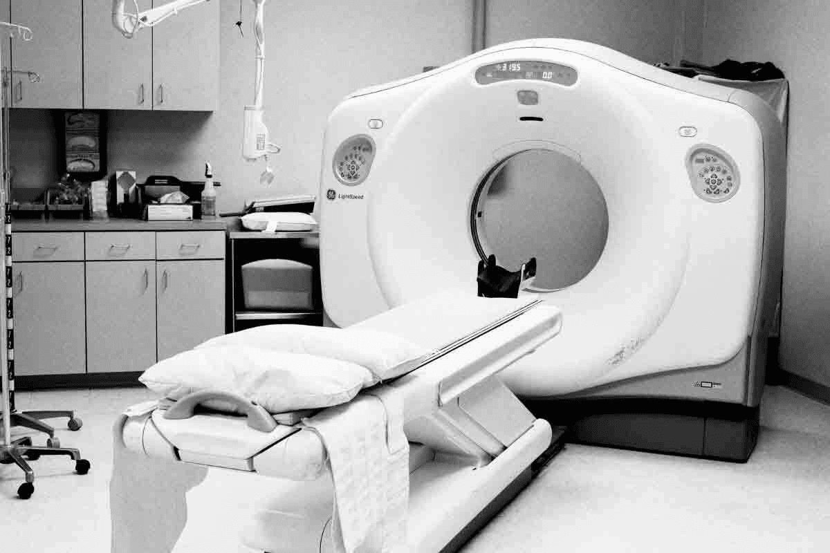
Imaging tests are critical in diagnosing and staging RMS. CT, MRI, and PET scans give important details about the tumor’s size, location, and spread.
CT scans are great for looking at tumors in the lungs, abdomen, and pelvis. MRI is better for soft tissue tumors and their relation to nearby structures. PET scans spot active tumor sites, helping with staging and treatment response.
Biopsy Techniques and Molecular Testing
A biopsy is key to confirming RMS. There are different biopsy methods, like needle and surgical biopsies. The choice depends on the tumor’s location and how easy it is to reach.
Molecular testing, like genetic analysis, is also vital. Tests like fluorescence in situ hybridization (FISH) and polymerase chain reaction (PCR) find specific genetic changes in RMS, like the PAX-FOXO1 fusion gene in alveolar RMS.
Together, these diagnostic steps help doctors accurately diagnose RMS. They then create a treatment plan based on the diagnosis and other factors.
Staging and Risk Stratification of RMS
Accurate staging and risk stratification are key for treating Rhabdomyosarcoma (RMS) patients. We use these methods to see how far the disease has spread. This helps us decide the best treatment for each patient.
The Clinical Group Classification System
The Clinical Group Classification System is a common way to stage RMS. It sorts patients by how much of the tumor is removed and if there’s metastasis. This system helps us understand how serious the disease is and plan the right treatment.
- Group I: Completely resected tumor
- Group II: Microscopic residual disease
- Group III: Gross residual disease
- Group IV: Distant metastatic disease
This classification is key because it affects the treatment plan and how well the patient might do.
TNM Staging for Rhabdomyosarcoma
TNM staging is another important system for evaluating RMS. It looks at three main things: the size and spread of the primary Tumor (T), if nearby Lymph Nodes (N) are involved, and if there are distant Metastasis (M).
TNM staging gives a detailed view of the disease. It helps us choose the best treatment plan for each patient.
How Risk Groups Determine Treatment Approaches
Risk stratification in RMS sorts patients into different groups. These groups are based on age, tumor location, size, and whether there’s metastasis. The groups are low, intermediate, and high risk.
For example, high-risk patients might need more intense treatments like aggressive chemotherapy and radiation. Patients in lower-risk groups might get less intense treatments. This approach aims to be effective while also reducing side effects.
By using the Clinical Group Classification System and TNM staging with other factors, we can group patients correctly. This guides our treatment plans.
Surgical Management of Rhabdomyosarcoma
Surgery is key in treating Rhabdomyosarcoma. It can cure the disease or greatly reduce the tumor size. Our team focuses on a detailed surgical plan for each patient.
Goals of Surgery: Complete Resection vs. Debulking
The main goal of surgery is to remove the tumor completely, if possible. But, if the tumor is too close to important areas, we might do debulking. This makes the tumor smaller, helping with other treatments like chemo and radiation.
Our surgeons work with other experts to find the best surgery plan. They aim to control the tumor while keeping function and looks in mind.
Surgical Approaches by Tumor Location and Type
The surgery method changes based on where the tumor is and its type. Tumors in different places need different approaches. Before surgery, we use imaging to see how big the tumor is and where it is.
At times, we give neoadjuvant chemotherapy to make the tumor smaller. Then, we decide the best surgery plan based on the patient’s health and the tumor’s details.
Minimizing Functional and Cosmetic Impact
Reducing the impact on the patient’s function and looks is important. We plan carefully and use advanced techniques like reconstructive surgery to help with this.
We aim to provide top-notch care for all patients, including those from abroad. We make sure every part of their care, from surgery to recovery, meets their unique needs.
Radiation Therapy in the Multimodal Treatment of RMS
Radiation therapy is a key part of treating RMS. It helps control tumor growth. We use it with surgery and chemotherapy to improve treatment results.
Indications for Radiation Based on RMS Type and Stage
Radiation therapy is used for RMS patients based on their disease type and stage. For example, those with alveolar RMS or unresectable embryonal RMS may benefit from it.
The choice to use radiation therapy also depends on the tumor’s location and the patient’s health. We look at the tumor’s size, its response to chemotherapy, and if it has spread.
Advanced Radiation Techniques for Pediatric Patients
Advanced radiation techniques like IMRT and PBT are safer for kids. They allow us to target the tumor more precisely.
These methods help us give more radiation to the tumor while protecting healthy tissues. This reduces the risk of long-term side effects.
Managing and Minimizing Long-term Radiation Effects
It’s important to manage long-term radiation effects to improve RMS survivors’ quality of life. We use regular follow-ups and monitoring for late effects like growth issues and secondary cancers.
| Treatment Modality | Local Control Rate | Long-term Side Effects |
| Conventional Radiation | 80% | High |
| IMRT | 85% | Moderate |
| PBT | 90% | Low |
Chemotherapy Protocols and Targeted Therapies
The treatment of Rhabdomyosarcoma has changed a lot. Chemotherapy and targeted therapies are now key parts of treatment. Chemotherapy is a mainstay, with different plans for different types and risk levels of RMS.
Standard Chemotherapy Regimens for Different RMS Types
Chemotherapy plans for Rhabdomyosarcoma depend on the type, stage, and risk. For Embryonal RMS, Vincristine, Actinomycin-D, and Cyclophosphamide (VAC) are often used. Alveolar RMS might need stronger treatments.
We mix different chemotherapy drugs to be effective yet safe. The right plan depends on the patient’s risk, based on age, tumor location, and disease spread.
Treatment Intensification for High-Risk Disease
High-risk Rhabdomyosarcoma needs more intense treatment. This means using stronger chemotherapy, sometimes with radiation or surgery too.
Patients at high risk might get treatments like Topotecan or Irinotecan. Ongoing clinical trials are checking if these stronger plans work better.
| Chemotherapy Regimen | RMS Type | Key Agents |
| VAC | Embryonal RMS | Vincristine, Actinomycin-D, Cyclophosphamide |
| Intensified VAC | Alveolar RMS, High-Risk | Vincristine, Actinomycin-D, Cyclophosphamide, Topotecan |
Emerging Targeted Therapies and Clinical Trials
New treatments for Rhabdomyosarcoma are coming. Targeted therapies aim at specific weaknesses in RMS cells. This could lead to better, safer treatments.
Clinical trials are looking at drugs like Tyrosine Kinase Inhibitors and mTOR Inhibitors for RMS. These studies are key to finding new ways to treat RMS and improve results.
We’re dedicated to keeping up with these new treatments. This way, our patients can get the best, most promising care.
Conclusion: Prognosis and Future Directions in RMS Treatment
Rhabdomyosarcoma (RMS) has different outcomes based on age, where the tumor is, and its stage. Thanks to new treatments, more people are surviving this aggressive cancer. This gives hope to those fighting RMS.
Each type of RMS, like embryonal and alveolar, needs its own treatment plan. Surgery, radiation, and chemotherapy are key. New targeted therapies and trials are also on the horizon. They could lead to better, less harsh treatments.
Research and innovation are key to improving RMS treatment. By understanding RMS better and tailoring treatments, we can help more patients. It’s important to find treatments that work well but also protect kids from long-term side effects.
We’re committed to top-notch healthcare for all, including international patients. As we keep working on RMS treatment, we aim to meet each patient’s unique needs. This way, we can help create a brighter future for RMS patients.
FAQ
What is Rhabdomyosarcoma (RMS)?
Rhabdomyosarcoma is a rare and aggressive cancer. It starts in the soft tissues, like muscles attached to bones. These muscles help us move.
What are the main types of Rhabdomyosarcoma?
There are two main types: Embryonal RMS (ERMS) and Alveolar RMS (ARMS). ERMS is more common, mainly in kids.
What is Embryonal RMS (ERMS)?
Embryonal RMS is common in children. It often appears in the head, neck, and genitourinary tract. It’s known for its cellular and histological features.
What is Alveolar RMS (ARMS)?
Alveolar RMS is more aggressive. It usually shows up in the extremities, chest, and abdomen. It has a FOXO1-PAX fusion, which affects its prognosis.
How is Rhabdomyosarcoma diagnosed?
Diagnosing RMS involves imaging like CT, MRI, and PET scans. A biopsy and molecular tests confirm the presence and type of RMS.
What is the role of surgery in treating Rhabdomyosarcoma?
Surgery is key in treating RMS. It aims to remove or reduce the tumor. This helps avoid long-term damage and keeps the patient’s appearance intact.
How is radiation therapy used in the treatment of RMS?
Radiation therapy is part of RMS treatment. It depends on the RMS type and stage. New radiation methods help reduce side effects.
What chemotherapy protocols are used for Rhabdomyosarcoma?
Different chemotherapy plans are used for RMS types. High-risk cases get more intense treatment. New targeted therapies and trials are also being tested.
What are the rare subtypes of Rhabdomyosarcoma?
There are rare subtypes like anaplastic RMS and spindle cell/sclerosing RMS. They have unique features and treatment challenges.
How is the prognosis for Rhabdomyosarcoma patients determined?
Prognosis depends on RMS type, stage, treatment response, and risk level. This guides treatment and predicts outcomes.
What is the significance of the FOXO1-PAX fusion in Alveolar RMS?
The FOXO1-PAX fusion is a genetic marker of Alveolar RMS. It’s important for prognosis and treatment planning.
Are there any new treatments being developed for Rhabdomyosarcoma?
Yes, new targeted therapies and clinical trials are being explored. They aim to improve outcomes for RMS patients, including those with high-risk or rare subtypes.
References
- Cho, J., et al. (2023). The evolving landscape of immunotherapy and targeted therapy in rhabdomyosarcoma. Journal of Pediatric Hematology/Oncology, 45(4), 210-222. https://pubmed.ncbi.nlm.nih.gov/36704431/


