Last Updated on November 27, 2025 by Bilal Hasdemir
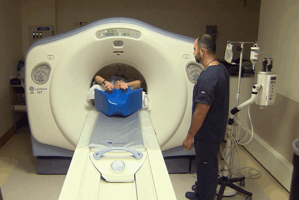
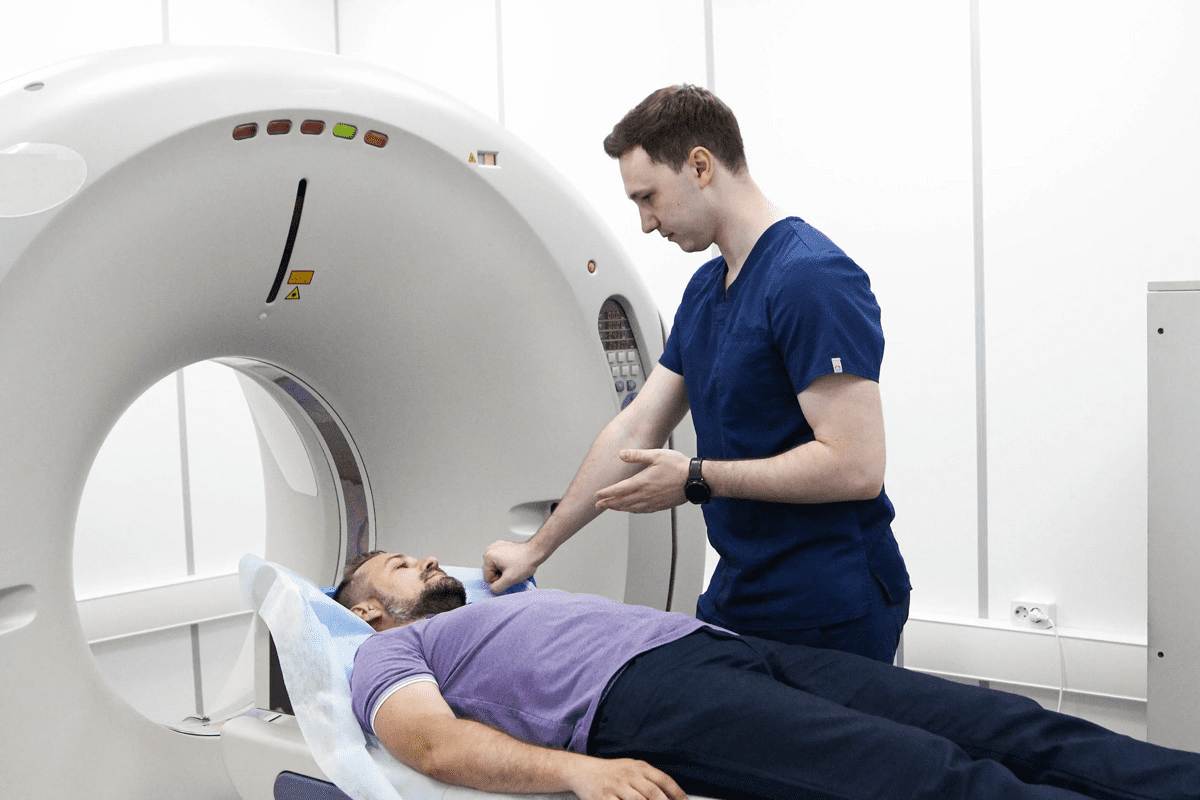
Positron Emission Tomography (PET) scans are key in cancer diagnosis. They help doctors find and understand cancer better. The scans measure Standardized Uptake Values (SUV), showing how much glucose the body uses.
A high SUV level might mean cancer cells are present. But what does this really mean? Knowing about FDG uptake and SUV levels is important for figuring out how serious cancer is and planning treatment.
The SUV level in PET scans is very important in oncology imaging. Getting it right can really change how well a patient does.
Key Takeaways
- PET scans measure SUV levels to detect cancerous cells.
- High SUV levels can indicate cancer, but require careful interpretation.
- Understanding SUV levels is vital for effective cancer diagnosis and treatment.
- SUV levels help determine the severity of cancer and guide treatment plans.
- Accurate interpretation of SUV levels significantly impacts patient outcomes.
Understanding SUV in PET Scan Imaging
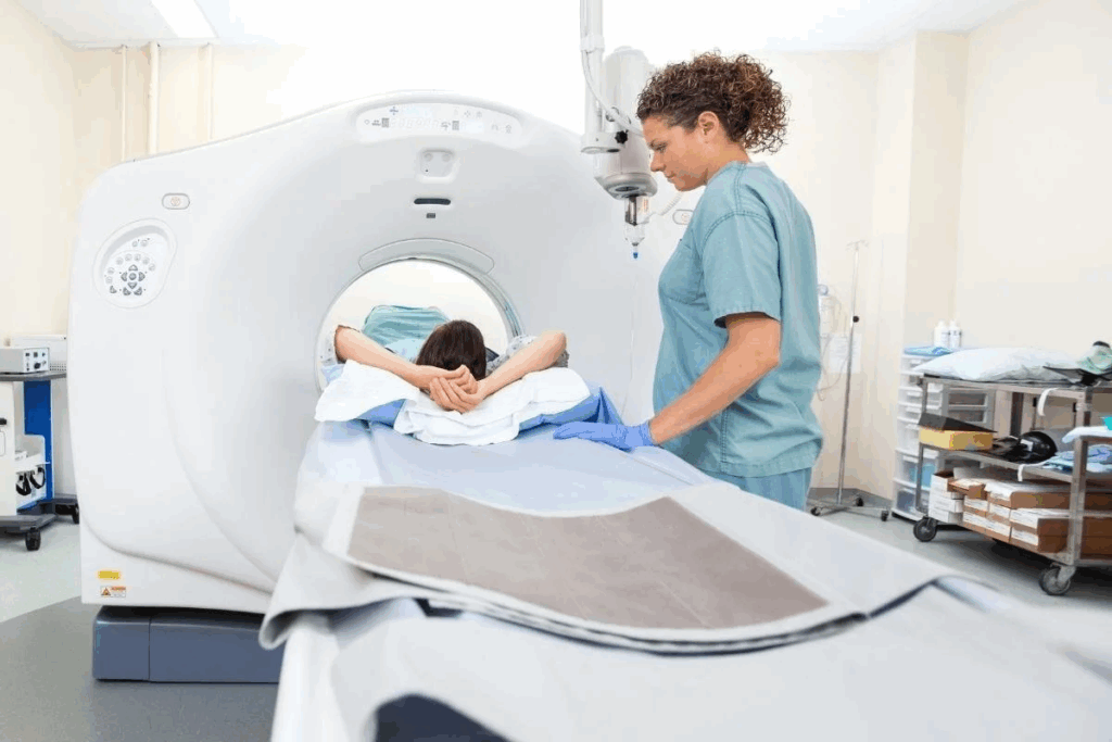
SUV, or Standardized Uptake Value, is key in PET scans. It shows how much radiotracers are taken up by the body. In PET scans, SUV helps measure tissue activity, mainly for cancer diagnosis and tracking.
Definition of Standardized Uptake Value (SUV)
The Standardized Uptake Value (SUV) is a way to measure radiotracer uptake. It compares the activity in a tissue area to the average body uptake. It’s figured out by looking at tissue activity, the dose given, and the patient’s weight.
Key aspects of SUV include:
- Quantification of radiotracer uptake
- Normalization to injected dose and body weight
- Useful in assessing metabolic activity
The Role of SUV in Medical Imaging
SUV is very important in medical imaging, like in cancer care. It helps in:
- Spotting cancer by showing high metabolic activity areas
- Tracking how treatments work by watching SUV changes
- Figuring out tumor aggressiveness based on metabolic activity
Knowing about SUV helps doctors make better choices for patient care.
The Science Behind FDG Uptake in Cancer Detection
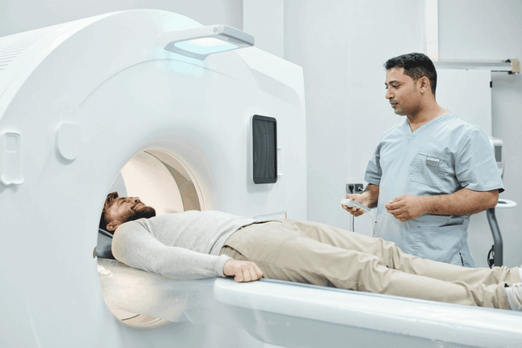
Cancer cells have a special way of using energy that PET scans can spot. This is because they use glucose differently than normal cells. This difference is known as the Warburg effect.
How FDG Mimics Glucose in the Body
FDG, or Fluorodeoxyglucose, acts like glucose in the body. It gets taken into cells the same way glucose does. But, unlike glucose, it can’t move forward in the energy-making process.
This means FDG stays in cells that use a lot of glucose. This includes cancer cells. So, PET scans can find these cells because they take up more FDG.
The Warburg Effect in Cancer Metabolism
The Warburg effect is when cancer cells use glycolysis for energy, even with oxygen around. This is different from normal cells, which use oxygen for energy. This change means cancer cells need more glucose, leading to more FDG uptake.
Why Cancer Cells Show Increased Glucose Consumption
Cancer cells need a lot of energy because they grow fast. The Warburg effect helps them use glycolysis for energy. They also need to make new parts quickly, which glycolysis helps with.
| Characteristics | Normal Cells | Cancer Cells |
| Primary Energy Source | Oxidative Phosphorylation | Glycolysis (Warburg Effect) |
| Glucose Consumption | Low | High |
| FDG Uptake | Low | High |
Knowing how FDG works in cancer detection is key to understanding PET scans. Cancer cells’ unique energy use makes FDG-PET scans very useful in finding cancer.
How SUV is Calculated in PET Imaging
It’s important to know how SUV is calculated to understand PET scan results. The SUV calculation uses a formula that considers several key factors.
Mathematical Formula for SUV Calculation
The SUV formula is: SUV = (tissue activity concentration) / (injected dose / body weight). This formula gives a semi-quantitative measure of the radiotracer uptake in the tissue of interest.
The tissue activity concentration comes from PET images. The injected dose is the radiotracer amount given to the patient, in megabecquerels (MBq). The body weight is in kilograms (kg).
Factors Affecting SUV Measurement Accuracy
Several factors can impact SUV measurement accuracy. These include:
- Patient factors: Blood glucose levels, body composition, and recent food intake can influence SUV values.
- Scanner factors: The PET scanner type, calibration, and reconstruction algorithm used can affect SUV measurements.
- Image acquisition and processing: The timing, duration, and attenuation correction methods used can also impact SUV accuracy.
Normalization Methods in SUV Calculation
To make SUV measurements more consistent, several normalization methods are used. These include:
- Body weight normalization: This is the most common method, where SUV is normalized to the patient’s body weight.
- Lean body mass normalization: This method normalizes SUV to lean body mass, which is more accurate for patients with a high BMI.
- Body surface area normalization: Some studies suggest normalizing SUV to body surface area may provide more consistent results across different patient populations.
Understanding these factors and normalization methods helps healthcare professionals accurately interpret SUV values in PET imaging. This leads to better diagnosis and treatment planning.
Types of SUV Measurements in Oncology
PET scans in oncology use several SUV metrics to understand cancer metabolism. These measurements help doctors see how severe and widespread cancer is in the body.
SUVmax: Maximum Standardized Uptake Value
SUVmax is the highest SUV value in a tumor area. It shows the most active part of the tumor. It’s a key metric for measuring cancer severity.
- Provides a snapshot of the most active tumor area
- Useful for monitoring treatment response
- Can be influenced by image noise and reconstruction algorithms
SUVmean: Average Uptake Value
SUVmean is the average SUV in a tumor area. It gives a broader view of tumor metabolism. It’s less affected by noise than SUVmax.
- Represents overall tumor metabolic activity
- Less affected by outliers and image noise
- Useful for assessing total tumor burden
SUVpeak and Other Measurement Types
SUVpeak averages SUV values in a small tumor area. Other metrics like SUVtotal and Metabolic Tumor Volume (MTV) also give insights into tumors.
These SUV measurements help doctors evaluate cancer metabolism. Each has its own benefits and drawbacks. Knowing these metrics is key for accurate diagnosis and treatment.
Patient Preparation and Its Impact on FDG Uptake
Getting ready for a PET scan is key to getting good results. It helps doctors make accurate diagnoses and treatment plans.
Fasting Requirements Before PET Scanning
Fasting is a big part of getting ready for a PET scan. Patients usually need to fast for 4 to 6 hours. This helps keep their blood sugar levels low, which is important for the scan.
Table: Fasting Guidelines for PET Scan
| Fasting Duration | Recommended Blood Glucose Level |
| 4-6 hours | < 200 mg/dL |
| > 6 hours | < 150 mg/dL |
Blood Glucose Level Management
Keeping blood sugar levels in check is vital for a good PET scan. High blood sugar can make FDG uptake lower. This might lead to wrong SUV measurements.
People with diabetes should talk to their doctor about managing their blood sugar before the scan.
Physical Activity Restrictions
Staying active can also affect FDG uptake. Patients are told to avoid hard exercise before the scan. This helps prevent FDG from being taken up by muscles.
Normal SUV Ranges in Different Body Tissues
Normal SUV ranges in body tissues help doctors diagnose and track cancer. Knowing these ranges is key for reading PET scans right.
Baseline SUV Values in Healthy Organs
Healthy organs have specific SUV values. These values can change a bit from person to person. For example, the liver’s high metabolic rate makes its SUV value high.
Liver SUV is a common reference because it’s consistent. It usually falls between 2.0 and 2.5. But, it can change based on blood sugar levels and the PET scan method.
Normal Liver SUV as Reference Standard
The liver’s SUV is a good standard for comparing other tissues. A normal liver SUV is between 2.0 and 2.5. This range can be affected by blood sugar levels and how long it takes to scan after FDG injection.
| Organ/Tissue | Typical SUV Range |
| Liver | 2.0 – 2.5 |
| Mediastinal Blood Pool | 1.5 – 2.0 |
| Brain | 6.0 – 8.0 |
Mediastinal Blood Pool Reference Values
The mediastinal blood pool is another key reference. It shows the average SUV in major blood vessels in the mediastinum. Its SUV range is usually 1.5 to 2.0. This helps doctors check the activity of mediastinal lesions.
Knowing these SUV ranges is essential for PET scan interpretation. It helps doctors tell the difference between harmless and possibly cancerous tissues.
What SUV Levels Are Considered Suspicious for Cancer?
Understanding SUV values is key to accurately diagnosing cancer. SUV levels in PET scans help tell apart cancerous and non-cancerous tumors. This information guides treatment plans.
General Threshold Values for Malignancy
There’s no one SUV value that means cancer. But, SUV values above 2.5 to 3.0 are often seen as cancerous. The exact value can change based on the PET/CT system and imaging method.
Key factors influencing SUV threshold interpretation include:
- The type of cancer being evaluated
- The location of the tumor
- Patient-specific factors such as blood glucose levels
Variation by Cancer Type and Location
SUV levels that suggest cancer can differ a lot. For example, aggressive lymphomas might show high SUV values. But, some prostate cancers might have lower SUV values.
Examples of SUV variation by cancer type:
- Lung nodules: SUV > 2.5 often considered suspicious
- Lymphoma: High SUV values common, often > 10
- Breast cancer: Variable SUV, with some types showing low uptake
The Concept of SUV Cutoff Values
The idea of SUV cutoff values helps standardize PET scan readings. But, finding one value for all cancers is hard. This is because tumors vary and so do PET scan methods.
“The use of SUV cutoff values must be tailored to the specific clinical context and cancer type, taking into account the limitations and variability of PET imaging.”
— Expert in Nuclear Medicine
It’s important to grasp these details for accurate cancer diagnosis and treatment planning. SUV levels from PET scans play a big role in this.
SUV Values Across Different Cancer Types
SUV values mean different things for different cancers. This affects how doctors diagnose and treat cancer. It’s key to understand these differences for the best care.
Lung Cancer SUV Patterns
Lung cancer is common, and PET scans help diagnose and stage it. SUV values in lung cancer can vary a lot. This depends on the tumor’s type and how aggressive it is. Generally, higher values mean more aggressive tumors.
A study showed lung adenocarcinomas have lower SUV values than squamous cell carcinomas. Knowing this helps doctors choose the right treatment.
Lymphoma SUV Characteristics
Lymphomas include Hodgkin lymphoma (HL) and non-Hodgkin lymphoma (NHL). SUV values in lymphoma help with staging, checking treatment success, and finding relapse. Lymphomas usually take up a lot of FDG, with SUV values over 10.
The amount of FDG uptake varies by lymphoma type. For example, HL and aggressive NHL have higher SUV values than indolent NHL.
Breast Cancer SUV Profiles
In breast cancer, SUV values tell us about tumor biology and how aggressive it is. Higher SUV values often mean more aggressive tumors. This includes higher histological grade and hormone receptor negativity.
Using SUV values in breast cancer is key for checking how well neoadjuvant chemotherapy works. It also helps predict patient outcomes.
Colorectal and Other GI Cancer SUV Patterns
Colorectal cancer, like other GI cancers, shows different SUV values. The primary tumor’s SUV value helps understand tumor aggressiveness and prognosis. SUV values also help find and track metastases in lymph nodes and distant organs.
In colorectal cancer, a higher SUV value in the primary tumor means a higher risk of recurrence and worse survival.
Knowing SUV patterns in various cancers is vital for accurate PET scan interpretation. It helps make better clinical decisions.
Non-Cancerous Causes of Elevated FDG Uptake
It’s important to know that high SUV values don’t always mean cancer. Many other things can cause FDG uptake to go up. Non-cancerous conditions can make glucose metabolism higher, leading to higher SUV readings on PET scans.
Inflammatory and Infectious Conditions
Inflammatory and infectious processes can greatly affect FDG uptake. For example, pneumonia, abscesses, and tuberculosis can cause SUV values to rise. This is because immune cells are working harder.
A patient with pneumonia might show high FDG uptake in the lungs. This could look like cancer. But, knowing the patient’s situation and other test results is key to telling them apart.
Metabolic Factors Influencing Uptake
Metabolic factors like diabetes and obesity can change how FDG is distributed. High blood sugar can make FDG uptake different because glucose and FDG compete for cells.
| Metabolic Condition | Effect on FDG Uptake |
| Diabetes | Alters FDG uptake due to high blood glucose |
| Obesity | Increased FDG uptake in fat tissues |
Medication Effects on SUV Readings
Some medicines can change how FDG uptake works. For instance, granulocyte-colony stimulating factor (G-CSF) can make bone marrow more active. This leads to more FDG uptake.
Post-Treatment Inflammatory Changes
Changes after treatment, like surgery or radiation, can also raise SUV values. These changes are part of the healing process. They might look like cancer if not seen in the right context.
Knowing about these non-cancerous reasons for high FDG uptake is vital for reading PET scans right. Doctors need to look at the whole picture of the patient. This includes symptoms, medical history, and other test results to make good decisions.
Interpreting PET Scan SUV Values in Clinical Practice
Understanding PET scan SUV values is key in medical practice. It needs a deep grasp of the technology and the patient’s health. Getting these values right is vital for spotting cancer, planning treatments, and keeping track of progress.
Qualitative vs. Quantitative Assessment
Reading PET scan SUV values involves both looking and measuring. Qualitative assessment is about seeing how much activity there is compared to the background. On the other hand, quantitative assessment uses SUV values to give a clearer picture of metabolic activity.
Qualitative methods can be hit-or-miss, depending on who’s looking. But, they’re quick. Quantitative methods offer a more precise look but can be affected by technical and biological factors.
The Role of Visual Interpretation
Looking at PET scans is a big part of the initial check-up. It helps doctors spot unusual activity and see how widespread the disease is. Visual assessment is great for spotting disease spread and possible metastases.
But, it’s important to also use SUV values for a more accurate reading.
Integration with Other Imaging Modalities
Reading PET scan SUV values often goes hand-in-hand with other scans like CT or MRI. Multi-modality imaging helps pinpoint where the abnormal activity is and how it relates to the body’s structures.
This combined method boosts accuracy and helps in planning treatments. It gives a fuller view of the disease.
SUV Changes During Cancer Treatment
SUV changes during treatment give us important clues about how well cancer therapy is working. By watching these changes, doctors can see if a patient is responding well to treatment.
Monitoring Treatment Response with Serial SUV Measurements
Serial SUV measurements are key in managing cancer treatment. By comparing SUV values before and after starting treatment, doctors can see if the therapy is effective. A big drop in SUV levels usually means the treatment is working well.
“Using serial PET scans to track SUV changes helps tailor treatment plans to each patient,” says a top oncologist. “It makes cancer care more personal.”
Predictive Value of Early SUV Changes
Early SUV changes can predict how well treatment will work. Studies show that patients with big SUV drops early on tend to live longer. This helps doctors make changes to treatment plans sooner.
- Early spotting of non-responders
- Adjusting treatment plans
- Better patient outcomes
Metabolic Complete Response vs. Partial Response
Metabolic response to treatment is split into complete and partial responses based on SUV changes. A complete response means SUV levels are back to normal, showing no active cancer. A partial response means SUV levels drop but not to normal, showing some cancer left.
Knowing the difference between these responses is key for deciding what to do next in patient care. A complete metabolic response usually means a better outlook.
Limitations of Using SUV for Cancer Diagnosis
Using SUV for cancer diagnosis has its limits. These need to be understood to get the most from PET scan results.
Technical Variability in SUV Measurement
There are many reasons for SUV measurement variability. These include differences in scanner hardware and software, image reconstruction, and patient preparation. For example, scanner calibration can impact SUV accuracy.
“Standardizing PET imaging protocols is key to reduce technical variability,” a study on SUV standardization highlights.
Biological Factors Affecting Interpretation
Biological factors greatly influence SUV value interpretation. Things like patient blood glucose levels, inflammation, and certain medications can affect FDG uptake. This can lead to SUV value misinterpretation.
For instance, high blood glucose can lower FDG uptake in cancer cells. This results in lower SUV values.
Scanner and Protocol Differences
Differences in scanner models and protocols also affect SUV measurements. Scanners vary in sensitivity and resolution, impacting FDG detection and quantification.
Also, image acquisition protocols like time per bed position and attenuation correction can influence SUV values. It’s vital to consider these when comparing SUV values across scans or institutions.
False Positives and False Negatives in SUV Interpretation
Getting SUV values right in PET scans is key for cancer diagnosis. But, false positives and false negatives often get in the way. It’s vital for doctors to grasp these issues to make better choices.
Common Causes of False Positive SUV Readings
False positive SUV readings can happen for many reasons. These include:
- Inflammatory processes: Conditions like sarcoidosis or post-surgical inflammation can lead to increased FDG uptake.
- Infectious diseases: Active infections can cause elevated SUV values, mimicking cancerous activity.
- Benign tumors: Certain non-cancerous growths can exhibit high metabolic activity.
- Physiological uptake: Normal physiological processes, such as uptake in brown adipose tissue, can be misinterpreted.
Scenarios Leading to False Negative Results
False negative results are also a big problem. They can lead to missed diagnoses or delayed treatment. Common scenarios include:
- Small tumor size: Tumors that are too small may not be detectable due to limitations in PET scan resolution.
- Low metabolic activity: Tumors with low glucose metabolism may not show significant FDG uptake.
- High blood glucose levels: Elevated blood glucose can competitively inhibit FDG uptake in tumor cells.
- Technical issues: Problems with the PET scanner or image reconstruction can lead to inaccurate SUV measurements.
Strategies to Minimize Misinterpretation
To cut down on false positives and false negatives, several strategies can be used:
- Clinical correlation: Integrating PET findings with clinical information and other diagnostic tests.
- Image fusion: Combining PET with CT or MRI to provide more accurate anatomical localization.
- Standardized protocols: Ensuring consistent patient preparation and imaging protocols.
- Quantitative analysis: Using metrics beyond SUV, such as metabolic tumor volume (MTV) and total lesion glycolysis (TLG).
By understanding and tackling the causes of false positives and false negatives, doctors can improve SUV interpretation in PET scans. This will help in better cancer diagnosis and treatment.
Advanced PET Metrics Beyond Standard SUV
PET imaging has evolved, introducing advanced metrics beyond SUV. These metrics give deeper insights into tumor biology. They are key for personalized cancer treatment, guiding treatment choices.
Metabolic Tumor Volume (MTV)
Metabolic Tumor Volume (MTV) shows the tumor tissue with high FDG uptake. Unlike SUV, MTV gives a detailed look at tumor size. It’s a strong predictor for cancers like lymphoma and lung cancer.
Calculating MTV involves setting a threshold for FDG uptake. This can be done automatically or manually. The choice of threshold is critical and is the focus of ongoing research.
Total Lesion Glycolysis (TLG)
Total Lesion Glycolysis (TLG) combines tumor volume and metabolic activity. It’s found by multiplying MTV by the average SUV in the tumor. TLG offers a broader view of tumor metabolism, possibly better than SUV alone.
Studies show TLG can predict treatment success and patient outcomes. A drop in TLG after starting treatment suggests a good response, even with small SUV changes.
Texture Analysis and Radiomics
Texture analysis and radiomics are at the forefront of PET imaging. They extract features from PET images, like heterogeneity and texture. This approach goes beyond traditional metrics, focusing on FDG distribution in tumors.
Radiomics, a wider field, turns imaging data into usable data for analysis. It aims to find new biomarkers linked to outcomes and tumor biology.
While these metrics are promising, challenges exist. Standardizing image and analysis protocols is essential for consistency. Yet, their use in clinical practice could greatly improve PET imaging in oncology.
What Patients Should Know About SUV Values
Knowing about SUV values is key for patients getting PET scans for cancer. SUV, or Standardized Uptake Value, shows how much glucose different tissues take up. This is important for finding, checking, and tracking cancer treatment.
Understanding Your PET Scan Report
Your PET scan report might have terms and numbers you don’t know. The SUV value is a main thing your doctor will check. A high SUV value often means more glucose uptake, which could mean cancer. But, your doctor also looks at the uptake pattern, where the issues are, and how they’ve changed over time.
It’s important to talk about your PET scan report with your doctor. They can explain what the SUV values mean for you. They’ll tell you if the values are normal or if there’s a problem.
Questions to Ask Your Doctor About SUV Results
It’s good to have questions ready when you talk to your doctor about PET scan results. Some important questions include:
- What does the SUV value for the area of concern mean?
- How does this SUV value compare to previous scans?
- Could other things like inflammation or infection affect the SUV value?
- What steps should we take next based on these SUV results?
Emotional Impact of SUV Findings
Getting PET scan results can be tough emotionally. If your SUV values are higher than expected, it can be really hard. Remember, SUV values are just one part of your diagnosis and treatment plan. Your healthcare team is there to support you, giving you context and guidance based on your health and history.
Being informed and asking questions can make you feel more in charge of your care. If you’re feeling overwhelmed, look into support groups or counseling. They can offer emotional support during this time.
Conclusion: The Clinical Value of SUV in Cancer Management
The Standardized Uptake Value (SUV) is key in cancer care. It gives vital info for diagnosis and treatment planning. It helps doctors see how active tumors are and how well treatments are working.
SUV shows how much glucose tumors use. This is important for finding cancer, tracking treatment, and predicting outcomes. As PET scan tech gets better, SUV’s role in cancer care will grow. This means more tailored care for patients.
Using SUV in care helps doctors make better diagnoses and plans. It leads to better patient results. As cancer research advances, SUV will keep being a big part of good patient care.
FAQ
What is SUV in PET scan imaging?
SUV stands for Standardized Uptake Value. It’s a way to measure how much a PET scan picks up in the body. This helps doctors see how active tissues are, like cancer cells.
How is SUV calculated in PET imaging?
To find SUV, a formula is used. It looks at the activity in tissues, the dose given, and the patient’s weight. Things like how the image is made and the patient’s prep can also affect the results.
What are the different types of SUV measurements used in oncology?
In oncology, SUV measurements include SUVmax, SUVmean, and SUVpeak. Each gives a different view of how tumors work.
How does patient preparation affect FDG uptake?
Getting ready for a PET scan is key. Patients need to fast, keep blood sugar levels right, and avoid hard exercise. This helps get accurate readings.
What are normal SUV ranges in different body tissues?
SUV values vary by tissue. For example, the liver is usually around 2-3, and blood pools in the chest are lower. Knowing these helps spot problems.
What SUV level is considered suspicious for cancer?
No single SUV number means cancer. But, values over 2.5-3 might suggest cancer, depending on the situation. Always look at the whole picture, not just the number.
Can non-cancerous conditions cause elevated FDG uptake?
Yes, things like infections or inflammation can make FDG uptake go up. It’s important to think about these when looking at PET scans to avoid mistakes.
How are PET scan SUV values interpreted in clinical practice?
Doctors look at SUV values in two ways: by eye and with numbers. They also use other tests and patient info to make a diagnosis and plan treatment.
What are the limitations of using SUV for cancer diagnosis?
SUV has its limits. Things like how the scan is done and the body’s own biology can affect the results. These can lead to wrong conclusions if not considered.
Are there advanced PET metrics beyond standard SUV?
Yes, there are. Metrics like Metabolic Tumor Volume (MTV) and Total Lesion Glycolysis (TLG) give more details on tumors. They help doctors understand tumors better.
What should patients know about SUV values?
Patients should know SUV values are part of their PET scan report. They should talk to their doctor about what their SUV values mean for their health and treatment.
Reference
- Liberti, M. V., & Locasale, J. W. (2016). The Warburg effect: How does it benefit cancer cells? Trends in Biochemical Sciences, 41(3), 211–218. https://pubmed.ncbi.nlm.nih.gov/26778478/


