Last Updated on November 27, 2025 by Bilal Hasdemir

At Liv Hospital, we use the latest nuclear medicine tech, like the gamma camera nuclear medicine system. It’s a special machine that catches gamma rays emitted from drugs given to patients. These rays help us see inside the body and provide functional images of organs and tissues for accurate diagnosis and treatment.
First, we give patients a small dose of a special drug. This drug sends out rays that the gamma camera picks up. This tool is key for checking how organs work and finding diseases.
It also helps us see how treatments are going and if cancer has spread. For more on gamma cameras,
Key Takeaways
- Gamma cameras detect gamma rays emitted by radiopharmaceuticals.
- They provide detailed information about organ function.
- The diagnostic process is painless and does not cause discomfort.
- Gamma cameras are used for scintigraphy scans.
- Nuclear medicine technologists operate the gamma camera.
The Fundamentals of Nuclear Medicine Imaging

Nuclear medicine imaging uses special substances to see inside the body. It helps doctors understand how the body works and what might be wrong. This method is key in today’s medicine.
Basic Principles of Medical Imaging
Nuclear medicine imaging finds radiation in the body. It uses tiny amounts of radioactive materials, or radiotracers. These are given to the body in different ways.
These materials go to certain parts of the body. Then, special cameras detect the radiation. This helps doctors see where the materials are and what they mean.
Role of Radioactive Tracers in Diagnostics
Radioactive tracers are very important in nuclear medicine. They help find specific parts of the body or how they work. Doctors choose the right tracer based on what they need to know.
These tracers are very good at finding certain things. For example, they can spot cancer or check how well the heart works. This makes diagnosing easier and more accurate.
Evolution of Nuclear Medicine Technology
Nuclear medicine technology has grown a lot. New cameras can see more clearly and accurately. This means doctors can make better diagnoses.
Now, nuclear medicine works with other imaging like CT and MRI. This combination, like SPECT/CT, gives doctors a full picture of what’s happening in the body. It’s a big step forward in understanding diseases.
What Is a Gamma Camera: Definition and Purpose

A gamma camera is a key tool in nuclear medicine. It helps us see how the body works. We use it to find and manage diseases.
Definition of Gamma Cameras in Medical Context
A gamma camera, also known as a scintillation camera or Anger camera, is used to see where radioactive material is in the body. It catches gamma radiation from a special drug given to the patient. This lets us make images that show how different parts of the body work.
Gamma cameras are very important in nuclear medicine. They help us see how organs and tissues work. This is key for diagnosing and tracking diseases.
Historical Development of Gamma Camera Technology
The gamma camera has changed a lot over the years. It was first made in the 1950s by Hal Anger. Back then, it could only detect gamma radiation but not very well.
Now, thanks to better technology, we have SPECT systems. These can make two-dimensional and three-dimensional images. This makes finding diseases easier and more accurate.
Advantages Over Other Imaging Modalities
Gamma cameras have big advantages over other imaging methods. They show how the body works, not just what it looks like. This is really helpful for diseases that affect how organs function.
Also, gamma cameras are very good at finding diseases early. They use special drugs to focus on certain parts of the body. This makes them very accurate for diagnosis.
| Imaging Modality | Primary Use | Key Benefits |
| Gamma Camera | Functional imaging, disease diagnosis | Early disease detection, functional information |
| X-ray/CT Scan | Anatomical imaging, structural abnormalities | High-resolution anatomical images |
| MRI | Soft tissue imaging, detailed anatomy | Excellent soft tissue contrast, detailed images |
Understanding gamma cameras helps us see their importance in nuclear medicine. They keep getting better, helping doctors diagnose and treat patients more effectively.
The Science of Gamma Radiation in Medical Imaging
Gamma rays are a type of electromagnetic radiation used in medical imaging. They help create detailed pictures of what’s inside our bodies. Gamma radiation is used in many tests to see how our bodies work and find health problems.
Understanding Gamma Rays and Their Properties
Gamma rays have a lot of energy and are very short. This makes them great for going through tissues and showing pictures of organs inside. Their special properties, like being emitted by certain isotopes, are key for medical tests.
We use radiopharmaceuticals, which are compounds with radioactive isotopes that give off gamma rays. We pick these isotopes for their right half-lives and energy levels. This makes sure the imaging is safe and works well.
Radiopharmaceuticals: The Source of Gamma Radiation
Radiopharmaceuticals are made to go to certain parts of the body. They build up in areas we want to see. As they break down, they send out gamma rays. These rays are caught by gamma cameras to make pictures for doctors.
Choosing the right radiopharmaceutical depends on what we want to find. Different isotopes and compounds are used for different health issues, like cancer, heart problems, and brain diseases.
How Gamma Rays Interact with Body Tissues
Gamma rays can be absorbed, scattered, or pass through tissues in different ways. The way they are detected outside the body tells us about where the radiopharmaceuticals are in the body. This is how we get images of what’s going on inside.
This interaction is key for gamma camera imaging. It lets us see how the body works and find health issues.
Core Components of a Gamma Camera Nuclear Medicine System
The gamma camera is a complex system used in nuclear medicine. It has several key parts that help create clear images. New technology and quality checks have made gamma scans more reliable and clear.
We will look at the main parts of a gamma camera system. We’ll see how each part helps make the system work well and images clear.
The Collimator: Gateway for Gamma Rays
The collimator is key for guiding gamma rays. It lets the camera know where the radiation is coming from in the body. It’s made of lead with holes that let gamma rays through but block others.
The collimator’s design is important for the camera’s resolution and sensitivity. Different collimators are used for different needs and gamma radiation energies.
Scintillation Crystal: Converting Radiation to Light
The scintillation crystal is vital for turning gamma radiation into light. When gamma rays hit the crystal, it lights up. This light is then detected and made stronger.
Thallium-activated sodium iodide (NaI(Tl)) is the most used crystal material. The crystal’s thickness and quality affect the camera’s performance.
Photomultiplier Tubes and Signal Amplification
Photomultiplier tubes (PMTs) detect and boost the light signals from the crystal. They are very good at catching faint light. This makes them perfect for this job.
The signals from PMTs are mixed and processed. This helps figure out where and how strong the gamma radiation is. This information is used to make an image.
Digital Processing Systems and Image Formation
The digital system turns the electrical signals into a digital image. It uses complex algorithms to fix issues like energy resolution. This makes a high-quality image.
Today’s gamma cameras have advanced digital systems. These systems can enhance and rebuild images. This makes the images even more useful for diagnosis.
In summary, the main parts of a gamma camera system work together. They capture, process, and create images of the body’s functions. Knowing about these parts helps us understand gamma camera imaging technology.
Types of Gamma Camera Systems and Techniques
Gamma cameras come in many types and techniques. We use them to get detailed info for many health issues.
Planar Imaging: Two-Dimensional Gamma Scans
Planar imaging is a key method in gamma cameras. It makes two-dimensional pictures of where radioactive substances are in the body. This helps us see how organs and tissues work and look.
This method, called scintigraphy, is safe and doesn’t hurt much. We give a radioactive substance to the patient. It goes to the area we want to check.
SPECT: Three-Dimensional Nuclear Imaging
SPECT imaging is a big step up in gamma camera tech. The camera moves around the patient to get images from different sides. Then, these images are turned into 3D pictures for better details.
SPECT is great for looking at complex body parts and how they work. It helps us understand and treat many diseases, like heart and brain problems.
Hybrid Systems: SPECT/CT and Other Combinations
Hybrid systems mix gamma camera info with details from CT or MRI. SPECT/CT is a top example. It shows how things work and where they are, thanks to both SPECT and CT.
These systems are getting better. Scientists are working on adding more imaging types to them. This will help us diagnose even better.
The Complete Gamma Camera Imaging Process
The gamma camera imaging process is complex, with many steps. We’ll cover the whole process, from patient prep to image improvement.
Patient Preparation Protocols
Patients must prepare well for a gamma camera scan. They might need to follow dietary rules, remove metal items, and stay hydrated. The exact prep steps depend on the study and the tracer used.
| Preparation Step | Description |
| Dietary Restrictions | Patients might need to fast or avoid certain foods and drinks before the scan. |
| Removal of Metallic Objects | They are asked to remove jewelry, glasses, or any other metallic objects that could interfere with the scan. |
| Hydration Instructions | They might be told to drink plenty of water to help the radiopharmaceutical distribute throughout the body. |
Radiopharmaceutical Administration and Uptake
The next step is giving a radiopharmaceutical, a compound with a radioactive tracer. This tracer targets specific body processes or organs. The way it’s given (intravenously, orally, or by inhalation) depends on the study.
After it’s given, the tracer spreads and builds up in the target area. How long it takes can range from a few minutes to hours, based on the tracer and the area being studied.
Image Acquisition Techniques and Parameters
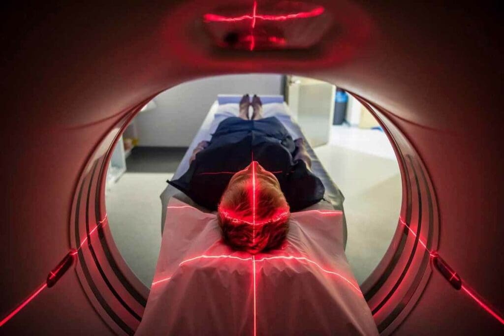
Once the tracer is in the body, the patient is placed under the gamma camera. The camera captures the gamma radiation from the tracer, showing detailed images of body processes. The camera settings, like energy window and collimator type, are chosen for the study.
Data Processing and Image Enhancement
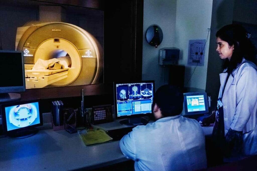
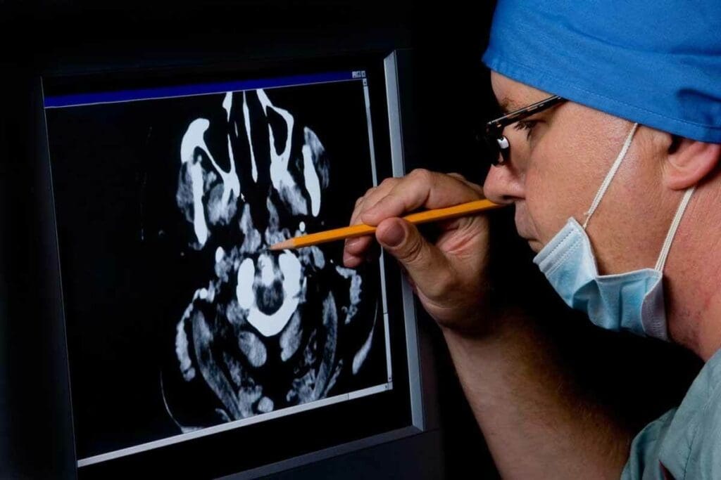
After capturing the images, the data is processed to improve image quality. Techniques like filtering and reconstruction are used to make clear and accurate images. These images are then checked by nuclear medicine experts to diagnose and monitor conditions.
Clinical Applications of Gamma Camera Imaging
Gamma cameras are key in modern medicine. They help doctors diagnose and track many conditions. These systems are used in many ways to improve patient care and accuracy.
Cardiac Function and Perfusion Studies
In cardiology, gamma cameras help check the heart’s function and blood flow. They use special drugs to see how blood moves through the heart. This is key for finding heart disease and planning treatment.
Myocardial perfusion imaging is a special use. It shows how the heart’s blood flow changes under stress and at rest. This helps spot patients at risk of heart problems.
Oncology: Tumor Detection and Staging
In cancer care, gamma cameras are vital for finding and checking tumors. They use drugs to see tumors and how active they are. This helps doctors know how far the cancer has spread and if treatment is working.
Sentinel lymph node imaging is another big use in cancer care. It finds the first lymph node cancer might spread to. This helps plan surgery and avoid removing too many lymph nodes.
Neurological Applications
Gamma cameras also help in neurology to find and track brain issues. They use drugs to see blood flow and find abnormal brain activity.
DaTSCAN is a special tool for finding Parkinson’s disease and other movement disorders. It shows where dopamine is in the brain.
Renal, Hepatobiliary, and Other Organ Studies
Gamma cameras also check how well other organs like the kidneys and liver work. They use special drugs to see how these organs function. This helps diagnose problems and check how well treatments are working.
Advancements in Gamma Imaging Technology
Gamma imaging camera technology has seen big improvements. These changes have made gamma scans more reliable and clear. This is a big step forward in personalized nuclear medicine.
Digital Detectors and Resolution Improvements
Digital detectors have changed gamma camera systems a lot. Digital detectors are more sensitive and have better resolution. They turn gamma radiation into digital signals for detailed body images.
Higher resolution is key for spotting small issues, like in cancer and heart disease. It helps doctors give better treatments and track patient progress better.
Software Innovations in Image Processing
Software has been a big help in gamma imaging. Advanced image processing algorithms make images clearer and more useful. They fix problems in the images, making them accurate.
Also, new software tools help doctors analyze images. These tools help measure important body functions. This helps doctors make better treatment plans.
Future Directions in Gamma Camera Technology
We’re expecting even more changes in gamma camera tech. Artificial intelligence (AI) and machine learning (ML) will likely be used. They could make image analysis faster and more accurate.
Future systems might also use new materials and detectors. This will make them even more sensitive and clear. These updates will keep improving nuclear medicine, leading to better treatments.
Conclusion
Gamma cameras are key in nuclear medicine. They give important info for diagnosing and treating diseases. These cameras turn radioactive energy from the body into images. This helps doctors understand and manage health issues.
Gamma cameras are important because they show how the body’s organs work. They use special drugs to spot problems in the heart, cancer, and brain. For more on gamma cameras, In short, gamma cameras are vital in nuclear medicine. They help doctors give better care by making detailed images. As tech gets better, gamma cameras will help even more, making patient care even better.
FAQ
What is a gamma camera in nuclear medicine?
A gamma camera is a tool in nuclear medicine. It detects gamma rays from radiopharmaceuticals. This gives important info about the body’s functions and structures.
How does a gamma camera work?
It detects gamma rays from radiopharmaceuticals in the body. A collimator directs these rays to a scintillation crystal. This crystal turns the radiation into light, which is then processed to create an image.
What is the role of radiopharmaceuticals in gamma camera imaging?
Radiopharmaceuticals emit gamma radiation. They are used in gamma camera imaging. This helps provide info about the body’s functions and structures, like blood flow and organ function.
What are the advantages of gamma camera imaging over other imaging modalities?
Gamma camera imaging has many benefits. It gives functional info about the body’s processes. It’s also very sensitive and can image the whole body or specific organs. This makes it a valuable tool for diagnosing and monitoring medical conditions.
What is SPECT imaging, and how does it differ from planar imaging?
SPECT imaging creates three-dimensional images of the body. Planar imaging makes two-dimensional images. SPECT gives more detailed info about the body’s structures and functions.
What are the clinical applications of gamma camera imaging?
Gamma camera imaging has many uses in medicine. It’s used for cardiac function studies, oncology, and neurological applications. It helps diagnose and monitor conditions like coronary artery disease, cancer, and neurological disorders.
How has gamma camera technology advanced in recent years?
Recent advances have improved image quality and sensitivity. They’ve also led to new applications, like hybrid systems that combine SPECT with CT or MRI. This enhances diagnostic capabilities.
What is the correct spelling for recording of radioactivity with gamma cameras?
The correct term is gamma camera imaging or scintigraphy.
What is a gamma scan?
A gamma scan is a test that uses a gamma camera. It detects gamma radiation from radiopharmaceuticals in the body. This provides info about the body’s functions and structures.
What is gamma imaging?
Gamma imaging is a medical imaging modality. It uses gamma cameras to detect gamma radiation from radiopharmaceuticals. This gives diagnostic info about the body’s functions and structures.
References
- Acharya, U. K., et al. (2023). Directed searching optimized texture-based adaptive gamma correction for medical image enhancement. Sensors, 23(12), Article 5678. https://pmc.ncbi.nlm.nih.gov/articles/PMC10239541/


