Last Updated on November 25, 2025 by Ugurkan Demir

At Liv Hospital, we know how important cardiac imaging is in today’s heart care. Techniques like CT and MRI are key for checking the structure and function of the heart. They help doctors make accurate diagnoses and create effective treatment plans.
We use the latest imaging tools to see the heart’s cross-sectional anatomy. This gives us important information about heart problems. By looking at heart anatomy on CT and MRI scans, our team can spot issues early. They then create treatment plans that fit each patient’s needs.

Cardiac imaging is key in today’s medicine. It lets doctors see the heart clearly. This helps a lot in diagnosing and treating heart diseases.
Cardiac imaging has come a long way. From simple X-rays to advanced scans like Cardiac CT and MRI, it has changed cardiology a lot.
It all started with X-rays, which showed little of the heart. Technological advancements brought us better tools like echocardiography, Cardiac CT, and MRI.
These new methods have changed how we diagnose heart diseases. They let us see the heart’s details, like chambers and valves. Cardiac CT and MRI are now key in cardiology, giving us deep insights into the heart.
Cross-sectional imaging, like Cardiac CT and MRI, is vital in heart diagnosis. They give us clear views of the heart’s anatomy. This helps doctors understand the heart better.
Seeing the heart from different angles has changed how we understand heart anatomy and problems. It’s key for spotting complex heart issues and planning treatments.
Cardiac MRI and CT angiography are big steps forward in heart imaging. They offer great detail and accuracy. These tools have made diagnosing and treating patients better.

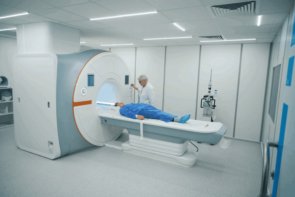
CT and MRI are key in cardiac imaging, each with its own strengths. We’ll look at how they differ and their roles in heart anatomy.
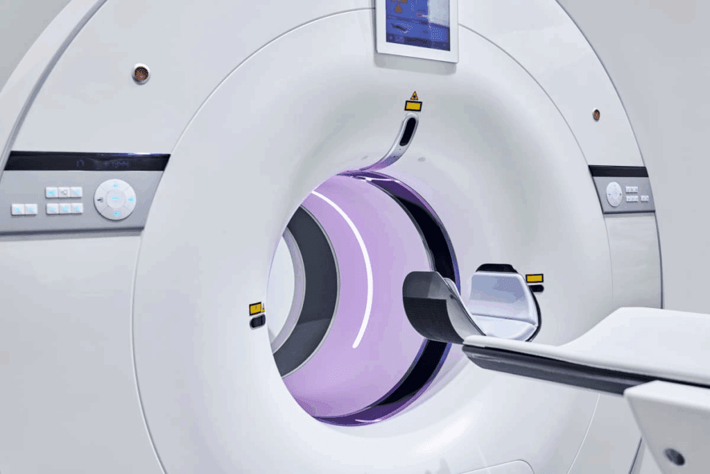
CT scans are great for spotting coronary artery disease and calcifications. They give clear images of the heart’s structure. This makes them perfect for seeing calcium in arteries, a sign of atherosclerosis.
Key benefits of CT scans include:
Cardiac MRI offers detailed images of the heart and its tissues. It helps check for heart damage, scar tissue, and blood flow issues. MRI’s soft tissue contrast is unmatched, making it essential for heart exams.
Key benefits of MRI include:
Choosing between CT and MRI depends on several factors. These include the clinical question, patient health, and what imaging options are available.
| Imaging Modality | Primary Use | Key Advantages |
| CT | Coronary artery disease assessment, calcification detection | High-speed, excellent for calcification visualization |
| MRI | Myocardial viability, scar tissue assessment, cardiac function evaluation | Superior soft tissue contrast, no ionizing radiation |
Knowing the strengths and weaknesses of each modality helps healthcare providers choose the best imaging for each patient.
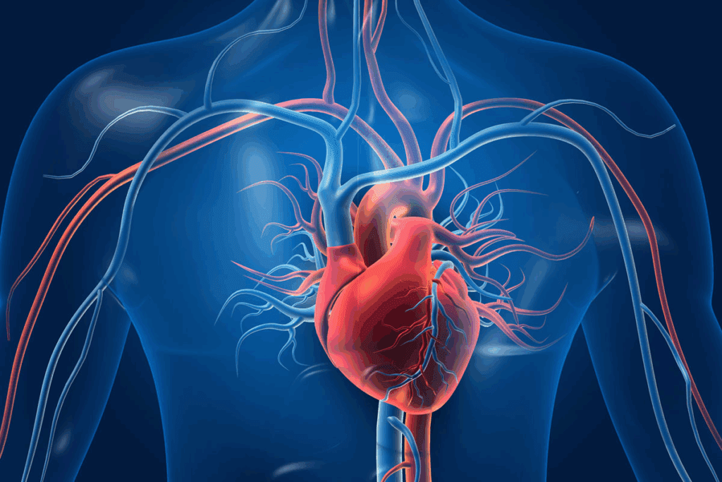
The heart’s cross-sectional anatomy is key in cardiovascular medicine. It’s vital for diagnosing and treating heart issues. Different imaging planes help us see the heart’s structure.
Axial views show the heart in a transverse plane. They help us see the heart’s chambers and major vessels.
Key structures visible in axial views include:
Coronal views give a frontal view of the heart. They show the heart’s structure and how its parts relate to each other.
Important landmarks in coronal views include:
| Plane | Key Visible Structures |
| Axial | Four chambers, major vessels, septum |
| Coronal | Cardiac silhouette, heart valves, coronary arteries |
| Sagittal | Heart’s anterior-posterior dimension, valve orientation |
Sagittal views show the heart’s length and how its parts relate to each other.
Sagittal views help in assessing:
By looking at the heart from different angles, doctors can understand it better. This is key for accurate diagnosis and treatment.
Cardiac CT technology has advanced, allowing us to see the heart’s anatomy with great detail. It uses ECG-gated multiplanar reconstructions to show the heart’s position and how it relates to nearby organs. This is key for a full cardiac check-up.
Cardiac CT scans are great for seeing coronary arteries and finding calcification. Calcification in the arteries is a sign of atherosclerosis. The amount of calcification can be measured with CT scans. This info is important for figuring out heart risk and deciding on treatments.
The four-chamber view from cardiac CT scans with contrast is very helpful. It shows the heart’s chambers and how the septum is doing. This view is key for checking the heart’s structure and function, spotting any issues or defects.
Cardiac CT scans also let us look closely at the heart valves. By seeing the valves at different times in the heart’s cycle, doctors can check their shape and how well they work. This is important for finding valve problems and planning the right treatment.
Lastly, cardiac CT scans help check the great vessels like the aorta and pulmonary arteries. These images are important for knowing the size, shape, and health of these big vessels. They help in diagnosing and managing problems with the great vessels.
Cardiac CT scans can help find out how bad coronary artery disease is, guide treatment choices, and see if interventions are needed. Looking at these four key images, doctors can fully understand the heart’s anatomy. This helps them make better decisions for patient care.
Magnetic Resonance Imaging (MRI) has changed how we look at the heart. It shows the heart’s soft tissues and how well it works. This helps us see the heart muscle and valves clearly.
We use MRI to check blood flow to the heart. It helps find areas where blood flow is low or blocked. It also spots scar tissue or damaged areas.
The four-chamber view is key in MRI. It shows the heart’s chambers and the wall between them. This view helps us see if the heart is the right size and working well.
It’s great for finding problems like holes in the heart wall or if the chambers are too big.
The short-axis view is important for looking at the heart muscle. It shows a cross-section of the heart. This helps us see if the muscle is thick enough and working right.
It’s also good for finding damaged areas or spots where blood flow is low.
The long-axis view shows the heart’s layout along its length. It helps us see how the chambers and valves work. This view is key for checking the mitral and tricuspid valves.
It also helps us look at the left ventricle’s outflow tract.
Cardiac MRI is great for checking how well the heart valves work. It can spot problems like valves that are too narrow or leak. By watching how the valves move, we can tell how well they’re working.
In short, these four views give us a full picture of the heart. MRI helps us understand the heart better. This leads to better care for patients.
Specialized views of the heart give us key insights into its structure and function. These advanced imaging methods are vital for diagnosing and treating heart conditions. We will look at four essential views in modern cardiology.
ECG-gated multiplanar reconstructions give precise heart images. They match image taking with the patient’s heart rhythm. This reduces motion blur, showing the heart’s structures clearly at certain times.
3D volume rendering shows the heart’s anatomy in full detail. It helps doctors see complex structures in three dimensions. This is great for planning surgeries and understanding how different parts of the heart relate to each other.
Perfusion imaging checks blood flow to the heart muscle. It’s key for spotting issues like ischemia. This method is essential for finding areas where blood flow is low, which might mean coronary artery disease.
Delayed enhancement imaging finds scar tissue in the heart muscle. It spots fibrosis, helping diagnose heart attacks and cardiomyopathy. This is a big help in understanding heart damage.
| Imaging Technique | Primary Use | Key Benefits |
| ECG-Gated Reconstructions | Minimizes motion artifacts | Clear images during specific cardiac phases |
| 3D Volume Rendering | Visualizes complex cardiac anatomy | Useful for pre-surgical planning |
| Perfusion Imaging | Assesses myocardial blood flow | Diagnoses ischemia and coronary artery disease |
| Delayed Enhancement | Detects scar tissue | Aids in diagnosing myocardial infarction and cardiomyopathy |
Heart radiology is key in modern heart care. It gives insights into the heart’s structure and function. Advances in CT and MRI have changed how we diagnose and treat heart diseases.
Heart radiology is mainly used to spot coronary artery disease (CAD). Cardiac CT scans can find calcium in arteries, a sign of CAD. CT angiography shows the arteries in detail, helping to find blockages and plan treatments.
Key benefits of cardiac CT in CAD diagnosis include:
Cardiac MRI is great for checking heart function and size. It gives precise measurements of heart chambers and how well they pump. This info is key for diagnosing heart failure and other heart problems.
| Parameter | Normal Value | Clinical Significance |
| Left Ventricular Ejection Fraction (LVEF) | 55-70% | Shows how well the left ventricle works |
| Left Ventricular End-Diastolic Volume (LVEDV) | 100-150 mL | Tells about the left ventricle’s preload |
| Right Ventricular Systolic Pressure (RVSP) | 15-25 mmHg | Sign of high blood pressure in the lungs |
Heart radiology is also important for planning surgeries and checking after them. It helps doctors plan complex surgeries like TAVR and see if patients can have surgery.
After surgery, imaging checks if the procedure worked and looks for any problems. For example, cardiac CT can check how well stents and valves work. MRI can see if the heart muscle is alive or scarred.
It’s key to know the difference between normal and abnormal heart anatomy for good patient care. When looking at heart images, we must spot both normal and abnormal signs to give the right diagnosis.
Normal heart images can sometimes look like problems. For example, a big crista terminalis or Eustachian valve might look like a mass or clot. Knowing what these normal parts look like is important for correct reading.
Cardiac CT and MRI can spot many issues, like scar tissue, inflammation, and heart defects. Cardiac ct scan anatomy is great for seeing coronary artery disease. MRI heart anatomy is best for soft tissue details, like checking if heart muscle is alive.
Some common problems include:
Consider a case where cross-sectional heart anatomy is key for diagnosis. A patient with chest pain gets a cardiac CT. The scan shows a big blockage in the left anterior descending coronary artery.
In another case, an MRI checks if heart muscle is alive. The MRI shows scarring in the inferior wall, meaning the heart muscle is damaged.
These examples show how important it is to read heart images right. Knowing normal and abnormal signs helps us give better diagnoses and treatment plans.
Cardiac imaging technology is getting better, helping us understand the heart better. The field is growing fast, thanks to new tech and ways to see the heart. This includes CT and MRI scans.
Heart radiology is getting better too, leading to more accurate diagnoses. This means doctors can treat patients more effectively. CT and MRI scans are key in checking the heart’s health early on.
As tech improves, so will our ability to diagnose and care for patients. The future of heart imaging looks bright, with ongoing research. This will make these technologies even better.
Using CT and MRI scans, doctors can give more precise diagnoses and treatments. This leads to better care for patients. It’s a step towards better health outcomes.
Heart anatomy images are key in checking the heart’s structure and function. They help doctors diagnose and treat heart conditions accurately.
Heart imaging has changed a lot. It moved from old methods to new ones like CT and MRI. These give detailed views of the heart.
CT scans show the heart’s details clearly. They help spot problems in the heart’s arteries and other issues. This makes them great for finding heart disease.
MRI shows the heart’s soft tissues well. It helps doctors see how the heart works and if it’s damaged. Plus, it doesn’t use harmful radiation.
Choosing between CT and MRI depends on what you need to know. CT is best for artery images. MRI is better for heart function and damage.
Important views include axial, coronal, and sagittal. Each view gives a different look at the heart. Together, they help doctors understand the heart fully.
Special views include ECG-gated images and 3D renderings. They also include perfusion and delayed enhancement scans. These help doctors make better decisions.
Heart images help find heart disease and check how well the heart works. They also help plan surgeries and check how well treatments work.
Challenges include telling normal from abnormal and spotting common problems. It takes skill and careful looking to get it right.
New imaging tech has made diagnosing better and care more precise. It helps doctors make better choices and treat patients more effectively.
Cardiac CT scans are key in seeing the heart’s arteries and finding blockages. This helps catch heart disease early and treat it.
MRI gives detailed info on the heart’s structure and how it works. It helps doctors see if the heart is damaged and how well it’s working. This guides treatment choices.
Subscribe to our e-newsletter to stay informed about the latest innovations in the world of health and exclusive offers!