Last Updated on November 4, 2025 by mcelik

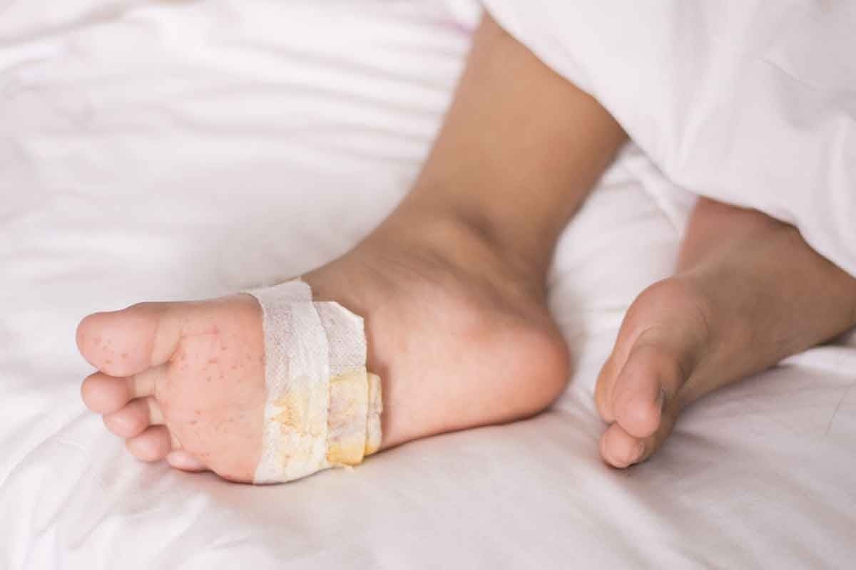
Did you know that hip pain affects millions of people worldwide? It really changes their life. Bursitis is a big reason for hip pain. It’s when the fluid-filled sacs around joints get inflamed. Learn how a Hip bursitis MRI reveals soft tissue damage, inflammation, and fluid buildup for accurate diagnosis.
These sacs, called bursae, help joints move smoothly. Knowing if you have hip bursitis is key to feeling better.
We use MRI to find out if you have hip bursitis. MRI for hip pain is very important. It shows detailed pictures of the soft tissues in your hip, like the bursae.
This allows for identification of trochanteric bursitis or other hip issues. This helps them decide the best treatment for you.
Key Takeaways
- MRI is a key tool for finding hip bursitis.
- Hip bursitis is when the bursae get inflamed, causing pain.
- Getting a correct diagnosis with an MRI is vital for treatment.
- Trochanteric bursitis is a specific type of hip bursitis.
- Advanced imaging helps understand hip pain causes.
To illustrate the impact of hip bursitis on daily life, we can examine some of the common challenges individuals face:

Hip bursitis is when the bursae in the hip get inflamed. Bursae are fluid-filled sacs that help bones and soft tissues move smoothly. Bursae play a key role in joint movement.
What is a Bursa and Its Function
A bursa is a small, fluid-filled sac that cushions bones, tendons, and muscles near joints. It reduces friction and allows for smooth movement. The hip has several bursae that help tendons and muscles glide over bony areas.
“The presence of bursae is essential for the normal functioning of the hip joint,” as they prevent irritation and inflammation of the surrounding tissues.
Common Types of Hip Bursitis
There are several types of hip bursitis, each affecting different bursae around the hip joint. The most common types include:
- Trochanteric bursitis: affecting the bursa near the greater trochanter
- Iliopsoas bursitis: involving the bursa located near the iliopsoas muscle
- Ischiogluteal bursitis: affecting the bursa near the ischial tuberosity
Each type of bursitis has its own symptoms and needs a specific diagnosis.
Risk Factors for Developing Hip Bursitis
Several factors increase the risk of developing hip bursitis, including:
- Repetitive motion or overuse of the hip joint
- Poor posture or biomechanics
- Direct trauma to the hip
- Underlying conditions such as arthritis or tendinopathy
Understanding these risk factors is crucial for early prevention and treatment of hip bursitis.
Symptoms and Clinical Presentation of Hip Bursitis

Hip bursitis shows up in many ways, making daily life tough. It’s caused by swelling in the bursae, which are fluid-filled sacs around the hip. This swelling can cause pain and make it hard to move.
This allows for identification of trochanteric bursitis or other hip issues.
Trochanteric bursitis is common and hurts the outer hip. The pain can be sharp and go down the thigh. Walking, climbing stairs, or lying on the side can make it worse.
People with this bursitis might feel tender when pressed on the outer hip. Sometimes, the pain is so bad it changes how they walk, causing a limp.
Iliopsoas Bursitis Symptoms
Iliopsoas bursitis hurts the groin or front of the hip. It’s often linked to activities like cycling or running. The pain feels deep and aching, sometimes with a snapping sound when moving the hip.
Ischiogluteal Bursitis Symptoms
Ischiogluteal bursitis hurts the buttock, getting worse with long sits. It makes it hard to stand up from sitting. The pain is usually near the ischial tuberosity.
Impact on Daily Activities
Hip bursitis symptoms can make everyday tasks hard. Even simple things like walking or getting dressed can be tough. It can also mess with sleep, as lying on the affected side hurts.
To illustrate the impact of hip bursitis on daily life, we can examine some of the common challenges individuals face:
| Activity | Common Challenges |
| Walking | Pain on the outer hip or groin area, limping |
| Climbing Stairs | Increased pain, specially with trochanteric bursitis |
| Sitting | Discomfort with ischiogluteal bursitis, pain when transitioning to standing |
| Sleeping | Pain when lying on the affected side |
Understanding these risk factors is crucial for early prevention and treatment of hip bursitis.
Initial Diagnosis of Hip Bursitis
Diagnosing hip bursitis starts with a detailed physical exam and patient history. A thorough clinical evaluation is key to accurately diagnose hip bursitis.
Physical Examination Techniques
A physical exam is essential for diagnosing hip bursitis. We assess hip mobility and pain using various techniques. These include palpation, range of motion tests, and specific maneuvers to reproduce symptoms.
Palpation checks for tenderness by applying pressure to the affected area. Range of motion tests show the hip’s mobility and identify painful movements.
Patient History Importance
Understanding the patient’s history is vital for diagnosing hip bursitis. We ask about symptom onset, previous injuries, and activities that worsen the condition.
A detailed patient history helps us identify risk factors and understand the patient’s health context.
Clinical Tests for Hip Bursitis
Specific clinical tests help diagnose hip bursitis. These tests help us tell hip bursitis apart from other hip conditions.
| Test | Description | Positive Finding |
| Ober’s Test | Assesses tightness in the iliotibial tract | Inability to adduct the leg |
| Freiberg’s Test | Reproduces pain with resisted hip internal rotation | Pain upon internal rotation |
| Pace Test | Reproduces pain with resisted hip abduction | Pain upon abduction |
These clinical tests, along with physical exam findings and patient history, help us accurately diagnose hip bursitis.
Hip Bursitis MRI: Fundamentals
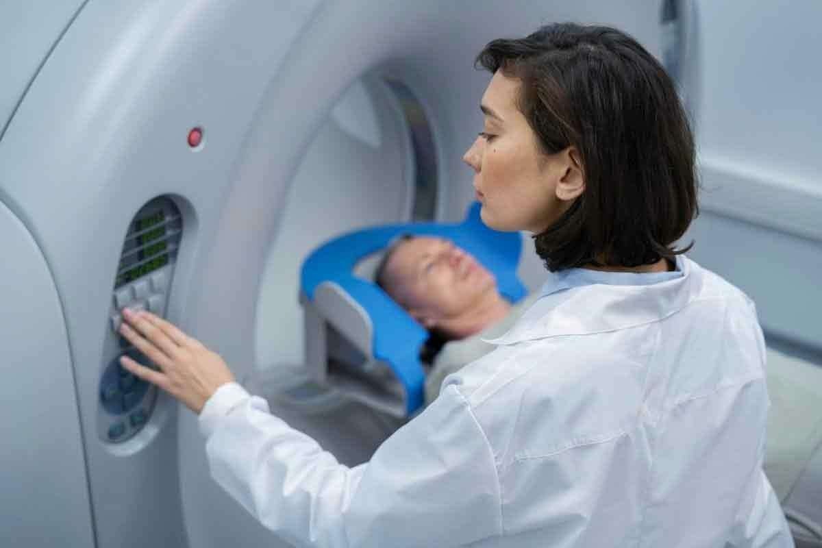
Magnetic Resonance Imaging (MRI) is key in diagnosing hip bursitis. It’s a non-invasive method that shows soft tissues around the hip. This helps doctors diagnose and plan treatments accurately.
How MRI Technology Works
MRI uses a strong magnetic field and radio waves to show body structures. For hip bursitis, MRI is great for seeing bursae and soft tissues. It works by aligning hydrogen atoms with a magnetic field, then using radio waves to create images.
MRI’s clear images are vital for spotting hip bursitis. It shows inflammation, fluid, and tissue changes.
Preparing for a Hip MRI
Getting ready for a hip MRI is simple. Remove metal items like jewelry or clothes with metal. Tell your doctor about any metal implants or devices.
Some MRIs use a contrast agent for better images. Talk to your doctor about any worries before the test.
What to Expect During the Procedure
In a hip MRI, you lie on a table that slides into the machine. The machine creates images with a magnetic field and radio waves. The test is usually painless, but some might feel claustrophobic or uncomfortable.
The MRI tech will talk to you through an intercom. They’ll guide you and keep you calm. The test lasts from 15 to 45 minutes.
MRI Findings in Hip Bursitis
MRI findings are key for diagnosing hip bursitis. They give us detailed images of the bursae and tissues around them. This helps us see the type and how severe the bursitis is.
Trochanteric Bursitis MRI Appearance
Trochanteric bursitis is when the bursa near the femur’s greater trochanter gets inflamed. On MRI, it shows as fluid in the bursa with soft tissue swelling around it. The images can show how bad the inflammation is.
Iliopsoas Bursitis MRI Characteristics
Iliopsoas bursitis is when the bursa between the iliopsoas muscle and the hip joint gets inflamed. MRI shows fluid in the bursa that might spread too far. The muscles nearby might also show signs of inflammation.
Ischiogluteal Bursitis MRI Features
Ischiogluteal bursitis is when the bursa between the ischial tuberosity and the gluteus maximus muscle gets inflamed. MRI finds fluid in the bursa and inflammation in nearby tissues. It helps tell if it’s ischiogluteal bursitis or something else causing hip pain.
Fluid Collection and Inflammation Markers
A big sign of hip bursitis on MRI is fluid in the bursa. We also look for soft tissue swelling and enhancement after contrast. These signs help confirm the diagnosis and how serious it is.
Greater Trochanteric Pain Syndrome on MRI
Diagnosing greater trochanteric pain syndrome on MRI is more than just spotting bursitis. It’s about checking the tendons and soft tissues around the greater trochanter. This condition includes trochanteric bursitis, gluteal tendon tears, and external coxa saltans (snapping hip syndrome).
Beyond Simple Bursitis
Trochanteric bursitis is part of GTPS, but it’s not the whole story. Gluteal tendon pathology is also key. MRI can look at both the bursae and tendons, giving a clearer picture than just a doctor’s check-up.
“MRI has changed how we see GTPS,” say top orthopedic doctors. It shows how bad the tendon damage is, if there’s bursitis, and other issues.
Gluteal Tendon Pathology
Gluteal tendon problems, like tendinosis and tears, are big parts of GTPS. MRI can spot these by looking at tendon thickness, signal, and fluid or swelling. High-resolution MRI is great for catching these small changes.
Distinguishing Features on Imaging
Spotting what causes GTPS on MRI means looking for specific signs. For example, trochanteric bursitis shows fluid in the bursa. Gluteal tendon tears have gaps or changes in the tendon. Knowing these signs is key for correct diagnosis and treatment.
Using MRI helps doctors understand GTPS better. This leads to more focused treatments. As MRI tech gets better, so will our ability to help patients with GTPS.
Accuracy of MRI for Diagnosing Hip Bursitis
Knowing how well MRI works for diagnosing hip bursitis is key for good patient care. MRI is a top choice for doctors because it shows soft tissues around the hip in detail.
Sensitivity and Specificity Rates
Research shows MRI is very good at spotting hip bursitis. It can catch trochanteric bursitis 80% to 90% of the time, depending on the study and the people involved. But, it’s important to look at the patient’s symptoms too.
“MRI is great at showing inflamed bursas and swelling,” a study on MRI for hip bursitis found. This is really helpful for diagnosing trochanteric bursitis.
Limitations of MRI in Bursitis Detection
Even though MRI is great, it has its limits. It can be hard to tell if hip pain is from bursitis or something else like tendinopathy or osteoarthritis. Also, sometimes, MRI might show fluid in the bursa even when the person doesn’t have pain.
Can MRI Miss Hip Bursitis?
Yes, MRI might not catch hip bursitis in some cases. If the bursitis is mild or if there’s a problem with the MRI, it might not show up. Other conditions can look like bursitis on MRI too, which can lead to wrong diagnoses.
Clinical Correlation Importance
When looking at MRI results for hip bursitis, it’s important to consider more than just the scan. Doctors need to think about the patient’s symptoms, medical history, and physical exam too. This helps make a correct diagnosis and plan the right treatment.
In short, MRI is a strong tool for finding hip bursitis, but its success depends on many things. By understanding these and matching MRI results with what the patient is experiencing, doctors can make better choices.
Alternative Diagnostic Imaging for Hip Bursitis
While MRI is a powerful tool for diagnosing hip bursitis, other imaging modalities can also provide valuable insights. In this section, we will explore alternative diagnostic imaging methods used for hip bursitis.
Ultrasound Evaluation
Ultrasound is a highly effective imaging modality for diagnosing hip bursitis, specially for superficial bursae like the trochanteric bursa. It offers several advantages, including real-time imaging, dynamic assessment, and the ability to perform ultrasound-guided injections.
Key benefits of ultrasound for hip bursitis include:
- High-resolution images of superficial structures
- Ability to assess bursae dynamically
- Guidance for therapeutic injections
- No radiation exposure
X-Ray Limitations
X-rays are often used as an initial imaging technique for hip pain but have limited utility in directly diagnosing hip bursitis. They can, though, help rule out other causes of hip pain such as fractures or degenerative joint disease.
Limitations of X-rays for hip bursitis diagnosis:
- Cannot directly visualize bursae or soft tissues
- Limited to assessing bone structures
- May not detect early or mild cases of bursitis
CT Scan Applications
CT scans provide detailed images of bone structures and can be useful in evaluating hip bursitis when there is suspected involvement of surrounding bony structures. They are also helpful in detecting calcifications within bursae.
CT scan benefits for hip bursitis:
- Excellent for visualizing bone pathology
- Can detect calcifications within bursae
- Quick imaging process
Comparative Effectiveness
When comparing the effectiveness of different imaging modalities for hip bursitis, several factors come into play. These include diagnostic accuracy, availability, cost, and patient factors.
| Imaging Modality | Diagnostic Accuracy | Cost | Availability |
| Ultrasound | High for superficial bursae | Moderate | High |
| X-ray | Limited for bursitis | Low | Very High |
| CT Scan | High for bone pathology | High | Moderate |
| MRI | Very High | High | Moderate |
In conclusion, while MRI remains a gold standard for diagnosing hip bursitis, other imaging modalities like ultrasound, X-ray, and CT scans have their own strengths. They can be used in various clinical scenarios or as complementary tools.
Differentiating Hip Conditions on MRI
Differentiating hip conditions is key for effective treatment. MRI is a vital tool in this process. Hip pain can stem from various conditions, and accurate diagnosis is essential for proper care.
Hip Tendonitis vs. Bursitis
Hip tendonitis and bursitis are common causes of hip pain. Tendonitis is inflammation of the tendons, which connect muscles to bones. Bursitis, on the other hand, is inflammation of the bursae, fluid-filled sacs that cushion joints.
On MRI, tendonitis shows as tendon thickening and changes in signal intensity. Bursitis is seen as a distended bursa with fluid and inflammation.
Labral Tears and Their Appearance
The labrum is a cartilage structure around the acetabulum, providing stability to the hip joint. Labral tears can occur due to trauma or degenerative changes. MRI identifies labral tears by showing irregularities in the labrum.
MR arthrography, which involves injecting contrast into the joint, can better show labral tears. It outlines the labrum and highlights any defects.
Arthritis vs. Bursitis on MRI
Arthritis and bursitis can both cause hip pain. Arthritis shows joint space narrowing and osteophyte formation on MRI. Bursitis involves inflammation of the bursae, appearing as fluid distension of the bursa.
Distinguishing between these conditions is important because their treatments differ. MRI provides a detailed look at both the joint and soft tissues, aiding in differentiation.
Bone Bruises and Stress Fractures
Bone bruises and stress fractures can cause hip pain, often from overuse or trauma. Bone bruises show bone marrow edema without a fracture line on MRI. Stress fractures have a clear fracture line and surrounding edema.
MRI is great for early detection of these injuries. It can show changes in bone marrow and cortex before X-rays or CT scans can.
Advanced MRI Techniques for Hip Evaluation
Advanced MRI techniques have changed how we check for hip bursitis and other hip issues. These new methods make it easier for doctors to find problems and fix them. This means better care for patients.
3T vs. 1.5T MRI Machines
There’s a big leap in MRI tech from 1.5T to 3T machines. The 3T machines give clearer images of the hip. This helps doctors spot hip bursitis and other issues more accurately.
Research shows 3T MRI is better at showing small details in the hip. It’s great for catching early signs of bursitis or other problems.
MR Arthrography Benefits
MR arthrography is a new way to check the hip. It involves putting a special dye into the joint before scanning. This dye makes the inside of the joint clearer, helping doctors see more.
The good things about MR arthrography are:
- It finds labral tears and cartilage problems better
- It shows more about the joint’s structures
- It helps see how much damage there is
Dynamic and Functional MRI
Dynamic and functional MRI are new tools for checking the hip. They let doctors see how the joint moves and works. This helps understand how different parts of the hip work together.
Dynamic MRI can spot problems that regular scans miss. This is key for planning the right treatment, like surgery or physical therapy.
Using these advanced MRI methods, doctors can understand hip problems better. This leads to more accurate diagnoses and better treatments.
When Is an MRI Necessary for Hip Pain?
Deciding when to use an MRI for hip pain is complex. It depends on several clinical factors. We will explain when an MRI is needed for hip pain.
Indications for Hip MRI
An MRI is often suggested for hip pain if initial checks show a serious issue. Indications include ongoing pain, major trauma, suspected fractures, or when other tests don’t give clear results. For example, after a bad fall, an MRI can show how bad the injury is. It can spot fractures or damage to soft tissues.
When Simpler Tests Are Sufficient
Not every hip pain case needs an MRI. First, simpler tests like X-rays or ultrasound are used. These can find obvious problems like fractures or dislocations. This might mean you don’t need an MRI right away.
Insurance Coverage Considerations
Insurance coverage is key in deciding on an MRI. We need to check the patient’s insurance and what’s needed for approval. Knowing this helps plan the right tests.
Cost-Benefit Analysis
Doing a cost-benefit analysis is important for MRI decisions. We compare the benefits of detailed tests to the costs and risks. This helps make choices that are safe and consider the patient’s finances.
Treatment Options Based on MRI Findings
After getting a hip bursitis diagnosis from an MRI, doctors can create a treatment plan just for you. This plan is based on your specific needs and condition.
Conservative Treatment Approaches
For hip bursitis, the first step is usually conservative management. This means resting and changing how you move. It also includes physical therapy to make your muscles stronger and more flexible.
Doctors might also suggest anti-inflammatory drugs to help with pain and swelling. Losing weight if you’re overweight is another important part of the treatment.
These steps can help manage your symptoms and improve how you move.
Interventional Procedures
If conservative treatments don’t work, doctors might suggest interventional procedures. These include injections of corticosteroids to reduce swelling and PRP therapy to help your body heal faster.
They might also do an aspiration to remove extra fluid from the bursa. These methods can offer a lot of relief for those with ongoing symptoms.
Surgical Options for Severe Cases
In very severe cases, surgery might be the best option. This could involve removing the inflamed bursa or repairing tendons if they’re damaged.
Surgery is usually considered when other treatments haven’t worked.
Rehabilitation Protocols
Rehabilitation is key, no matter the treatment you start with. It includes slowly getting back to your activities, doing strengthening exercises, and improving flexibility and mobility.
A good rehabilitation plan helps prevent the condition from coming back and improves your overall outcome.
| Treatment Approach | Description | Indications |
| Conservative Management | Rest, physical therapy, anti-inflammatory medications | To illustrate the impact of hip bursitis on daily life, we can examine some of the common challenges individuals face: |
| Interventional Procedures | Corticosteroid injections, PRP therapy, aspiration | Moderate to severe hip bursitis not responding to conservative management |
| Surgical Options | Bursectomy, tendon repair | Severe or refractory hip bursitis |
Conclusion
Getting a correct diagnosis is key to treating hip bursitis well. We’ve seen how MRI technology is important in diagnosing hip bursitis. It gives detailed images that help doctors see the problem and how bad it is.
Knowing the different types of hip bursitis and their symptoms is important. This helps doctors choose the right treatment. From simple treatments to more serious surgeries, the right choice depends on the diagnosis.
There are many ways to treat hip bursitis, like physical therapy and medicine. The best treatment is one that matches the patient’s needs. Using advanced tools like MRI helps doctors give better care and improve patients’ lives.
FAQ
What is hip bursitis and how is it diagnosed?
Hip bursitis is when the bursae around the hip joint get inflamed. Doctors use a physical exam, patient history, and imaging like MRI to diagnose it.
Can an MRI detect hip bursitis?
Yes, MRI is very good at finding hip bursitis. It shows the inflamed bursae and changes in the surrounding tissue.
What are the common symptoms of hip bursitis?
Symptoms include pain on the outer hip, thigh, or groin. This pain gets worse with activity. It also hurts when lying on the affected side or getting up from sitting.
How does MRI differentiate between hip bursitis and other hip conditions?
MRI can tell the difference between hip bursitis, tendonitis, labral tears, and arthritis. It looks at the affected structures and markers like fluid collection and inflammation.
Are there alternative imaging methods for diagnosing hip bursitis?
Yes, you can use ultrasound, X-ray, and CT scans. But MRI is the best because it’s very sensitive and specific for soft tissue issues.
What are the treatment options for hip bursitis based on MRI findings?
Treatment can be conservative, like physical therapy and pain management. Or it can include interventional procedures like corticosteroid injections. In severe cases, surgery might be needed.
Can MRI miss hip bursitis?
While MRI is very sensitive, it’s not perfect. It’s important to match MRI findings with clinical symptoms to avoid false negatives or misinterpretation.
When is an MRI necessary for evaluating hip pain?
You might need an MRI if hip pain doesn’t go away with conservative treatment. Or if there’s a suspicion of serious damage like labral tears or severe bursitis.
What advanced MRI techniques are used for hip evaluation?
Advanced techniques include higher field strength MRI (3T) and MR arthrography for better joint and labrum visualization. Dynamic/functional MRI assesses the hip under different conditions.
How does the diagnosis of greater trochanteric pain syndrome differ on MRI?
Greater trochanteric pain syndrome involves bursitis and gluteal tendon pathology. MRI can show tendon thickening, tears, or tendinosis in addition to bursitis.
Reference
- Pianka, M. A., Serino, J., DeFroda, S. F., & Bodendorfer, B. M. (2021). Greater trochanteric pain syndrome: Evaluation and management of a wide spectrum of pathology. SAGE Open Medicine, 9, 20503121211022582. https://www.ncbi.nlm.nih.gov/pmc/articles/PMC8182177/

