Last Updated on October 20, 2025 by
Hepatoblastoma is a rare and serious liver condition. It mainly affects young children, with most cases found before they are 3 years old.
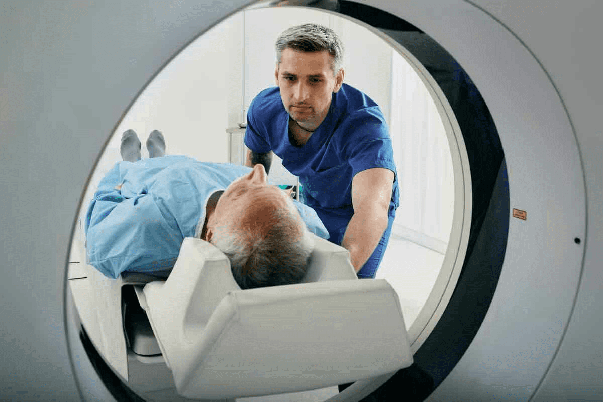
We are committed to giving top-notch care to kids with this condition. Tests like CT and MRI scans show what liver tumors look like. They find well-defined, mixed masses that might have dead tissue, bleeding, or hard spots.

Kids with this condition often have a painless lump in their belly. Blood tests usually show high levels of alpha-fetoprotein (AFP). This is a key sign of the disease. At our place, we use CT or MRI scans in different phases to spot and handle hepatic tumors well.
Key Takeaways
- Hepatoblastoma is a rare malignant liver tumor mainly found in kids under 5.
- Imaging techniques like CT and MRI are key for finding the disease.
- High alpha-fetoprotein (AFP) levels are a major sign of the disease.
- Symptoms often include a painless lump in the belly.
- Multiphase CT or MRI is used for effective diagnosis and management.
Hepatoblastoma: Clinical Characteristics and Presentation
Hepatoblastoma often shows up as a painless lump in the belly. This is usually found during routine check-ups or by parents. Spotting it early is key to treating it.
Painless Abdominal Mass: The Primary Sign
A painless abdominal mass is the main sign of hepatoblastoma. It’s different from other belly problems that hurt. By the time it’s found, the tumor is usually big, showing it’s been growing for a while.
Finding this mass early can really help patients. Regular doctor visits are important for catching it early.

A hepatic mass on liver can cause belly swelling and discomfort. Knowing about hepatoblastoma’s signs is key to treating it well.
Things like liver cancer survival rate by age and the patient’s health matter a lot. A tumor on liver needs quick medical care to avoid more problems.
At our place, we focus on full care for hepatoblastoma patients. Our team works hard to give the right diagnosis and treatment plans for each patient.
Visual Appearance of Hepatoblastoma on Physical Examination
During a physical exam, signs of hepatoblastoma include abdominal distension. This is often the first sign of a liver tumor.

Abdominal Distension Patterns
The way the abdomen swells can change based on the tumor’s size and where it is. Sometimes, the swelling is even all over. Other times, it’s more focused in one spot.
Along with swelling, other signs like weight loss, feeling tired, and not wanting to eat can show up. These signs show how the tumor is affecting the body’s health.
External Visual Cues and Associated Findings
Visible signs outside the body can hint at hepatoblastoma. These signs include:
- Visible swelling in the abdomen
- A mass in the abdomen that can be felt
- Jaundice, or yellow skin and eyes
Stanford Children’s Health notes that jaundice is common in kids with liver tumors, like hepatoblastoma.
Other Laboratory Findings in Hepatoblastoma
Labs are key in diagnosing hepatoblastoma. Key findings include:
| Laboratory Test | Typical Finding |
| Alpha-fetoprotein (AFP) levels | Elevated |
| Liver function tests | Abnormal |
| Complete Blood Count (CBC) | May show anemia or other abnormalities |
Lab results, along with physical exams and imaging, help us diagnose and treat hepatoblastoma well.
Ultrasound Appearance of Hepatoblastoma
Ultrasound imaging is often the first step in identifying hepatoblastoma. It gives us valuable information about the tumor’s size, location, and characteristics. On ultrasound, hepatoblastoma typically appears as a large, heterogeneous mass with variable echogenicity.
The ultrasound characteristics can vary, but often, hepatoblastoma is seen as a well-defined mass. It has both hyperechoic and hypoechoic areas, showing the tumor’s complex internal structure. Doppler ultrasound can also be used to assess the vascularity of the tumor.
Multiphase CT Findings in Hepatoblastoma
Multiphase CT scans are key for further evaluating hepatoblastoma after initial ultrasound detection. CT scans give detailed information about the tumor’s extent and its relationship with surrounding structures. They also help spot any metastases.
The typical CT appearance of hepatoblastoma includes:
- A large, well-defined mass with heterogeneous enhancement
- Variable attenuation due to necrosis, hemorrhage, or calcifications
- Potential vascular invasion or compression of adjacent structures
A study published on Biomed Central shows CT scans are vital in preoperative assessment of hepatoblastoma. They help determine if the tumor can be removed.
By combining ultrasound and CT findings, we can accurately diagnose and stage hepatoblastoma. This is key for developing an effective treatment plan.
MRI Features That Define Hepatoblastoma
Magnetic Resonance Imaging (MRI) is key in spotting hepatoblastoma, a liver hepatic tumor or growth on the liver. It gives detailed images of the tumor. This helps doctors diagnose and plan treatment.
The MRI shows unique features of hepatoblastoma. It helps us understand the definition of the lesion on the liver. Usually, it looks like a well-defined mass on MRI. The look can change based on the tumor’s makeup.
On MRI, hepatoblastoma often has mixed signal intensities. This is because it can have necrotic and hemorrhagic areas. The T1 and T2-weighted images give clues about its nature.
We use MRI to see how big the tumor is and how it affects nearby areas. It helps us plan for surgery or other treatments. The detailed MRI info is key for a good treatment plan for patients with hepatoblastoma.
At our place, we’re experts in reading MRI results for hepatoblastoma. We create treatment plans that fit each patient’s needs.
Distinctive Visual Elements: Calcifications, Necrosis, and Hemorrhage
When doctors look at the liver for hepatoblastoma, they see special signs. These signs help plan treatment. They also tell us about the tumor’s makeup and how it might act.
Necrotic and Hemorrhagic Components
Hepatoblastoma can show necrosis and hemorrhage in scans. Necrosis means dead tissue in the tumor. Hemorrhage is bleeding inside the tumor. Both are key to understanding the tumor’s growth and how to treat it.
Calcifications are also a key sign of hepatoblastoma. These are calcium deposits seen on CT scans. They help doctors know more about the tumor and confirm the diagnosis.
It’s vital for doctors to know about these signs. They help in diagnosing and treating hepatoblastoma. By looking at these features, doctors can make a treatment plan that fits the patient’s needs.
Hepatoblastoma Prognosis and Survival Rates
The chances of survival for hepatoblastoma depend on several things. These include the tumor’s size and where it is on the liver. The child’s overall health is also important. Knowing the liver cancer survival rate by age helps decide the best treatment.
We have made big strides in treating liver tumors, like hepatoblastoma. Finding the tumor early and giving complete care are key to better results. We help families get the best care for their child. This is based on the tumor’s details and the child’s health.
We aim to give the best care for kids with hepatoblastoma. Our team focuses on both physical and emotional needs. We want to help our patients and their families in every way we can.
FAQ
What is hepatoblastoma?
Hepatoblastoma is a rare and serious liver tumor. It mainly affects kids under 5 years old.
What are the symptoms of hepatoblastoma?
The main sign is a painless lump in the belly. This can make the belly look bigger. Kids might also lose weight, feel tired, or not want to eat.
How is hepatoblastoma diagnosed?
Doctors use imaging like ultrasound, CT, and MRI to find the tumor. They also check blood for alpha-fetoprotein (AFP) levels.
What does hepatoblastoma look like on imaging studies?
On scans, it looks like a big, mixed-up mass with spots and bleeding. Ultrasound, CT, and MRI help see its size and details.
What is the prognosis for hepatoblastoma?
The outlook depends on the tumor’s size, where it is, and the child’s health. Finding it early and getting good care can help a lot.
What are the treatment options for hepatoblastoma?
Treatments include surgery, chemo, and sometimes a liver transplant. The best plan is made for each child based on their needs and the tumor.
What is the survival rate for hepatoblastoma?
Survival rates have gone up a lot. Now, 70% to 90% or more kids can live, depending on the stage and treatment.
Can adults get hepatoblastoma?
Yes, though it’s very rare in adults. Adults often face a different outlook and treatment than kids.
How common is liver cancer in children?
Liver cancer is rare in kids. Hepatoblastoma is the most common type in those under 5.
What is the difference between hepatoblastoma and other liver tumors?
Hepatoblastoma is a special liver tumor for young kids. Other liver tumors, like hepatocellular carcinoma, can happen in kids and adults but are different.
References
Centers for Disease Control and Prevention. (2024). Facts about birth defects. https://www.cdc.gov/ncbddd/birthdefects/facts.html
Hockenberry, M. J., Wilson, D., & Rodgers, C. C. (2019). Wong’s nursing care of infants and children (11th ed.). Mosby. https://www.elsevier.com/books/wongs-nursing-care-of-infants-and-children/hockenberry/978-0-323-54939-5
The Society of American Gastrointestinal and Endoscopic Surgeons (SAGES). (2023). Patient information on hernia repair. https://www.sages.org/publications/patient-information/patient-information-for-hernia-repair-from-sages/


