Last Updated on November 27, 2025 by Bilal Hasdemir

At Liv Hospital, we know you might worry about imaging tests and radiation. Chest X-rays use a small amount of radiation, about 0.1 millisieverts (mSv) per scan. This is less than the natural radiation most people in the U.S. get in a year, which is about 3 mSv. Many patients often ask how many chest x rays are safe in a year, and while there’s no exact limit, doctors ensure that X-rays are only done when medically necessary to minimize risk.
Many ask about the safety of having many chest X-rays and how they compare to CT scans. A chest X-ray is like 10 days of natural background radiation. But a CT scan of the chest is like 2 years of natural background radiation. Knowing these risks helps keep you healthy and at ease.
Key Takeaways
- The average annual background radiation dose is about 3 mSv.
- One chest X-ray equals 0.1 mSv, equivalent to 10 days of natural background radiation.
- A CT scan of the chest equals 6.1 mSv, equivalent to 2 years of natural background radiation.
- Theoretically, up to 30 chest X-rays can be done in a year without exceeding the average annual background radiation dose.
- CT scans have a significantly higher radiation dose compared to chest X-rays.
Understanding Medical Radiation Basics
To understand the risks of chest X-rays and CT scans, we need to know about medical radiation. Medical imaging uses ionizing radiation to see inside the body. This radiation has enough energy to remove electrons from atoms, creating ions.
What Is Ionizing Radiation?
Ionizing radiation is a type of energy that comes in waves or particles. In medical imaging, it helps create detailed images of the body. X-rays and gamma rays are common types used. They can go through tissues and show different structures inside the body.
How Radiation Is Measured in Medical Imaging
The amount of radiation the body absorbs is measured in grays (Gy) or sieverts (Sv). The effective dose, in sieverts, considers how different tissues react to radiation. This helps us understand the risks of radiation from medical tests. For example, a chest X-ray has a very low dose, about 0.01 millisieverts (mSv).
Here’s a simple comparison of radiation doses from common tests:
| Imaging Procedure | Typical Effective Dose (mSv) |
| Chest X-ray | 0.01 |
| CT Scan (Chest) | 7 |
| Mammogram | 0.4 |
Natural vs. Medical Radiation Exposure
We are always exposed to radiation from nature, like cosmic rays and radon. The average natural background radiation is about 2.4 mSv a year. Medical radiation, on the other hand, is a big part of our total exposure, thanks to more imaging tests. A report by the National Council on Radiation Protection and Measurements (NCRP) says medical imaging now makes up nearly half of our total radiation exposure in the U.S.
“The increasing use of medical imaging has led to a significant rise in radiation exposure from medical procedures. It’s essential to balance the benefits against the risks.” –
NCRP Report
Knowing about ionizing radiation and its use in medical imaging helps us understand the need for careful use. It shows why we should use the least amount of radiation needed for tests. This is known as ALARA (As Low As Reasonably Achievable).
Chest X-Ray Radiation: What You Need to Know

Chest X-rays are a common way to see inside the body. They use ionizing radiation, so it’s important to know more about it. We’ll look at how chest X-rays work, the amount of radiation they use, and what can change that amount.
How X-Ray Technology Works
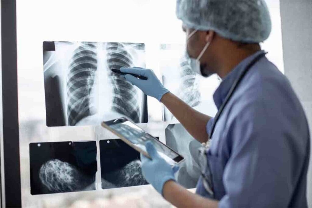
X-rays use ionizing radiation to show what’s inside the body, like the chest. When an X-ray beam hits the chest, different parts absorb different amounts of radiation. This makes it possible to see things like bones, lungs, and the heart.
Typical Radiation Dose from a Single Chest X-Ray
A chest X-ray usually gives off about 0.1 mSv of radiation. This is a small amount, similar to what we get from natural background radiation over a few days.
Factors Affecting Radiation Exposure During X-Rays
Many things can change how much radiation a patient gets from a chest X-ray. These include the X-ray machine, the settings used, the patient’s size and body type, and what the doctor needs to see.
| Factor | Effect on Radiation Dose |
| X-ray Equipment | Modern digital X-ray machines can reduce radiation doses compared to older models. |
| Patient Size | Larger patients may require higher doses to achieve adequate image quality. |
| Imaging Settings | Adjusting the X-ray beam energy and exposure time can impact the radiation dose. |
Knowing about these factors helps us understand how to keep radiation low while getting good images.
How Many Chest X-Rays Are Safe in a Year?
There’s no single answer to how many chest X-rays are safe. It’s important to know about radiation exposure. The safety of chest X-rays depends on many things, not just how many you have in a year.
Why There’s No Strict Annual Limit
Global guidelines don’t set a strict limit on chest X-rays per year. This is because the risk from radiation builds up over time. It changes based on your age, health, and past radiation exposure.
“The risk of radiation-induced harm is more closely related to the total dose accumulated over a lifetime than the number of procedures per year.”
Cumulative Exposure Considerations
Cumulative exposure is the total radiation a person gets over time. This is key to understanding the risks of chest X-rays. Radiation effects can last long after exposure.
| Age Group | Cumulative Dose (mSv) | Risk Level |
| 0-20 | 0-5 | Low |
| 21-50 | 5-20 | Moderate |
| 51+ | 20+ | Higher |
Risk Factors That Influence Safety Thresholds
Many factors affect the safety of chest X-rays. These include your age, health, and past radiation exposure. For example, children are more vulnerable to radiation because their bodies are growing.
Knowing these factors helps doctors decide when chest X-rays are needed. They also try to reduce radiation exposure.
Lifetime Radiation Exposure Guidelines
Medical imaging is getting more common. It’s key to know about lifetime radiation exposure guidelines. These guidelines help doctors and patients make smart choices about using radiation in tests.
The 100 mSv Lifetime Exposure Benchmark
There’s a talk about a lifetime radiation limit. A common number is 100 mSv. This is seen as a point where health risks from radiation start to rise.
100 mSv is like getting 10,000 chest X-rays. This shows how safe most X-rays are.
American College of Radiology Recommendations
The American College of Radiology (ACR) has guidelines for radiation. There’s no strict yearly limit for medical imaging radiation. But, the ACR suggests keeping doses As Low As Reasonably Achievable (ALARA).
This rule helps make imaging safer. It means using the least amount of radiation needed for good results.
Putting 10,000 Chest X-Rays in Perspective
Let’s look at 10,000 chest X-rays:
| Imaging Procedure | Typical Radiation Dose (mSv) | Equivalent Number of Chest X-Rays |
| Chest X-Ray | 0.01 | 1 |
| CT Scan (Chest) | 7 | 700 |
| 100 mSv Dose | 100 | 10,000 |
This table shows that while one chest X-ray is low in radiation, many can add up.
It’s vital to know about lifetime radiation exposure guidelines. This helps balance the good of medical imaging with its risks. By following the American College of Radiology’s advice, we can help patients get the info they need safely.
CT Scan Technology and Radiation
CT scans have changed how we diagnose diseases, but they also raise concerns about radiation. It’s important to know how CT scans work and how they compare to X-rays. This helps us understand the safety of these scans for patients.
How CT Scans Differ from X-Rays
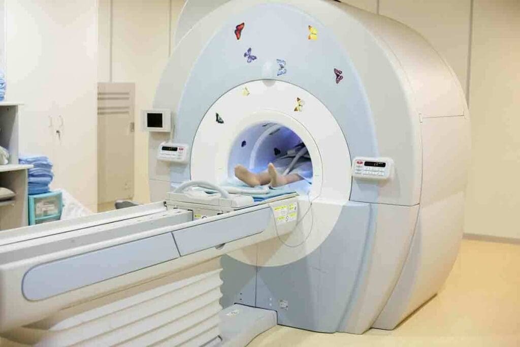
CT scans use X-rays and computer tech to show detailed images of the body. Unlike X-rays, which show only two dimensions, CT scans give us a three-dimensional view. This is because they take many X-ray measurements from different angles and then put them together into one image.
Key differences between CT scans and X-rays include:
- Multi-dimensional imaging: CT scans provide detailed images of internal structures from various angles.
- Higher resolution: CT scans offer higher resolution images compared to traditional X-rays.
- Ability to detect smaller abnormalities: The detailed images from CT scans allow for the detection of smaller issues that might not be visible on an X-ray.
Why CT Scans Use More Radiation
CT scans need more radiation than X-rays because they take many X-ray measurements. The amount of radiation used can change based on the type of CT scan and the body part being scanned. Even though there’s more radiation, the benefits of CT scans often outweigh the risks for many patients.
The trade-off between diagnostic accuracy and radiation exposure is a critical consideration in medical imaging.
Types of CT Scans and Their Radiation Levels
There are many types of CT scans, each with its own uses and radiation levels. Some common types include:
| Type of CT Scan | Typical Radiation Dose (mSv) | Common Applications |
| Head CT | 1-2 | Diagnosing head injuries, strokes, and tumors |
| Chest CT | 5-7 | Examining lung diseases, cancers, and cardiovascular conditions |
| Abdomen/Pelvis CT | 8-12 | Investigating abdominal pain, cancers, and infections |
Knowing about the different CT scans and their radiation levels helps both patients and doctors make better choices about imaging tests.
CT Scan Radiation vs. X-Ray Radiation: A Detailed Comparison
Understanding the differences between CT scan and X-ray radiation is key for patients and doctors. Both are important for diagnosing and tracking health issues. Yet, they differ in how much radiation they use and what they can show.
Radiation Dose Differences
CT scans use more radiation than X-rays. A chest CT scan’s dose is like 100 to 400 chest X-rays. This is because CT scans use many X-rays from different angles to create detailed images.
CT scans have a higher dose because they show more details inside the body. X-rays are good for seeing bones and some lung issues. But CT scans are better for soft tissues, organs, and complex structures.
Diagnostic Benefits That Justify Higher Exposure
Even with more radiation, CT scans are often needed for complex diagnoses. The benefits of CT scans make the extra radiation worth it in many cases. They are key in emergencies for quick injury checks, spotting internal bleeding, or finding pulmonary embolism.
In cancer care, CT scans help stage cancer, track treatment, and find if cancer comes back. They give doctors detailed images for accurate diagnoses and treatment plans.
When CT Scans Are Necessary Despite Radiation Concerns
Though radiation is a worry, sometimes CT scans are the best choice. Doctors consider the need for CT scans and the risks, mainly for those needing many scans.
When MRI or ultrasound can’t be used, CT scans are preferred. For example, they’re used for patients with metal implants or pacemakers who can’t have MRI.
Knowing the differences between CT scan and X-ray radiation helps patients understand their doctor’s choices. It also shows why talking about radiation worries with doctors is important.
Health Risks Associated with Medical Radiation
Medical imaging technology is getting better, but we need to know about radiation risks. Medical radiation is used in many tests, and it’s mostly safe. But, there are risks, mainly with repeated use.
Short-Term vs. Long-Term Effects
Medical radiation can have short-term and long-term effects. Short-term effects include nausea, fatigue, and skin burns, but they’re rare. Long-term effects, like cancer or genetic mutations, are a bigger worry.
It’s important to think about the total amount of radiation a person gets over their lifetime. The risk of cancer from medical radiation is small but real. The American Cancer Society says the risk is small but not zero, for those who get many tests.
Cancer Risk from Repeated Imaging
Studies show that getting many CT scans can slightly increase cancer risk. CT scans give off more radiation than X-rays. We need to think about the benefits and risks of these tests, for those who need them often.
Special Considerations for Children and Pregnant Women
Children and pregnant women are more at risk from medical radiation. Kids are more sensitive to radiation because their bodies are growing. Pregnant women should avoid radiation to protect their babies, and ultrasound is often a safer choice.
| Population | Special Considerations | Alternative Imaging |
| Children | More sensitive to radiation, longer life expectancy | Ultrasound, MRI |
| Pregnant Women | Avoid fetal exposure, careful dose management | Ultrasound, MRI |
Knowing about these risks helps us make better choices about medical imaging. We can find a balance between getting the right diagnosis and keeping radiation exposure low. This way, we can give patients the best care while protecting them from radiation.
Balancing Diagnostic Benefits Against Radiation Risks
Medical imaging is a big deal in healthcare. We must think about the good it does and the risks of radiation. X-rays and CT scans help doctors a lot. But, we need to be careful with radiation to keep patients safe.
The ALARA Principle
The ALARA principle is key in medical imaging. It means we should use as little radiation as we can. This way, we get good images without harming patients too much.
To follow ALARA, we do a few things:
- We use the least amount of radiation needed for good images.
- We adjust images based on the patient’s size and age.
- New imaging tech helps reduce radiation.
- We keep an eye on radiation doses and change them if needed.
Questions to Ask Your Doctor Before Imaging
Talking to your doctor before imaging is important. Here are some questions to ask:
- Is this imaging really needed for my care?
- Are there other ways to get the same info without radiation?
- What are the risks of radiation from this test?
- How do the benefits of this test outweigh the risks?
- Can we use less radiation or adjust the test to lower exposure?
Alternatives to Radiation-Based Imaging
There are imaging options that don’t use radiation. For example:
- Ultrasound: Uses sound waves to see inside the body.
- Magnetic Resonance Imaging (MRI): Uses magnetic fields and radio waves for detailed images.
These options can help without the radiation risks. But, they depend on what the doctor needs to see.
In short, finding the right balance in medical imaging is tough. We need to know about ALARA, ask the right questions, and look at other options. This way, we make sure imaging is safe and helpful for everyone.
Radiation Safety Protocols in Modern Medical Imaging
Modern medical imaging has made big strides, focusing on safety. We balance the need for clear images with the risk of radiation. Hospitals take steps to cut down on radiation exposure.
How Hospitals Minimize Unnecessary Exposure
Hospitals follow the ALARA principle to lower radiation. This means using the least amount of radiation needed for good images. They do this by choosing the right patients, fine-tuning imaging methods, and using new tech that cuts down on radiation.
For example, hospitals:
- Set strict rules for when to use imaging tests
- Check imaging equipment regularly
- Train techs on how to use less radiation
Technological Advances Reducing Radiation Doses
New tech has been key in lowering radiation doses. For example, modern CT scanners use new methods to make clear images with less radiation. Also, X-ray tech has improved, making detectors better and images clearer.
| Technological Advance | Description | Impact on Radiation Dose |
| Iterative Reconstruction | Improved image reconstruction technique | Reduces dose by up to 50% |
| Advanced X-ray Detectors | More efficient detection of X-rays | Lowers dose by improving image quality |
| Image Processing Algorithms | Enhanced image processing | Optimizes image quality at lower doses |
Patient Protection Measures During Imaging
Keeping patients safe during imaging is our top priority. We make sure patients are in the right spot and only the needed area is scanned. We also use shields to protect sensitive parts.
By using these steps, we greatly reduce the risks of radiation from medical imaging. Our focus on safety means patients get the images they need without too much radiation.
Myths About Radiation “Detox” and Protection
Many patients worry about radiation after medical imaging. They look for ways to ‘detox’ or protect themselves. But, it’s important to know the truth about these practices.
Why You Can’t “Flush Out” Radiation After Exposure
Radiation from medical imaging worries many. The idea of ‘detoxing’ from it is tempting. But, the body can’t quickly get rid of radiation.
Radiation exposure is not like a toxin that can be flushed out. Once radiation goes through the body, it’s done. The body doesn’t hold onto radiation like it does toxins.
Debunking Common Misconceptions About Radiation Protection
Many myths surround radiation protection. Some think certain supplements or diets can help. But, there’s no scientific proof for these claims.
- Antioxidants are sometimes thought to fight radiation effects. But, their role is more complex.
- Some diets claim to ‘detox’ the body. But, there’s no proof they can remove radiation.
What Actually Helps Your Body After Radiation Exposure
While there’s no ‘detox’ for radiation, some health practices help. They support your body’s recovery and well-being.
- Eating a healthy diet with fruits, vegetables, and whole grains.
- Drinking plenty of water to help your body’s natural processes.
- Following your healthcare provider’s advice for after the imaging.
It’s key to trust evidence-based info on radiation exposure and protection. Talking to healthcare professionals can offer clarity and reassurance.
Conclusion: Making Informed Decisions About Medical Imaging
It’s important to know the risks and benefits of medical imaging. This knowledge helps us make smart choices about our health. We’ve looked at the risks of radiation from chest X-rays and CT scans. We’ve also talked about how they compare and what affects their safety.
Medical imaging is key for diagnosing and treating diseases. But, it’s not without risks. The goal is to weigh the good against the bad, focusing on radiation exposure. Knowing about medical radiation and the different imaging methods helps us make better choices.
When choosing our healthcare, talking openly with doctors is key. We should discuss if imaging is really needed, the risks, and other options. This way, we can make choices that are best for our health and well-being.
FAQ
Does a CT scan emit radiation?
Yes, CT scans use X-rays to create detailed images of the body’s internal structures. They emit a higher dose of radiation compared to standard X-rays.
How does the radiation from a CT scan compare to a chest X-ray?
CT scans expose patients to a much higher dose of radiation than chest X-rays. A chest X-ray might expose a patient to about 0.1 mSv of radiation. On the other hand, a CT scan can expose a patient to anywhere from 2 to 10 mSv or more, depending on the type of scan and the body part being imaged.
Is there a safe limit for radiation exposure from medical imaging?
There is no strict annual limit for radiation exposure from medical imaging. Guidelines suggest that cumulative exposure should be monitored and kept as low as reasonably achievable (ALARA) to minimize risks.
What are the health risks associated with radiation from CT scans and X-rays?
The primary health risk associated with radiation from CT scans and X-rays is an increased risk of developing cancer. This risk is higher for children and individuals who undergo multiple scans. But, the benefits of medically necessary imaging usually outweigh the risks.
Can radiation exposure from medical imaging be reduced?
Yes, there are several ways to reduce radiation exposure from medical imaging. These include using alternative imaging modalities like ultrasound or MRI when possible. Adjusting CT scan protocols to use the lowest necessary dose is also helpful. Implementing dose-tracking systems to monitor cumulative exposure is another way.
Are there any special considerations for children and pregnant women regarding radiation exposure?
Yes, children and pregnant women are considered more vulnerable to the effects of radiation. For children, the long-term risks are higher due to their developing tissues. For pregnant women, the concern is not just for the mother but also for the fetus. Imaging decisions for these groups are made with extra caution, often opting for alternative methods when feasible.
How can I minimize my radiation exposure from medical imaging?
To minimize radiation exposure, ask your healthcare provider about the necessity of the imaging. Find out if alternative imaging methods are available. Also, check if the imaging facility follows the ALARA principle. Keeping a record of your medical imaging history can also help healthcare providers avoid unnecessary repeat exams.
Is it possible to “detox” from radiation after a CT scan or X-ray?
No, it’s not possible to “detox” from radiation in the sense of removing it from the body after exposure. The body does not retain radiation in a way that can be “flushed out.” Instead, the focus is on minimizing exposure in the first place and managing the risks associated with necessary imaging.
What is the ALARA principle in medical imaging?
The ALARA principle stands for “As Low As Reasonably Achievable.” It is a guiding principle in medical imaging. It aims to ensure radiation doses are kept as low as possible while achieving the necessary diagnostic image quality.
How do hospitals minimize unnecessary radiation exposure?
Hospitals minimize unnecessary radiation exposure by implementing strict protocols for imaging. They use the lowest effective dose for diagnostic purposes. They also employ technology that reduces radiation exposure. Hospitals educate staff and patients on the importance of minimizing exposure.
Reference
- Mahmoud, A. M. (2024). A Comparative Study of Radiation Dose From Chest CT Scans. PubMed. https://pubmed.ncbi.nlm.nih.gov/39759664/
- Atlı, E., Üresin, Y., & Atlı, M. (2020). Radiation doses from head, neck, chest, and abdominal CT examinations: A single-center experience. PLoS ONE, 15(12), e0243711. https://pmc.ncbi.nlm.nih.gov/articles/PMC7837727/


