Last Updated on November 27, 2025 by Bilal Hasdemir

At Liv Hospital, we understand the importance of effective and compassionate care for patients with basal cell skin cancer. Basal cell carcinoma is a common skin cancer. Removing it is key to prevent further problems. We use advanced surgical methods to ensure high cure rates and less scarring.
Modern treatments for basal cell carcinoma have cure rates from 95% to 99%. Our experienced medical team uses different methods. These include surgical excision, Mohs micrographic surgery, and curettage and electrodessication. We focus on preserving tissue and achieving good looks, mainly for facial lesions.
Key Takeaways
- Effective surgical procedures are key for removing basal cell skin cancer.
- Modern treatments have high cure rates, from 95% to 99%.
- Various methods are used, including surgical excision and Mohs micrographic surgery.
- Tissue preservation and minimal scarring are our top priorities.
- We provide personalized care based on each patient’s needs.
Understanding Basal Cell Carcinoma
Basal cell carcinoma is the most common skin cancer. It’s important to know about it to manage it well. We’ll look at its characteristics, where it often shows up, and what increases the risk. We’ll also talk about how to prevent it.
What is Basal Cell Carcinoma?
Basal cell carcinoma starts in the basal cell layer of the skin. It grows in a way that can spread to other tissues if not treated. This cancer is not usually deadly but can damage the area a lot if not treated quickly.
Common Locations and Appearance
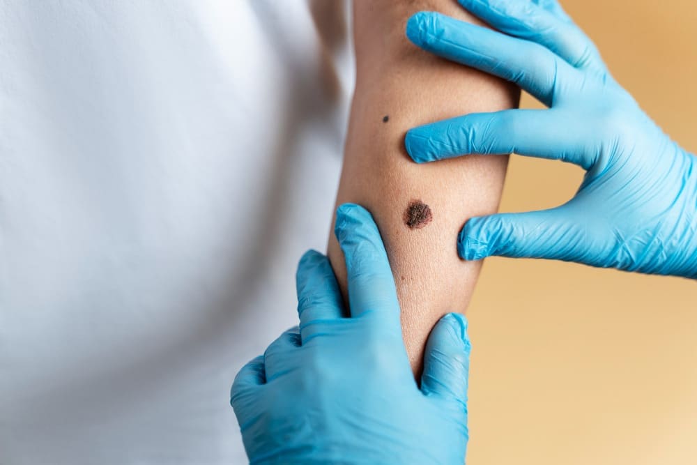
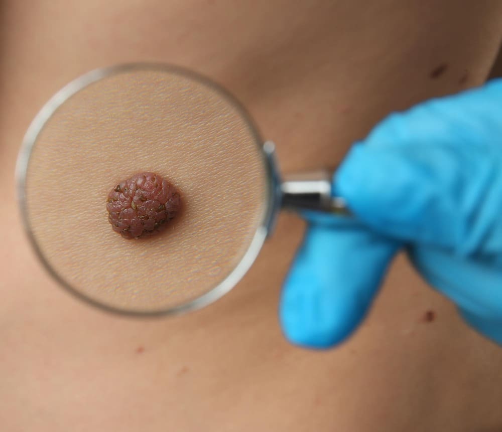
It often shows up on sun-exposed areas like the face, ears, and neck. It can look different, such as:
- Nodular lesions
- Pigmented lesions
- Sclerodermiform lesions
- Superficial lesions
These spots can be different colors and might bleed or ooze. Finding it early is key to treating it well.
Risk Factors and Prevention
Several things can make you more likely to get basal cell carcinoma, including:
| Risk Factor | Description |
|---|---|
| UV Exposure | Too much UV from the sun or tanning beds raises the risk. |
| Fair Skin | People with fair skin are more at risk because they have less melanin. |
| Genetic Predisposition | Having a family history of skin cancer can up your risk. |
| Previous Skin Cancer | If you’ve had skin cancer before, you’re more likely to get basal cell carcinoma. |
To prevent it, use sun protection, avoid tanning beds, and check your skin regularly.
Knowing about basal cell carcinoma and its risks helps us prevent and catch it early. This improves how well we can treat it.
Diagnosis and Pre-Surgical Assessment
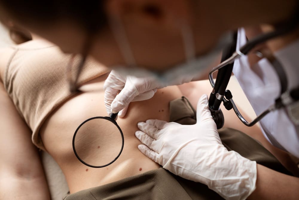
To diagnose basal cell carcinoma, we use a mix of clinical checks and biopsies. First, we visually examine the skin to spot signs of basal cell carcinoma.
Clinical Examination Process
Identifying basal cell carcinoma starts with a detailed skin check. We look for lesions that are shiny, firm, and pale. They often have visible blood vessels. We note the size, shape, and where the lesion is to plan treatment.
Biopsy Procedures
A biopsy is key to confirming basal cell carcinoma. There are various biopsy methods, like shave and punch biopsies. Each is chosen based on the lesion’s features.
Shave biopsies remove the top part of the lesion with a razor or scalpel. Punch biopsies take a small, round sample with a special tool. The right biopsy depends on the lesion’s depth and type.
Determining Appropriate Treatment Method
After confirming the diagnosis, we look at the tumor’s size, location, and risk factors. We also consider the patient’s health and preferences.
Choosing the right treatment involves guidelines and our clinical judgment. For example, surgical excision works well for small, defined tumors. Mohs micrographic surgery is better for sensitive or high-risk areas.
| Tumor Characteristic | Recommended Treatment | Rationale |
|---|---|---|
| Small, well-defined tumor | Surgical Excision | Effective for removing tumor completely with clear margins |
| Tumor in cosmetically sensitive area | Mohs Micrographic Surgery | Preserves tissue, ensuring best cosmetic outcome |
| High-risk tumor | Mohs Micrographic Surgery or Radiation Therapy | Offers highest cure rate for complex cases |
For more detailed information on basal cell carcinoma, including its causes and symptoms, you can visit Verywell Health.
Surgical Excision: The Standard Approach
Surgical excision is a common way to treat basal cell skin cancer. It involves taking out the tumor and some healthy tissue around it. This makes sure all cancer cells are removed.
Procedure Overview and Preparation
Before surgery, we check how big the tumor is. We tell patients what to expect and how they will feel during the surgery. The surgery is done under local anesthesia to keep them comfortable.
We remove the tumor and some healthy tissue. The amount of tissue we take out depends on the tumor’s size and type.
Margin Requirements for Different Tumor Types
The amount of tissue we remove varies by tumor type. For small, low-risk tumors, we take 3-5 mm of tissue. But for bigger, riskier tumors, we might need 5-10 mm or more. We decide based on the tumor and the patient’s health.
Wound Closure Techniques
After removing the tumor, we close the wound. For small wounds, we use simple closure. But for bigger wounds, we might use skin grafts or flaps. The choice depends on the wound’s size and the patient’s health.
We choose techniques that help the wound heal well and reduce scarring. Sometimes, we use sutures or staples. Other times, we use tissue adhesives.
Mohs Micrographic Surgery Explained
Mohs surgery is a detailed surgical method that removes all skin cancer cells. It’s known for its accuracy and ability to save healthy tissue.
The Layer-by-Layer Approach
The Mohs surgery process removes the tumor layer by layer. Each layer is checked under a microscope until no cancer is found. This layer-by-layer approach helps remove cancer while keeping healthy tissue.
We take off a layer of tissue and check it right away under a microscope. If cancer is seen, we take off another layer and keep checking until it’s cancer-free. This careful method is great for areas that are important for looks.
Tissue Mapping and Immediate Pathological Assessment
Tissue mapping is key in Mohs surgery. It makes a detailed map of the tissue to find cancer cells. This, along with immediate pathological assessment, helps us remove all cancerous tissue.
Checking the tissue right away is important. It lets us make sure all cancer is gone before closing the wound. This quick feedback is a big plus of Mohs surgery.
Benefits for Complex and Facial Cases
Mohs surgery is great for tough and facial cases. It has a high success rate and saves a lot of tissue. For facial lesions, keeping healthy tissue is key to look and function.
| Benefits | Description |
|---|---|
| High Cure Rate | Mohs surgery has a high cure rate for skin cancers, even for hard and recurring cases. |
| Tissue Preservation | The layer-by-layer method makes sure only bad tissue is taken out, saving healthy tissue. |
| Cosmetic Outcomes | By saving healthy tissue, Mohs surgery leads to better looks, mainly on the face. |
In summary, Mohs micrographic surgery is a top choice for skin cancer. It’s precise and saves tissue, making it perfect for complex and facial cases.
Curettage and Electrodessication Technique
The curettage and electrodessication technique involves scraping out the tumor and then cauterizing the base. It’s useful for some basal cell carcinoma types. This method is straightforward and effective.
The Scraping and Cauterizing Process
This technique is a two-step process. First, a curette is used to scrape out the tumor. The curette has a ring-shaped blade.
After scraping, electrodessication is done. This uses electric current to cauterize the area. It kills any remaining cancer cells and stops bleeding.
Key Steps in the Process:
- Tumor localization and preparation
- Curettage to remove the tumor
- Electrodessication to cauterize the base
- Wound care and follow-up
Ideal Candidates and Tumor Types
This method is best for small, superficial basal cell carcinomas. It works well for tumors on the trunk or extremities.
| Tumor Characteristic | Ideal for Curettage and Electrodessication | Not Ideal for Curettage and Electrodessication |
|---|---|---|
| Tumor Size | Small ( | Large (>2 cm) |
| Tumor Depth | Superficial | Deep or invasive |
| Tumor Location | Trunk, extremities | Face, high-risk areas |
Limitations and Healing Considerations
While useful, curettage and electrodessication has its limits. It’s not good for large, deeply invasive tumors or those in sensitive areas. Healing can also vary, leading to scarring or skin color changes.
“Curettage and electrodessication is a simple and effective method for treating certain basal cell carcinomas, but it requires careful patient selection and skilled execution to achieve optimal outcomes.”
We carefully choose the best treatment for each patient. We consider the tumor’s characteristics and the patient’s preferences.
Basal Cell Removal: Complete Procedure Guide
Learning about basal cell removal can ease worries and get you ready for the process. We’ll walk you through the whole thing, making sure you know what to expect.
Pre-Surgical Preparation Steps
Before we start, we do a few key things to make sure everything goes well. These include:
- Looking over your medical history to spot any possible risks or issues.
- Examining the tumor closely to figure out the best way to remove it.
- Talking to you about the procedure, risks, and what you can expect.
- Getting ready the tools and team needed for the surgery.
Anesthesia Administration
We use anesthesia to make sure you’re comfortable during the procedure. The type and amount depend on the tumor’s size, location, and your health. We make sure you’re pain-free and comfortable.
Surgical Execution and Tissue Handling
When we start the surgery, we remove the tumor and some healthy tissue around it. This is to make sure we get all the cancer. We handle the tissue carefully to keep it good for tests. Our goal is to damage as little as possible and help you heal fast.
Confirming Complete Cancer Removal
After taking out the tumor, we check the tissue to make sure we got all the cancer. This is a key step to make sure the surgery was a success. We use the latest techniques to check if we removed all the cancer cells.
By following these steps, we aim for a successful basal cell removal with low risk of problems. Our team is committed to giving you the best care every step of the way.
Facial Basal Cell Cancer Removal Considerations
Removing basal cell cancer from the face is a delicate task. Surgeons aim to remove the cancer while keeping scarring to a minimum. Mohs surgery is the best method for this, as it offers high cure rates and great cosmetic results.
Tissue Preservation Techniques
Surgeons use careful techniques to save tissue during facial basal cell cancer removal. Mohs micrographic surgery is very effective. It lets surgeons check every part of the tumor, ensuring only the needed tissue is taken out.
Other methods include:
- Planning the surgery site to match natural skin lines
- Using precise tools to avoid harming nearby tissue
- Applying reconstructive methods to make the area look natural again
Minimizing Scarring Approaches
It’s important to reduce scarring in facial basal cell cancer removal. Surgeons use several methods to make scars less visible, such as:
- Special suturing techniques for better wound closure
- Topical treatments to help wounds heal and reduce scars
- Laser treatments or other post-surgery methods to lessen scar appearance
Good communication between the surgeon and patient is also vital. It helps manage expectations and get the best cosmetic results.
Mohs Surgery Benefits for Facial Lesions
Mohs surgery is great for removing basal cell cancer from facial lesions. It has a high success rate and helps save tissue. The process involves:
- Removing and examining the tumor layer by layer
- Checking the tumor right away to make sure all cancer cells are gone
- Fixing the area to get the best look
Using Mohs surgery, surgeons can get excellent cure rates while keeping the patient’s appearance intact. It’s a top choice for removing facial basal cell cancer.
Squamous Cell Carcinoma Excision Specifics
Squamous cell carcinoma excision is a detailed process. It focuses on margin requirements and depth considerations. Knowing these specifics helps ensure the tumor is fully removed and reduces the chance of it coming back.
Margin Requirements for Effective Excision
The margin for squamous cell carcinoma excision is usually 4 to 6 mm. This ensures all tumor cells are removed, lowering the risk of it coming back. The exact margin needed can vary based on the tumor’s size, location, and how it looks under a microscope.
Key Considerations for Margin Requirements:
- Tumor size and location
- Histological grade of the tumor
- Presence of perineural invasion
Depth Considerations into Subcutaneous Fat
Depth is also key in squamous cell carcinoma excision. The depth of removal may go into the subcutaneous fat, depending on how deep the tumor is. It’s important to know the depth before surgery to plan it right.
Factors Influencing Depth of Excision:
- Pre-operative assessment using imaging techniques
- Clinical evaluation of tumor thickness
- Histological examination of biopsy samples
Approaches for High-Risk vs. Low-Risk Tumors
The way squamous cell carcinoma is excised differs for high-risk and low-risk tumors. High-risk tumors, with aggressive features or in critical areas, need wider margins and deeper removal. Low-risk tumors can often be treated with less extensive surgery.
Differentiating Between High-Risk and Low-Risk Tumors:
| Characteristics | High-Risk Tumors | Low-Risk Tumors |
|---|---|---|
| Tumor Size | Large (>2 cm) | Small ( |
| Location | Critical areas (e.g., near eyes, nose) | Non-critical areas |
| Histological Grade | Poorly differentiated | Well-differentiated |
By understanding these specifics, we can tailor the excision approach to the individual patient’s needs. This optimizes outcomes and minimizes the risk of recurrence.
Post-Surgical Care and Recovery
Proper care after basal cell skin cancer removal is key to healing. It helps you recover smoothly and lowers the chance of problems.
Wound Care Instructions
We give you detailed instructions for wound care after surgery. It’s important to keep the wound clean and dry. Wash the area with mild soap and water, then dry it gently.
Apply any ointments as told by your doctor to help it heal.
Key wound care steps include:
- Cleaning the wound gently with mild soap and water
- Applying topical ointments as prescribed
- Covering the wound with a bandage to protect it
- Monitoring for signs of infection, such as redness, swelling, or increased pain
Pain Management and Activity Restrictions
Managing pain is key for a comfortable recovery. We might give you pain medicine or suggest over-the-counter options. Always follow the dosage and talk to us if you have side effects.
It’s also important to avoid strenuous activities and heavy lifting. These steps help the wound heal right and prevent bleeding or pain.
| Activity | Recommended Restriction |
|---|---|
| Strenuous Exercise | Avoid for 2-3 weeks |
| Heavy Lifting | Avoid for 1-2 weeks |
| Bending or Straining | Minimize for 1 week |
Follow-up Appointments and Monitoring
Follow-up visits are vital for checking on your healing and catching any issues early. We’ll look at the wound, remove any stitches or staples, and talk about any worries you have.
Make sure to go to all your follow-up appointments. If you notice anything odd or have questions, reach out to us.
Potential Complications and Management
After basal cell carcinoma removal, patients may face several complications. These need careful management. The procedure is generally safe and effective, but knowing these risks is key for a good recovery.
Infection Prevention and Treatment
Infection is a possible complication after basal cell carcinoma removal. Proper wound care is vital to prevent infection. Keep the wound clean and dry, and follow any instructions from your healthcare team.
If infection happens, prompt treatment with antibiotics is usually effective. Watch the wound for signs of infection, like increased redness, swelling, or discharge.
Scarring Concerns and Minimization
Scarring is another possible complication, more so if the tumor is large or in a visible spot. Advanced wound closure techniques can help reduce scarring. Talk to your healthcare provider about scar management options, like silicone gel or sheeting, to improve the scar’s look over time.
“The key to minimizing scarring is proper wound care and follow-up with your healthcare provider.”
Recurrence Monitoring Protocol
Recurrence of basal cell carcinoma is a big concern. Regular follow-up appointments with your dermatologist or surgeon are key for early detection of recurrence. Be vigilant about monitoring the site of the removed tumor and report any new or suspicious changes to your healthcare provider.
By understanding these complications and taking proactive steps, patients can reduce risks and achieve the best outcomes after basal cell carcinoma removal.
Advanced and Alternative Treatment Options
New treatments for basal cell carcinoma offer hope to patients, even for tough cases. Medical research keeps growing, bringing new options beyond surgery.
Radiation Therapy Applications
Radiation therapy is a good choice for basal cell carcinoma. It’s great for those who can’t have surgery or have tumors in sensitive spots. We use the latest radiation methods to hit the tumor right, keeping healthy tissue safe.
This treatment is non-invasive and can tackle tumors surgery can’t. But, it’s key to talk about possible side effects and plan treatment carefully.
| Treatment Type | Benefits | Considerations |
|---|---|---|
| Radiation Therapy | Non-invasive, precise targeting | Potential side effects, treatment planning |
| Topical Medications | Less invasive, can be used for superficial tumors | Limited to superficial or early-stage tumors |
| Immunotherapy | Targets cancer cells, may have fewer side effects | Not for everyone, research ongoing |
Topical Medications and Immunotherapy
Topical treatments like imiquimod and fluorouracil are less invasive. They work by boosting the immune system or killing cancer cells directly.
Immunotherapy is a big hope for basal cell carcinoma treatment. It uses the body’s immune system to target cancer cells. Early trials show promising results, mainly for tough or recurring cases.
For more on cancer treatment, including immunotherapy, check out Regeneron’s press release on advances.
Emerging Treatments and Clinical Trials
The field of basal cell carcinoma treatment is always changing. New treatments and trials are coming up. They include targeted therapies and immunotherapies that might lead to better results and fewer side effects.
We suggest talking to your doctor about clinical trials. Joining one can give you access to new treatments not yet widely used.
Conclusion: Modern Success Rates and Long-Term Outlook
Modern treatments for basal cell carcinoma have greatly improved. They offer high success rates and a good outlook for patients. Cure rates are between 95% and 99%, making these treatments reliable for many.
We’ve looked at different surgical methods, like Mohs micrographic surgery and curettage. Each has its own benefits and is best for certain types and locations of basal cell carcinoma. The right treatment depends on the tumor’s size, location, and depth, and the patient’s health.
Most patients do well after treatment, with a good chance of full recovery. But, it’s important to follow up to watch for any signs of the cancer coming back. Protecting your skin from the sun is also key to preventing new cancers.
Knowing your treatment options and following care instructions can greatly improve your outcome. Our team is dedicated to giving you the best care and support. We aim to ensure the best results for our patients.
FAQ
What is basal cell carcinoma and how is it diagnosed?
Basal cell carcinoma is a common skin cancer found on sun-exposed areas. Doctors diagnose it through a physical check and a biopsy to confirm cancer cells.
How deep do they cut for squamous cell carcinoma?
The cut for squamous cell carcinoma varies based on the tumor’s size and where it is. Doctors usually cut deep enough to reach the fat under the skin to remove it all.
What are the different methods used for basal cell removal?
There are several ways to remove basal cells, including surgery, Mohs surgery, and curettage with electrodessication. The choice depends on the tumor’s size, location, and the patient’s health.
How is Mohs micrographic surgery different from standard surgical excision?
Mohs surgery removes tissue layer by layer and checks it right away to make sure all cancer is gone. It’s great for complex or facial tumors.
What is the curettage and electrodessication technique?
This method uses a curette to scrape away the tumor and then cauterizes the area to kill any left-over cancer cells. It works for some tumors and locations.
How can scarring be minimized after basal cell removal?
To reduce scarring, doctors handle the tissue carefully and close the wound precisely. Mohs surgery is also effective for facial lesions.
What are the recommended margin requirements for squamous cell carcinoma excision?
For squamous cell carcinoma, the margins needed are usually 4-6mm. This depends on the tumor’s type and where it is.
What post-surgical care is required after basal cell removal?
After surgery, you need to follow wound care instructions and manage pain. You’ll also have to limit your activities. Regular check-ups are important to watch for healing and any complications.
What post-surgical care is required after basal cell removal?
After surgery, you need to follow wound care instructions and manage pain. You’ll also have to limit your activities. Regular check-ups are important to watch for healing and any complications.
What are the possible complications after basal cell removal?
Complications can include infection, scarring, and the cancer coming back. To prevent and manage these, follow wound care, watch for infection signs, and go to regular check-ups.
Are there alternative treatments for basal cell carcinoma?
Yes, there are alternatives like radiation, topical treatments, and immunotherapy. New treatments and clinical trials are also being looked into for more serious cases.
How can recurrence be monitored and managed?
To monitor for recurrence, you need regular check-ups and physical exams. Catching it early is key. Treatment options may include more surgery, radiation, or other methods.
References
Skin Cancer Foundation. Mohs Surgery. https://www.skincancer.org/treatment-resources/mohs-surgery/


