Last Updated on November 27, 2025 by Bilal Hasdemir

Doctors use imaging tests and biopsies to find brain tumors. At LivHospital, we focus on accurate diagnosis and care that puts the patient first.
An increased uptake on a technetium-99m bone scan might show tumor areas. Our team helps understand these signs and guides patients through diagnosis.
We’ll look at how to test for brain cancer, including imaging and biopsies. Imaging tests such as MRI, CT scans, and PET scans help identify the size, location, and grade of brain tumors. Biopsies, either through surgery or needle procedures, provide tissue samples for lab analysis to confirm cancer type and guide treatment. We want to help patients understand their diagnosis and treatment choices with clear information about these critical tests.
Key Takeaways
- Doctors use a combination of imaging tests and biopsy procedures to diagnose brain tumors.
- Increased uptake on a technetium-99m bone scan can indicate areas of elevated bone metabolism.
- Accurate diagnosis is key for good treatment and care.
- Liv Hospital’s team offers reliable advice and care that focuses on the patient.
- Knowing the diagnostic steps helps patients make informed health choices.
Understanding Brain Cancer: Types and Symptoms

Brain cancer comes in different types and has symptoms and risk factors. Knowing these is key for early treatment. At Liv Hospital, we offer top-notch care and support for international patients. We focus on the latest in medical technology.
Common Types of Brain Tumors
Brain tumors are either primary or metastatic. Primary tumors start in the brain, while metastatic ones spread from other parts. There are over 30 types of brain tumors, each unique.
The most common primary brain tumors include:
- Gliomas: These tumors come from brain cells and can be mild or severe.
- Meningiomas: Usually not cancerous, these tumors grow in the brain’s protective membranes.
- Medulloblastomas: Found mostly in kids, these tumors are aggressive and start in the cerebellum.
- Pituitary Adenomas: These non-cancerous tumors in the pituitary gland can affect hormone levels.
Warning Signs and Symptoms
Symptoms of brain cancer depend on the tumor’s type, size, and where it is. Common signs include:
- Headaches: Often worse in the morning, they can signal increased pressure in the brain.
- Seizures: New seizures in adults can be a sign of a brain tumor.
- Cognitive Changes: Memory loss, confusion, and trouble concentrating are symptoms.
- Motor Symptoms: Weakness, numbness, or paralysis in body parts.
- Personality Changes: Mood swings, irritability, and changes in personality can be signs.
Risk Factors for Brain Cancer
Several factors increase the risk of brain cancer, though its exact causes are not known:
- Age: The risk goes up with age.
- Family History: A family history of brain tumors or certain genetic conditions raises risk.
- Exposure to Radiation: Past radiation exposure is a risk factor.
- Genetic Syndromes: Certain genetic syndromes, like Li-Fraumeni, increase brain tumor risk.
Knowing these risk factors and symptoms is key for early detection and treatment. At Liv Hospital, we aim to provide top care and support for brain cancer patients. We use the latest medical technology and treatments.
Initial Assessment and Neurological Examination

The first step in checking for brain cancer is a detailed neurological exam. A thorough neurological examination is key to checking brain function and spotting any problems that need more looking into.
Physical Examination Procedures
Doctors do a series of tests during the first check-up to find any neurological issues. They check reflexes, muscle strength, and sensation. They also look at coordination and balance to see if there are any motor control problems.
They look at the patient’s medical history and do a physical exam for signs of neurological problems. This includes checking for weakness or numbness in limbs, vision changes, or speech and language issues.
Cognitive and Neurological Testing
Cognitive and neurological tests are important parts of the first check-up. They check the patient’s memory, attention, and language skills. They also see how well the patient can do daily tasks and their level of awareness.
“A neurological exam tests different parts of the brain to see how they’re working,” which is key to understanding any brain damage or tumor impact.
When to Seek Medical Attention
It’s important for patients to know when to get medical help. Symptoms like persistent headaches, seizures, or changes in cognitive function need immediate medical attention. We tell patients to watch their health closely and seek help for any unusual or lasting symptoms.
“Early detection and diagnosis are critical in managing brain cancer effectively.” – Medical Expert
Advanced Imaging Techniques for Brain Tumor Detection
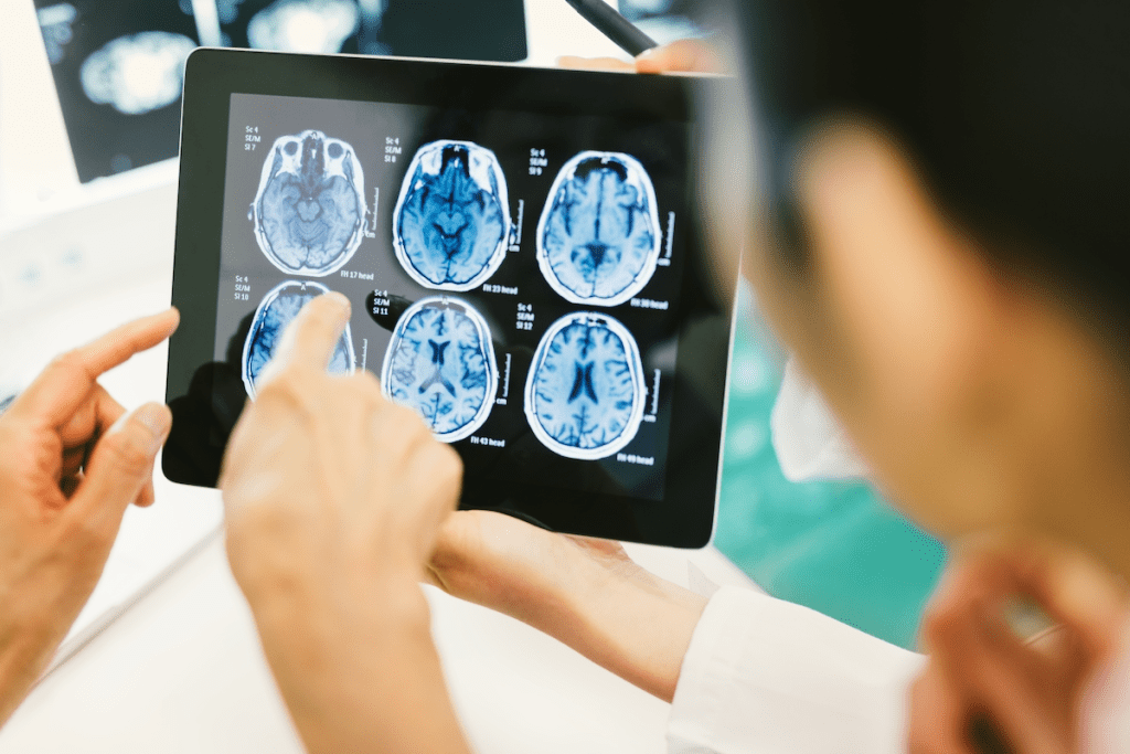
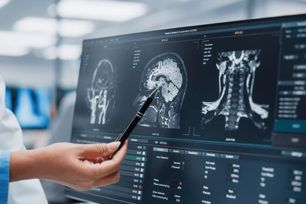
Diagnosing brain cancer relies heavily on advanced imaging. These methods help see tumors clearly. They show the tumor’s size, location, and type, which is key for treatment planning.
Magnetic Resonance Imaging (MRI)
MRI is top for brain tumor imaging. It’s very sensitive and shows soft tissues well. It’s great for seeing the brain’s details and finding tumors in hard-to-reach spots.
Key Benefits of MRI:
- High-resolution images of soft tissues
- Ability to detect tumors in sensitive areas
- No radiation exposure
Computed Tomography (CT) Scans
CT scans are also key for finding brain tumors. They’re fast and useful in emergencies. They show how big a tumor is and how it affects nearby areas.
Advantages of CT Scans:
- Quick and widely available
- Useful in emergency situations
- Provides information on bone structures
| Imaging Technique | Key Features | Clinical Use |
| MRI | High-resolution soft tissue imaging, no radiation | Detailed brain anatomy, tumor detection |
| CT Scans | Quick, widely available, bone structure information | Emergency situations, assessing tumor impact |
| PET Scans | Metabolic activity information, tumor grading | Tumor malignancy assessment, treatment planning |
Positron Emission Tomography (PET) Scans
PET scans give insight into tumor activity. This helps figure out how bad a tumor is and what treatment to use.
Benefits of PET Scans:
- Provides metabolic information
- Aids in tumor grading and treatment planning
- Useful in assessing treatment response
In conclusion, MRI, CT scans, and PET scans are essential for finding and diagnosing brain tumors. Each has its own strengths and weaknesses. Using them together improves accuracy and helps decide the best treatment.
Specialized Brain Imaging Methods
Specialized brain imaging methods have changed neuro-oncology a lot. They give detailed views of brain tumors and how they work. This helps doctors diagnose and treat brain cancer better.
Functional MRI (fMRI)
Functional MRI (fMRI) is a non-invasive way to see brain function. It finds changes in blood flow. fMRI is great for finding important brain areas like speech, movement, and vision. This is key for surgery, helping keep important brain functions safe.
fMRI shows which brain parts control speaking and moving. This helps surgeons plan and predict outcomes after surgery.
Magnetic Resonance Spectroscopy
Magnetic Resonance Spectroscopy (MRS) gives metabolic info on brain tumors. MRS can tell different brain lesions apart by their chemistry. This helps diagnose tumor types and how bad they are.
- Identifies metabolic changes in brain tumors
- Helps differentiate between tumor types
- Assists in assessing tumor aggressiveness
Perfusion and Diffusion MRI
Perfusion MRI looks at blood flow to tumors, showing how aggressive they might be. High blood flow often means more aggressive tumors. Diffusion MRI checks water movement in the brain. It finds areas where water can’t move well, which might mean a tumor.
Contrast Enhancement Techniques
Contrast enhancement uses agents to make brain areas stand out during imaging. These agents go to tumors, making them easier to see on scans. It’s good for seeing tumor edges and how well they’re responding to treatment.
Using these brain imaging methods together, doctors get a full picture of brain tumors. This leads to better diagnosis and treatment plans.
How to Test for Brain Cancer Through Biopsy Procedures
A biopsy is a key tool for diagnosing brain cancer. It lets doctors look at tumor tissue under a microscope. This helps figure out the type and how serious the cancer is, which guides treatment.
Stereotactic Biopsy
A stereotactic biopsy is a small procedure. It uses a special frame and imaging to find and take a sample of tumor tissue. It’s great for tumors in hard-to-reach brain spots.
- Precision: Stereotactic biopsy allows for accurate targeting of the tumor.
- Minimally Invasive: This procedure involves smaller incisions compared to open surgery.
- Diagnostic Accuracy: Provides tissue samples for detailed pathological examination.
Open Surgical Biopsy
An open surgical biopsy means opening the skull to get to the tumor. It’s used for bigger tumors or when more tissue is needed.
This method gets a bigger tissue sample, which is good for detailed analysis. But, it’s more invasive than stereotactic biopsy.
Analyzing Biopsy Results
After getting the biopsy sample, it goes to a lab for analysis. Pathologists look at it under a microscope to find cancer cells, figure out the tumor type, and its grade.
Key aspects of biopsy analysis include:
- Tumor type identification
- Grade of the tumor
- Presence of specific genetic markers
Risks and Recovery
Biopsies have risks like infection, bleeding, and neurological problems. The risks depend on the biopsy method and where the tumor is.
Recovery time varies. Stereotactic biopsies have quick recovery, while open surgical biopsies take longer. Our medical team will give you all the care instructions you need for a smooth recovery.
Can Blood Tests Detect Brain Tumors? Current Capabilities and Limitations
Traditional blood tests can’t directly find brain tumors. But, new tech is being made to help. Right now, blood tests can’t spot brain tumors because they don’t release markers into the blood.
“A blood test for brain cancer would be a big step,” it would be easier and less scary. Scientists are working hard to find new ways to detect tumors.
Existing Blood-Based Biomarkers
Blood biomarkers are signs in the blood that show disease. For brain tumors, researchers look at proteins, DNA, and more. They hope to find markers like GFAP to spot tumors.
Liquid Biopsy Technology
Liquid biopsy looks at blood for tumor DNA. It’s a new way to check cancer, including brain tumors. It’s less scary than regular biopsies.
Studies say liquid biopsies and DNA tests might soon help find tumors easily. This could mean less pain for patients.
Challenges in Blood-Based Brain Tumor Detection
But, finding brain tumors in blood is hard. The blood-brain barrier stops many markers. Also, brain tumors vary a lot, making it tough to find one marker for all.
- The blood-brain barrier blocks many molecules.
- Brain tumors are different, making it hard to find one marker.
- Current tech needs more work to be sure and standard.
As science gets better, we might see better blood tests for brain tumors. New biomarkers and better liquid biopsies could help a lot.
Cerebrospinal Fluid Analysis for Brain Cancer
Cerebrospinal fluid (CSF) analysis is key in finding brain cancer and other central nervous system issues. It looks at the fluid around the brain and spinal cord. This gives clues about infections, inflammation, or cancer cells.
Lumbar Puncture Procedure
A lumbar puncture, or spinal tap, is how CSF is collected. A needle is inserted between vertebrae in the lower spine. This is done under local anesthesia to reduce pain.
The procedure is quick and done in a clinic. It’s important for patients to stay very calm during it to avoid problems.
What CSF Analysis Can Reveal
CSF analysis can show a lot about the central nervous system’s health. For brain cancer, it can spot cancer cells in the CSF. This means there might be a tumor or cancer spread.
It can also find odd protein levels, glucose changes, and other signs of cancer or neurological issues.
Key findings from CSF analysis include:
- Presence of cancer cells
- Abnormal protein levels
- Changes in glucose concentrations
- Signs of infection or inflammation
When CSF Testing Is Recommended
CSF testing is suggested when brain cancer or other central nervous system issues are suspected. It’s vital for diagnosing and treating conditions like meningitis, multiple sclerosis, and certain brain tumors.
Doctors might suggest it for symptoms like severe headaches, confusion, or neurological problems. These could point to serious health issues.
Technetium-99m Bone Scans in Cancer Diagnosis
Technetium-99m bone scans have changed how we find and treat cancer. They help spot bone metastases and see how far cancer has spread.
How Technetium-99m Bone Scintigraphy Works
A tiny amount of radioactive tracer, technetium-99m methylene diphosphonate (Tc-99m MDP), is injected into the blood. This tracer goes to areas with lots of bone activity, like bone metastases. This makes them show up on the scan.
Key aspects of technetium-99m bone scintigraphy include:
- High sensitivity for detecting bone metastases
- Ability to image the entire skeleton
- Useful for monitoring response to treatment
Interpreting Increased Uptake on Bone Scans
An increased uptake on a bone scan means there’s high bone activity. This can be from cancer, fractures, or infections. The scan’s pattern helps figure out the cause.
Factors influencing the interpretation include:
- The intensity and location of the uptake
- The patient’s clinical history and symptoms
- Comparison with previous scans
Planar vs. SPECT Imaging Techniques
Technetium-99m bone scans can be done with planar or SPECT imaging. Planar gives two-dimensional views. SPECT creates three-dimensional images, helping spot and understand lesions better.
SPECT imaging advantages:
- Improved lesion detection and localization
- Better differentiation between bone and soft tissue uptake
- Enhanced diagnostic accuracy
Radionuclide Bone Scanning for Metastatic Brain Cancer
We use radionuclide bone scanning to find bone metastases in brain cancer patients. This method involves injecting a radioactive isotope, like Technetium-99m, into a vein. The isotope goes to areas where bone is changing.
The Role of Skeletal Scintigraphy in Cancer Staging
Skeletal scintigraphy, or radionuclide bone scanning, is a key tool in cancer staging. It helps find bone metastases early. This is important for knowing how far the cancer has spread and for planning treatment.
Skeletal scintigraphy is great because it can scan the whole skeleton at once. This makes it efficient for finding metastatic disease.
Radioactive Isotope Distribution Patterns
The way the radioactive isotope spreads can tell us a lot. Areas with more isotope uptake often have bone turnover. This can mean there are metastases.
It’s important to understand these patterns to read scan results right. For example, a focal area of increased uptake might show a metastatic lesion. But diffuse uptake could mean a metabolic bone disease.
Distinguishing Benign from Malignant Conditions
Radionuclide bone scanning is good at finding bone metastases but can’t tell if it’s cancer. So, it’s key to tell the difference between benign and malignant conditions.
We often need to use other imaging like MRI or CT scans to confirm what the bone scan shows. By using different diagnostic methods together, we can better diagnose and stage metastatic brain cancer.
Genetic and Molecular Testing for Brain Tumors
Genetic testing helps find specific mutations in brain tumors, guiding treatment. We’re seeing big changes in diagnosing and treating brain tumors. These tests tell us about the tumor’s genetic makeup, helping choose treatments and predict outcomes.
Identifying Genetic Mutations
Genetic mutations are key in brain tumor growth and spread. Next-generation sequencing (NGS) is a key tool for finding these mutations. It analyzes the tumor’s genetic material to spot growth drivers.
For example, IDH1 and IDH2 gene mutations are common in some gliomas. Finding these mutations helps us understand the tumor type and plan treatment.
Molecular Markers and Their Significance
Molecular markers give clues about tumor behavior and treatment response. MGMT promoter methylation is a marker linked to better chemotherapy response.
The presence or absence of certain markers greatly affects treatment plans. For instance, tumors with MGMT promoter methylation might better respond to temozolomide, a common glioblastoma treatment.
Personalized Treatment Based on Genetic Testing
Genetic testing lets us tailor treatments to each patient’s tumor. This personalized medicine is changing neuro-oncology.
Knowing a tumor’s genetic and molecular profile helps choose the best treatments. This targeted approach boosts patient outcomes and lowers side effect risks.
As Dr. Jane Smith, a leading neuro-oncologist, says,
“Genetic testing has changed how we treat brain tumors. We now tailor treatments to each patient’s unique genetic profile.”
Emerging Technologies in Brain Cancer Detection
New technologies are changing how we find brain cancer, giving hope to patients. These new ways to diagnose are helping us spot tumors sooner and more accurately.
Advanced Blood-Based Diagnostic Methods
New blood tests are being made to find brain cancer without surgery. Liquid biopsies and methylation profiling are promising. They look for tumor DNA and biomarkers in blood, a gentler way than biopsies.
Experts think these tests could soon be key for catching brain cancer early. They help doctors tailor treatments by finding specific genetic signs in the blood.
AI and Machine Learning Applications
Artificial Intelligence (AI) and machine learning are making brain cancer detection better. They can sift through imaging data, spot patterns, and help find tumors. AI can also tell different tumors apart and guess how they might grow.
Machine learning gets smarter with more data. It combines medical records with imaging to help doctors make better choices for patients.
Novel Imaging Techniques
New imaging tools are being made to see brain tumors better. Magnetic resonance imaging (MRI) and positron emission tomography (PET) scans are getting better. They show tumors in more detail and how they work.
These advanced scans are key for finding tumors right, planning treatments, and keeping an eye on them. They give important info on where tumors are, how big they are, and how they might affect the brain.
Conclusion: The Future of Brain Cancer Diagnostics
As we wrap up our look at brain cancer diagnostics, it’s clear that research and innovation are key. New imaging, genetic testing, and tech are making diagnosis and treatment better.
The outlook for brain cancer diagnostics is bright. We’re moving towards more accurate and tailored diagnostic methods. This means doctors can create better treatment plans for each patient.
New tech, like advanced blood tests and AI in imaging, is on the horizon. These tools will make diagnosis faster and more accurate. They’ll help us understand brain cancer better and improve patient care.
As we learn more about brain cancer, diagnostics will keep getting better. Genetic and molecular testing are already making a difference. They help doctors tailor treatments to each patient’s needs.
We’re dedicated to using medical innovation to help brain cancer patients. By pushing the boundaries of diagnostics, we’re hopeful for better treatment results. This will bring hope to those fighting this disease.
FAQ
What is a technetium-99m bone scan, and how does it work?
A technetium-99m bone scan, also known as bone scintigraphy, is a test that uses a small amount of radioactive material. This material, technetium-99m, is injected into the bloodstream. It then accumulates in areas of high bone activity.
This allows doctors to see where bone metabolism is high. It helps them find areas that might need attention.
What does increased uptake on a bone scan mean?
Increased uptake on a bone scan means there’s high bone activity. This can be a sign of many things, like cancer, fractures, or infections. The level of uptake helps doctors tell if it’s something serious or not.
How do doctors test for brain cancer?
Doctors use MRI, CT scans, and PET scans to test for brain cancer. They also do biopsies to get tissue samples. A full check-up of the brain and nervous system is done too.
Can blood tests detect brain tumors?
Right now, blood tests can’t find brain tumors directly. But scientists are working on new tests. They want to find biomarkers in the blood that show if a tumor is there.
What is the role of cerebrospinal fluid analysis in brain cancer diagnosis?
Cerebrospinal fluid (CSF) analysis is key in diagnosing brain cancer. A procedure called a lumbar puncture is used to get CSF. Then, it’s checked for abnormal cells or proteins.
How is genetic testing used in brain tumor diagnosis and treatment?
Genetic testing helps find specific mutations in brain tumors. This information helps doctors plan treatment. It makes treatment more targeted and effective.
What are the emerging technologies in brain cancer detection?
New technologies are coming for brain cancer detection. These include better blood tests, AI, and new imaging methods. They’re making diagnosis and treatment better.
What is the difference between planar and SPECT imaging techniques in bone scans?
Planar imaging shows a two-dimensional view of bones. SPECT imaging, on the other hand, shows a three-dimensional view. SPECT gives a clearer picture of bone activity.
How do radionuclide bone scans help in diagnosing metastatic brain cancer?
Radionuclide bone scans, like technetium-99m scans, spot areas of high bone activity. This can mean metastatic brain cancer. The pattern of the radioactive material helps doctors tell if it’s cancer or not.
What are the common types of brain tumors?
There are many types of brain tumors. Gliomas, meningiomas, and acoustic neuromas are some examples. Each has its own signs and symptoms.
References
- Louis, D. N., Perry, A., Wesseling, P., Brat, D. J., Cree, I. A., Figarella-Branger, D., Hawkins, C., Ng, H. K., Pfister, S. M., Reifenberger, G., Soffietti, R., Tachibana, O., von Deimling, A., & Ellison, D. W. (2021). The 2021 WHO Classification of Tumors of the Central Nervous System: a summary. Neuro-Oncology, 23(8), 1231-1251.https://pubmed.ncbi.nlm.nih.gov/34185072/


