Last Updated on November 27, 2025 by Bilal Hasdemir

Choosing the right tool for early disease detection is key. At LivHospital, we help you understand the difference between PET and MRI scans. This way, you can pick the best imaging technique for your health.
A PET scan and an MRI scan show different things in different ways. PET scans use a radioactive tracer to see how cells work. They’re great for finding cancer early and tracking cell changes. On the other hand, MRI scans use magnets and radio waves to show detailed images of soft tissues. They do this without using radiation.
Key Takeaways
- PET scans assess metabolic activity using a radioactive tracer.
- MRI scans use strong magnets and radio waves for high-resolution images.
- PET scans are valuable for detecting cancer and monitoring cellular changes.
- MRI scans are ideal for visualizing soft tissues like muscle and fat.
- The choice between PET and MRI scans depends on the specific health needs.
Understanding Medical Imaging Technologies
Medical imaging has made huge strides, with PET and MRI scans at the forefront. At Liv Hospital, we focus on cutting-edge, team-based healthcare. Advanced scanning tech is key to this.
Medical imaging has grown a lot, starting with X-rays in 1895. Now, we have many advanced tools. These include PET and MRI scans, which help us diagnose and treat diseases better.
The Evolution of Diagnostic Imaging
Diagnostic imaging has changed a lot over the years. New tech and our knowledge of the body have driven this change. Now, we can see the body’s inner workings in great detail thanks to PET and MRI scans.
PET scans let us see how the body’s cells work. This is very helpful for finding and managing cancer. MRI scans, on the other hand, give us clear pictures of soft tissues. This is great for checking the brain, spine, and muscles.
The Role of Advanced Scanning in Modern Medicine
PET and MRI scans are key in today’s medicine. They help doctors find diseases early and accurately. This is very important for good treatment.
In cancer care, PET scans show how far the disease has spread. MRI scans give detailed views of tumors and their surroundings. At Liv Hospital, we use these techs to give top-notch care. Our teams work together to understand imaging results and plan treatments for each patient.
| Imaging Modality | Primary Use | Key Benefits |
| PET Scan | Cancer diagnosis and staging, neurological disorders | Assesses metabolic activity, with high sensitivity for detecting disease |
| MRI Scan | Soft tissue imaging, brain and spinal cord assessment | High-resolution images, excellent for soft tissue visualization |
We pick the best imaging tool for each patient based on their needs. This is how we ensure personalized, high-quality care.
How PET Scans Work
Understanding PET scans means looking into the science behind them. They are a key tool in medical imaging. They show how the body’s cells work.
The Science Behind Positron Emission Tomography
PET scans use a special method to see how active the body’s cells are. They use radioactive tracers to find out. These tracers go to areas where cells are very active, like in cancer.
We use these tracers to see how different body parts work. For example, fluorodeoxyglucose (FDG) is a common tracer. It’s a radioactive sugar that cancer cells love more than normal cells.
Radioactive Tracers and Their Function
The type of tracer used depends on what we want to find out. FDG is often used for cancer. Other tracers help with heart or brain issues. These tracers send out positrons, which create gamma rays. The PET scanner catches these rays to make images.
The PET Scanning Process
The scan starts with a tracer injection into the patient’s blood. Then, the patient waits for the tracer to reach the right spots. Next, they lie down in the PET scanner.
The scanner picks up the gamma rays from the tracer. It uses this info to make detailed pictures of the body’s activity.
How MRI Scans Work
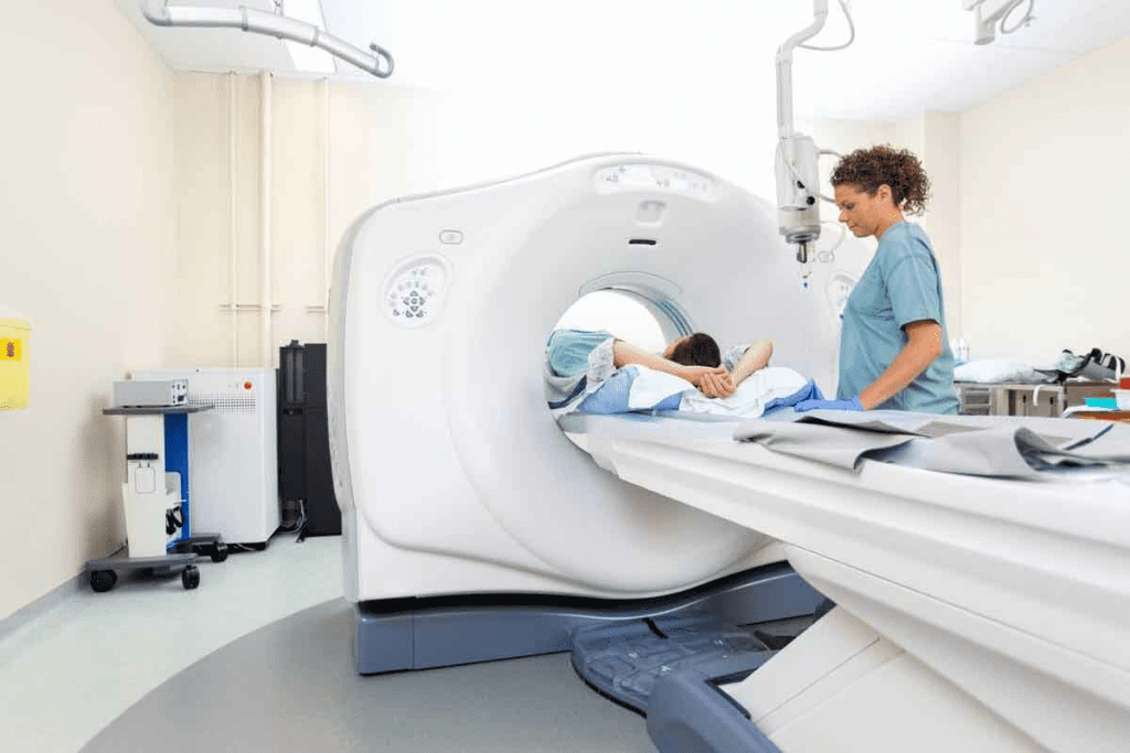
MRI scans use magnetic resonance to create detailed images of the body. They don’t use harmful radiation. This makes them a key tool in medical imaging.
The Principles of Magnetic Resonance Imaging
MRI scans work by aligning hydrogen atoms in the body with a strong magnetic field. Then, a radiofrequency pulse makes these atoms send signals. These signals help create detailed images.
First, the patient gets into the MRI machine, a big magnet. This magnet aligns the hydrogen nuclei in the body. Next, a radiofrequency pulse disturbs them, causing them to send signals as they return to their aligned state.
Magnets, Radio Waves, and Tissue Visualization
The magnetic field and radio waves work together to see different tissues in the body. The signals from different tissues are captured by the MRI machine. These signals help create images that show different soft tissues, like organs and tendons.
MRI scans are great for seeing soft tissues. They are very useful for checking the brain, spinal cord, and muscles. They can find problems that other scans can’t see.
The MRI Scanning Process
The MRI scanning process has several steps. First, patients remove metal objects and change into a special gown. This is to make sure they are safe and the images are clear.
| Step | Description |
| Preparation | Patient preparation includes removing metal objects and changing into an MRI-compatible gown. |
| Positioning | The patient is positioned on the MRI table, which is then moved into the MRI machine. |
| Scanning | The MRI machine captures images of the body’s internal structures using magnetic fields and radio waves. |
| Image Reconstruction | The captured data is reconstructed into detailed images that can be reviewed by healthcare professionals. |
Understanding MRI scans shows their importance in medicine. They give doctors the information they need to make better treatment plans. This helps patients get better faster.
Is a PET Scan Like an MRI? Key Differences Explained
PET scans and MRI scans are different because of their technology and what they show. Both are used to help doctors diagnose, but they do it in different ways.
Fundamental Operational Differences
PET scans use a special tracer to see how body tissues work. This tracer is given through the blood. MRI scans, on the other hand, use strong magnets and radio waves to show the body’s soft tissues in detail.
The main difference is what each scan shows. PET scans look at how tissues work, like their metabolic rate. MRI scans show the body’s structure in high detail.
What Each Scan Actually Measures
PET scans check how cells work, which helps find cancer. MRI scans are great for seeing soft tissues, like the brain and muscles.
PET scans show how tissues function, while MRI scans show their structure. This is why they are used differently in medicine.
Radiation Exposure Comparison
PET scans use a small amount of radiation from the tracer. MRI scans don’t use radiation, using magnets and radio waves instead.
MRI scans are safer for people who need many tests or are worried about radiation. Butthe right scan depends on what the doctor needs to know.
Clinical Applications of PET Scans
PET scans are very useful in diagnosing and managing many medical conditions. They give important information to help doctors make the best decisions for their patients.
Cancer Detection and Staging
PET scans are great for finding and figuring out how far cancer has spread. They help doctors know the best treatment plan. PET scans for cancer also check if the cancer is getting better with treatment.
- Detecting cancer at an early stage
- Staging cancer to understand its spread
- Monitoring cancer response to treatment
Neurological Disorders Assessment
PET scans are also used for neurological disorders like Alzheimer’s disease and dementia. They show how the brain works and help find problems early.
Some key uses are:
- Diagnosing Alzheimer’s disease and other dementias
- Assessing brain function in epilepsy
- Evaluating brain tumors
Cardiac Function Evaluation
In cardiology, PET scans check how well the heart works and find coronary artery disease. They see how blood flows to the heart muscle, spotting any damage or risk.
- Assessing myocardial viability
- Diagnosing coronary artery disease
- Evaluating heart function after a heart attack
PET scans give a special look at the body’s metabolic processes. They work well with other imaging l,,ike MRI. Knowing when to use PET scans vs MRI is key tothe best care.
Clinical Applications of MRI Scans
MRI scans are key in modern medicine, giving deep insights into the body. They show detailed images of soft tissues and organs. This makes them essential for many diagnoses.
Soft Tissue and Organ Imaging
MRI scans excel at showing soft tissues like organs and muscles. They are vital for spotting tumors, injuries, and vascular diseases. For example, they can find liver lesions or see heart damage after a heart attack.
Brain and Spinal Cord Assessment
MRI scans clearly show the brain and spinal cord. They help diagnose conditions like multiple sclerosis and spinal cord injuries. They also help plan brain surgeries by showing the brain’s layout.
Musculoskeletal System Evaluation
MRI scans are used to check injuries in the musculoskeletal system. They can spot torn ligaments, herniated discs, and bone tumors. This is helpful for both diagnosing and planning treatments.
Let’s look at how MRI scans are used in different parts of the body:
| Body System | Common Conditions Diagnosed | Benefits of MRI |
| Soft Tissues and Organs | Tumors, vascular diseases, orand gan damage | High-resolution imaging of soft tissues |
| Brain and Spinal Cord | Multiple sclerosis, spinal cord injuries, and dementia | Detailed anatomy for neurosurgical planning |
| Musculoskeletal System | Torn ligaments, herniated discs, and bone tumors | Non-invasive assessment of injury extent |
Advantages and Limitations of Each Technology
Understanding PET and MRI scans is key tomaking smart choices in medicine. Both have changed how we see the body. They give us new ways to understand health.
PET Scan Strengths and Weaknesses
PET scans show how active the body’s cells are. They’re great for finding cancer, studying the brain, and checking the heart. They highlight very active areas, which can mean disease.
But, PET scans have downsides. They don’t show where problems are as clearly as MRI scans. Also, they use a bit of radiation, which worries some people.
MRI Scan Strengths and Weaknesses
MRI scans give detailed pictures of the body’s soft parts. They’re best for looking at the brain, spine, and joints. MRI scans are very good at showing what’s going on inside these areas.
But, MRI scans don’t show how active the body’s cells are, like PET scans do. They can give some clues about how the body works, but not as much as PET scans.
Comparing Image Resolution and Detail
MRI scans win when it comes to clear images. They show the body’s parts very well. PET scans are not as clear, but they’re getting better with new tech.
In short, both PET and MRI scans have good points and bad points. The right choice depends on what you need to know. Knowing what each scan can do helps doctors pick the best one for each patient.
PET vs. MRI: When Is Each Scan Preferred?
Knowing when to use a PET scan versus an MRI is key toaccurate diagnosis and treatment planning. We will look at the factors that decide this choice. This ensures patients get the right imaging for their needs.
Disease-Specific Considerations
The choice between a PET scan and an MRI depends on the disease or condition. PET scans are great for finding and staging cancer. They show how active tissues are metabolically. This is why they’re useful for seeing how cancer has spread and how it’s responding to treatment.
MRI scans are better for soft tissues and organs. They’re perfect for checking the brain, spinal cord, and muscles. MRI’s clear images help spot structural problems and injuries that PET scans might miss.
Patient-Specific Factors
What’s best for a patient also matters. For example, people with metal implants or pacemakers can’t have an MRI because of the magnetic fields. In these cases, a PET scan might be better.
Also, patients with claustrophobia might struggle with MRan I because they need to stay in a small space for a long time. PET scans are done in a more open area, which might be easier for some.
Diagnostic Goals and Outcomes
The goals of the diagnosis and what’s expected are important in choosing between a PET scan and an MRI. If you want to see how active a tumor is, a PET scan is better. But if you need detailed images of structural damage or disease growth, an MRI is better.
In the end, picking between a PET scan and an MRI depends on a full review of the patient’s situation. This includes looking at the disease and the patient’s needs. By picking the right imaging, doctors can make accurate diagnoses and plan effective treatments.
Combined PET/MRI Technology: The Best of Both Worlds
PET/MRI technology is a big step forward in medical imaging. It gives a detailed look at the body’s inside and how it works. This mix of PET scans and MRI images is a powerful tool for doctors.
Hybrid Imaging Mechanism
The PET/MRI system combines PET and MRI scanners into one. This lets it get PET and MRI data at the same time. Then, it can show a full picture of the body’s inner workings.
Key components of PET/MRI technology include:
- Advanced PET detectors that can work in the MRI’s strong magnetic field
- High-field MRI magnets for detailed body images
- Smart software for combining images
Benefits of Simultaneous Imaging
Getting PET and MRI data at once has many benefits. It makes diagnoses more accurate and saves time for patients. It also helps doctors understand complex health issues better.
Some of the key advantages include:
- More accurate diagnoses by linking body function and structure
- Less need for many scans, making patients happier and saving money
- Can spot diseases early and track treatments better
Clinical Applications
PET/MRI technology is used in many areas, like cancer, brain, and heart health. It helps stage cancers, check brain diseases, and see how well the heart works.
| Clinical Area | Application of PET/MRI | Benefits |
| Oncology | Tumor staging and treatment response assessment | Improved accuracy in cancer staging and monitoring |
| Neurology | Assessment of neurodegenerative diseases | Enhanced understanding of disease progression |
| Cardiology | Evaluation of cardiac viability and function | Better assessment of cardiac health and viability |
As PET/MRI tech gets better, we’ll see new uses in medical imaging. This will help us diagnose and treat diseases even better.
Conclusion: Making Informed Decisions About Medical Imaging
It’s key for patients to know the difference between PET scans and MRI scans. This knowledge helps them make better choices about their health care. We’ve looked at the basics, how they’re used in medicine, and what each offers.
When deciding between PET scans vs MRI or MRI versus PET scan, it’s important to understand their strengths and weaknesses. PET scans are great for spotting cancer and brain issues because they show how cells work. MRI scans, on the other hand, are better for seeing soft tissues and injuries because they show detailed images.
The main difference between a PET scan and mri is their technology and what they reveal. Knowing this helps patients make smarter choices about their health care.
Medical imaging is always getting better, with new tools like PET/MRI scans. By keeping up with these advances, patients can be more involved in their health care. This way, they can use the best tools available to help them.
FAQ
What is the main difference between a PET scan and an MRI scan?
PET scans use radioactive tracers to show how cells work. MRI scans use strong magnets and radio waves to show soft tissues in detail. They don’t use radiation.
Is a PET scan better than an MRI for detecting cancer?
PET scans are great for finding cancer early and tracking cell changes. MRI scans are good for detailed soft tissue images. It depends on the case.
What is the difference between a PET scan and an MRI in terms of radiation exposure?
PET scans use radioactive tracers, which means some radiation. MRI scans don’t use radiation at all.
Can PET and MRI scans be used together?
Yes, PET/MRI technology combines both. It gives a full view of body structures and functions at the same time.
How do I choose between a PET scan and an MRI scan for my medical imaging needs?
Choosing depends on your medical condition, what you want to find out, and your personal needs.
What are the clinical applications of PET scans?
PET scans help find and stage cancer, check neurological disorders, and look at heart function. They have many uses.
What are the advantages of MRI scans in medical imaging?
MRI scans are great for soft tissues and organs, the brain and spinal cord, and muscles. They give detailed images without radiation.
How do PET scans work?
PET scans use radioactive tracers to see how cells work. They show the body’s internal functions.
What is the role of strong magnets and radio waves in MRI scans?
MRI uses strong magnets and radio waves to create detailed images. This lets us see inside the body.
Are there any limitations to using PET or MRI scans?
Both PET and MRI scans have their own strengths and weaknesses. The right choice depends on the situation and what you need to find out.
What is the difference between a PET scan and an MRI scan in terms of image resolution and detail?
MRI scans usually have better image quality, showing soft tissues well. PET scans show metabolic activity and cell changes.
Can combined PET/MRI technology improve diagnostic outcomes?
Yes, PET/MRI technology can give a better understanding of the body. This could lead to better diagnosis and treatment.
References
- Chan, B. Y., Crawford, A. M., Kobes, P. H., et al. (2020). Septic Arthritis: An Evidence-Based Review of Diagnosis and Image-Guided Aspiration. AJR American Journal of Roentgenology, 215(3), 568-581. https://pubmed.ncbi.nlm.nih.gov/32783556/


