Last Updated on November 27, 2025 by Bilal Hasdemir

At Liv Hospital, we use PET scans to find out how active tissues are. This helps us spot and keep an eye on different health issues, like cancer. The ‘glow’ on a PET scan shows how active the tissues are. Less intense ‘glow’ means lower activity.Learn what less glow on a PET scan means, understand SUV and uptake values, and interpret PET scan results correctly.
This activity is measured with a special tracer and a number called SUV (Standardized Uptake Value).
It’s key to know about SUV and tracer uptake to understand PET scan results. Our team is here to help you get it. We want to make sure you know your diagnosis and treatment options. We aim to give you top-notch healthcare with full support.
Key Takeaways
- PET scans measure metabolic activity using a radioactive tracer.
- A less intense ‘glow’ on a PET scan indicates lower metabolic activity.
- SUV is a quantitative measure of tracer uptake in tissues.
- Understanding PET scan results is key to diagnosis and treatment.
- Our team provides expert guidance and support throughout the process.
The Basics of PET Scan Technology

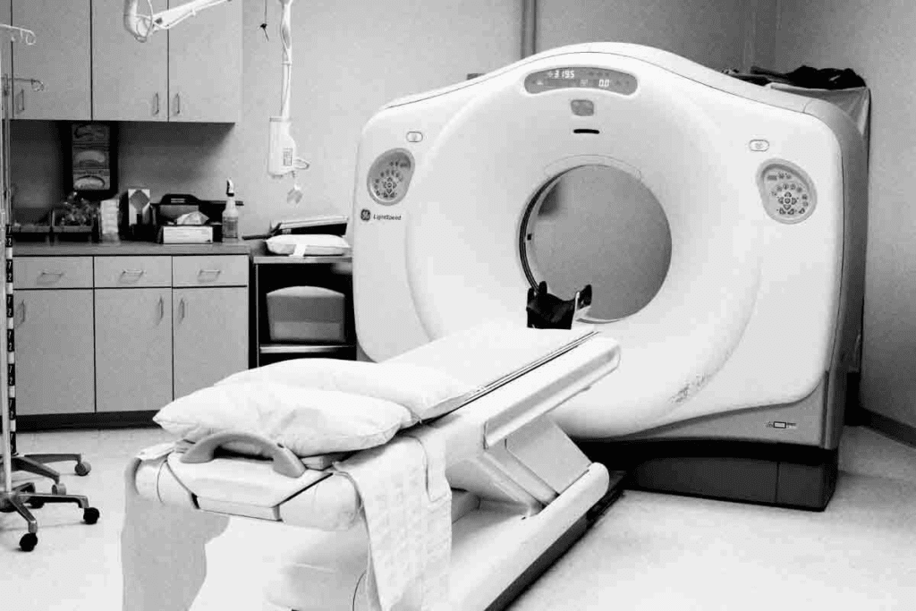
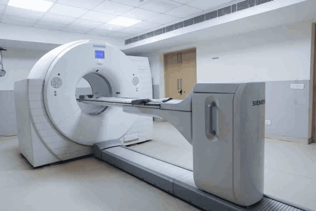
The PET scan, or Positron Emission Tomography scan, is a cutting-edge medical tool. It uses nuclear medicine to see how the body’s cells work. This tech is key to finding and tracking many health issues, like cancer.
How PET Scans Work
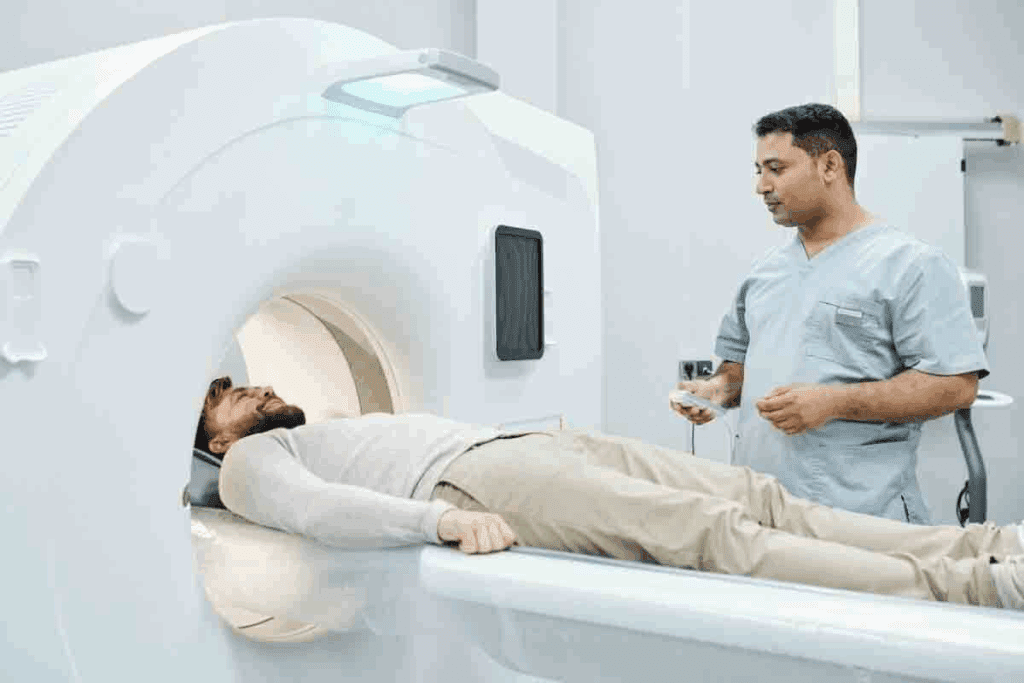
PET scans spot how active cells are in the body. We inject a slightly radioactive liquid, called a tracer, into the body. This liquid goes to areas where cells are very active, like in growing tumors.
The PET scan catches the energy from the tracer. It makes pictures of where the tracer is. These pictures show us where the body’s cells are most active. This helps us figure out what’s going on and plan treatments.
The Role of Radioactive Tracers
Radioactive tracers are the core of PET scans. They are made to be taken by cells, mainly those that are very active. The most used tracer is FDG (Fluorodeoxyglucose), a special sugar that’s been made radioactive.
Key traits of radioactive tracers include:
- They target specific cells or processes
- They can be seen by the PET scanner
- They have a short life to keep radiation low
Metabolic Activity and Imaging
Metabolic activity is key in PET imaging. Cells that are very active, like cancer cells, grab more of the tracer. This makes them show up clearly on PET scan images. It helps us spot areas where something’s not right, like disease.
Understanding PET scans and tracers shows their importance in health care. The info from PET scans is vital for making treatment plans and checking how patients are doing.
Understanding the “Glow” in PET Scan Images

The ‘glow’ in PET scan images shows how active tissues are in the body. It’s not just a visual trick. It helps doctors see how tissues work, which is key for diagnosing and tracking diseases like cancer.
What Causes the Glow Effect
The glow in PET scans comes from radioactive tracers in tissues. When a patient gets a tracer like Fluorodeoxyglucose (FDG), it goes to busy areas. This tracer makes gamma rays, which the scanner picks up, showing where it is.
A medical expert, a top nuclear medicine expert, says, “The tracer’s amount shows how active the tissue is. This makes PET scans great for seeing disease severity and spread.”
“The glow in PET scans is a window into the metabolic activity of tissues, providing critical information for diagnosis and treatment planning.” -A medical expert, Nuclear Medicine Expert
Interpreting Different Intensities
Different glow levels in PET scans mean different activity levels. Brighter areas have more activity, while darker ones have less. This helps spot problems like cancer, which is more active than normal tissue.
In cancer patients, bra ight glow means active cancer. Less bright might mean the cancer is shrinking or is less aggressive. When we look at PET scans, we must think about the whole picture and other tests, too.
Color Mapping in PET Imaging
Color mapping in PET scans makes it easier to see tracer uptake. It uses colors to show activity levels. For example, low activity is black and blue, and high activity is white and red.
This helps doctors quickly see what’s going on in the body. It’s very useful in tricky cases or when tracking changes over time.
What Less Glow on a PET Scan Means
Less glow on a PET scan often means lower metabolic activity. This can happen in many clinical situations. It’s important to know why this happens and what it means.
This decrease in activity can be due to several reasons. It could be because of normal tissue or treated lesions. It could also be because of non-aggressive lesions.
Reduced Metabolic Activity
Less glow on a PET scan often means cells are not as active. PET scans measure how active cells are. If cells are less active, it can mean different things.
For example, if a tumor is treated well, it might show less activity. This is because the treatment has worked.
Common Causes of Decreased Uptake
There are several reasons why a PET scan might show less activity. Here are some common ones:
- Treatment response: Tumors might show less activity after treatment.
- Benign conditions: Some non-cancerous conditions can also show less activity.
- Scar tissue: Scarred areas might have lower activity than active lesions.
- Non-aggressive lesions: Some lesions might naturally have lower activity.
Clinical Significance of Less Glow
The meaning of less glow on a PET scan varies. In cancer treatment, it can mean the treatment is working. But it’s important to look at all the information before making conclusions.
Distinguishing Normal from Abnormal Low Uptake
Telling normal from abnormal low uptake on a PET scan is tricky. The patient’s history, the PET scan details, and other tests are key. Doctors need to consider these to understand the scan results correctly.
Standardized Uptake Value (SUV): A Quantitative Measure
In PET scan imaging, the Standardized Uptake Value (SUV) is a key measure. It shows how much radioactive tracers are taken up by tissues. This helps doctors understand PET scan results and make better decisions.
What Is an SUV in PET Scan Analysis
The Standardized Uptake Value (SUV) is a number that shows the tracer uptake in tissues. SUV helps doctors tell normal from abnormal uptake. This is important for diagnosing and understanding disease stages.
How SUV Is Calculated
SUV is found using a formula: SUV = (tissue activity concentration)/(injected dose/body weight). This formula looks at the tracer uptake in tissues compared to the dose given and the patient’s weight. Getting the SUV right is key to accurate PET scan results.
Factors Affecting SUV Measurements
Several things can change SUV measurements, including:
- Patient-related factors like blood glucose levels and body composition
- Technical factors like scanner calibration and image reconstruction algorithms
- Procedural factors, including the time between tracer injection and imaging
Knowing these factors is important for correct SUV value interpretation.
The Importance of SUV in Clinical Decision-Making
SUV is very important in making clinical decisions. It gives a number to look at disease severity and how well treatments work. It helps doctors diagnose, stage, and treat diseases. This guides treatment plans.
Understanding SUV and its meaning helps doctors make better choices. This leads to better patient care.
Interpreting SUV Values in Clinical Context
Understanding SUV values is key to accurate PET scan results. SUV, or Standardized Uptake Value, measures how much a radioactive tracer is taken up. This helps doctors see how active tissues are and make better patient care plans.
Low vs. High SUV Values
SUV values change based on the tissue and condition being looked at. Higher SUV values mean more metabolic activity, often seen in cancer or inflammation. Lower SUV values might mean the tissue is not active or is benign. But it’s important to look at the whole clinical picture when reading SUV values.
In cancer diagnosis, a high SUV value might mean a tumor is aggressive. A low value could suggest a less aggressive or possibly harmless lesion. Knowing these differences is key to accurate diagnosis and treatment.
Normal vs. Abnormal Low Uptake
Telling normal from abnormal low uptake on PET scans can be tricky. Normal low uptake is seen in tissues with low activity, like some brain areas or inactive scars. Abnormally high uptake is seen in tissues that should be more active but aren’t.
For example, a low SUV lesion might be benign if it fits a non-active process. But, if a lesion should be active (like a tumor), low uptake could mean it’s responding to treatment or changing. More tests or imaging might be needed to figure out why uptake is low.
By understanding SUV values in the right clinical context, doctors can make more precise diagnoses and treatment plans. This detailed knowledge of SUV values is vital in modern medicine, mainly in oncology and other fields where PET scans are used.
Mild Uptake on PET Scans: Clinical Implications
Understanding mild uptake on PET scans is key to accurate diagnosis and treatment planning. Mild uptake shows a slight increase in metabolic activity. This can mean anything from a benign condition to early signs of disease.
Defining Mild Uptake
Mild uptake on a PET scan shows a slight increase in the Standardized Uptake Value (SUV). The SUV measures metabolic activity in the body. A mild uptake is an SUV value slightly above normal but not too high.
In cancer diagnosis, an SUV value between 2 and 4 might be mild uptake. This can vary based on the cancer type and organ involved. It’s important to look at these values in the patient’s overall health.
Differentiating Benign from Malignant Mild Uptake
Telling benign from malignant causes of mild uptake on PET scans is hard. Benign conditions like inflammation or normal processes can cause it. But it can also be an early sign of cancer.
Doctors use the patient’s history, imaging, and sometimes more tests to figure it out. The uptake pattern, location, and whether it’s focal or diffuse help. Also, comparing PET scans with CT or MRI can be useful.
Common Causes of Mild Uptake
Mild uptake on PET scans can come from many sources. Common benign causes include:
- Inflammatory processes
- Infections
- Post-surgical changes
- Normal physiological uptake in certain tissues
Malignant causes might include early cancers or tumors with low activity. It’s vital to consider the patient’s health and risk factors when seeing mild uptake.
| Cause | Description | Clinical Context |
| Inflammation | Increased metabolic activity due to inflammatory processes | Recent surgery, infection, or inflammatory disease |
| Early-stage cancer | Mild uptake is indicative of low metabolic activity in tumors | Patients with risk factors for cancer or a known history of malignancy |
| Normal physiological uptake | Mild uptake in certain tissues under normal physiological conditions | Variations in normal anatomy and physiology |
Follow-up Recommendations for Mild Uptake
When mild uptake is seen on a PET scan, more evaluation is needed. Follow-up might include:
- More imaging studies (e.g., CT, MRI) are needed to better understand the uptake.
- Looking at the patient’s symptoms and history.
- Lab tests to check for infection or inflammation.
- Biopsy if there’s a worry about cancer.
Regular check-ups and more PET scans might be needed to watch for changes. The follow-up plan should fit the patient’s specific situation and risk factors.
SUVmax: Understanding Maximum Standardized Uptake Value
Understanding SUVmax is key to diagnosing and tracking diseases with PET scans. SUVmax, or maximum standardized uptake value, shows the most active spot in a lesion.
Definition and Measurement of SUVmax
SUVmax is the highest value of a radioactive tracer in a tumor or lesion. It’s found by measuring activity in a voxel and adjusting for the dose and body weight.
Clinical Significance of SUVmax
SUVmax shows the metabolic activity in a lesion. A high value means aggressive disease, while a low value suggests less activity. This info is vital for diagnosing, staging, and tracking cancer treatment.
SUVmax in Different Types of Cancer
SUVmax is used in various cancers to gauge severity and track disease progression. For example, in lymphoma, a high SUVmax signals aggressive disease. In lung cancer, it helps assess tumor activity. Knowing SUVmax for different cancers helps doctors make better decisions.
Limitations of SUVmax as a Diagnostic Tool
Though useful, SUVmax has its limits. Patient prep, scanner setup, and scan timing can influence values. It also doesn’t show tumor heterogeneity. So, it’s best used with other tools and clinical checks for a full disease picture.
Variables Affecting PET Scan Interpretation
Understanding what affects PET scan results is key to accurate diagnosis and treatment. PET scans are complex tools. Their results depend on many factors.
Patient-Related Factors
Patient factors greatly influence PET scan results. Blood glucose levels are a big factor. High blood glucose levels can skew results. “Patients with diabetes need special prep for PET scans to get accurate results,” experts say.
Body weight and composition, and fasting status before the sn a, also matter. These can change how the tracer is taken up. This affects the scan’s findings.
Technical and Procedural Variables
Technical and procedural aspects are also critical. The PET scanner type, the scan protocol, and timing all play a role. For example, different scanners can detect small lesions or measure SUVs differently.
The algorithm used for image reconstruction also matters. “The choice of reconstruction parameters can significantly impact SUV values,” a study found. This can affect treatment decisions.
Standard Ranges Across Different Populations
Knowing standard PET scan ranges for different groups is vital. Age, sex, and ethnicity can change what’s considered normal or abnormal. SUV values can differ between age groups and sexes.
Setting standards for each population can make PET scan interpretations more accurate. As research advances, we learn more about these differences. This helps us improve clinical practice.
Improving the Accuracy of PET Scan Readings
To make PET scan readings more accurate, standardizing protocols is key. This means proper patient prep, the right tracer dose, and the right timing for the scan.
Also, keeping healthcare professionals up-to-date with the latest research is essential. This ensures PET scan results are reliable. It leads to better care for patients.
Conclusion: Putting PET Scan Results in Perspective
Understanding PET scan results is complex. It involves looking at SUV, uptake, and the patient’s overall health. We’ve seen how PET scans work and how SUV values help doctors make decisions.
When looking at PET scan results, the patient’s health is key. Maximal SUV values and PET scan results are important. But they must be seen with other health findings.
Healthcare providers need to understand PET scan results well. This helps them make the best care plans for patients. It’s about looking at all the information to give accurate diagnoses and treatments.
PET scan results are just part of the health puzzle. By understanding them, we can give patients the care they need. This helps them through their treatment journey.
FAQ
What does SUV mean on a PET scan?
SUV stands for Standardized Uptake Value. It measures how much radioactive tracer is taken up by tissues in a PET scan. This helps doctors see how active tissues are metabolically.
How is SUV calculated in PET scan analysis?
To calculate SUV, the PET scan measures activity in a certain area. It then normalizes this to the dose given and the patient’s body weight or lean body mass.
What causes the “glow” effect in PET scan images?
The “glow” effect comes from tissues taking up radioactive tracers. These tracers emit positrons that destroy electrons, creating gamma rays. The PET scanner picks up these rays.
What does less glow on a PET scan mean?
Less glow usually means lower metabolic activity. This can be normal or could point to disease, depending on the context.
How do you distinguish normal from abnormal low uptake on a PET scan?
To tell normal from abnormal low uptake, look at the clinical context and the area’s pattern. Compare it with known normal and abnormal patterns.
What is SUVmax, and how is it measured?
SUVmax is the highest metabolic activity in a region. It’s found by looking for the voxel with the highest uptake in the area of interest.
What are the clinical implications of mild uptake on a PET scan?
Mild uptake can mean either benign or malignant conditions. The exact meaning depends on the patient’s history, symptoms, and other test results.
How do patient-related factors affect PET scan interpretation?
Factors like blood sugar, body composition, and some medications can change how PET scans are read. They affect how tracers are taken up and spread.
What technical variables can impact PET scan readings?
Scanner type, image algorithms, and acquisition methods can change PET scan results. They affect image quality and SUV values.
How can the accuracy of PET scan readings be improved?
To improve accuracy, standardize imaging, use the right algorithms, and consider the patient’s situation and factors.
What is the significance of SUV in clinical decision-making?
SUV is key in making decisions because it quantifies metabolic activity. It helps diagnose, stage, and track treatment in diseases like cancer.
How do you interpret SUV values in a clinical context?
When interpreting SUV values, think about the question being asked, the tracer used, and the tissue or lesion’s characteristics. Compare with normal tissues and past scans.
References
- UK Government. (2025, July 7). National Diagnostic Reference Levels (NDRLs) for Radiology. Retrieved from https://www.gov.uk/government/publications/diagnostic-radiology-national-diagnostic-reference-levels-ndrls/ndrl


