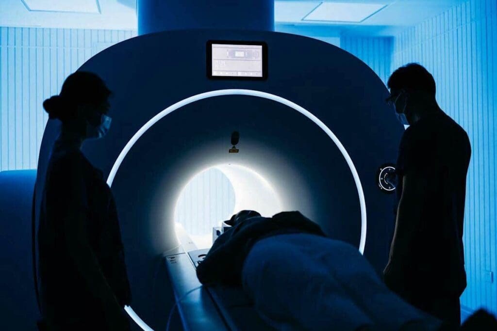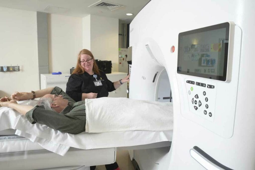
PET/CT scans are essential for assessing how far lung cancer has spread. Sometimes, lymph nodes lit up on PET scan because they are more metabolically active. While this can indicate cancer, it can also occur due to non-cancerous conditions like inflammation or infection.
At Liv Hospital, we use FDG uptake to identify potential cancerous areas. However, other factors can mimic cancer on scans, so careful interpretation is critical. Understanding why lymph nodes light up on a PET scan helps doctors make accurate diagnoses and provide the best treatment plans.
Key Takeaways
- PET/CT scans are vital for assessing lymph node involvement in lung cancer.
- Increased FDG uptake can indicate malignancy, but is not exclusive to cancer.
- Benign conditions can cause false positives on PET scans.
- Careful interpretation of PET scan results is essential for accurate diagnosis.
- Understanding the causes of false positives can improve diagnostic accuracy.
Understanding PET Scans in Lung Cancer Detection

PET scans have changed how we find lung cancer. They show us how tumors work. This helps us plan treatments.
The Science Behind PET Imaging Technology
PET scans use a special tracer to see how the body works. FDG is a glucose molecule with a radioactive atom. It goes to places with lots of glucose, like cancer cells.
The PET scanner picks up the radiation from FDG. It makes pictures of where the body is most active. This helps us find cancer and see how serious it is.
How FDG Uptake Reveals Metabolic Activity
FDG uptake shows how active tumors are. Cancer cells use more glucose than normal cells. This is why PET scans are great for finding and checking lung cancer.
The SUV value shows how active a tumor is. A higher SUV means the tumor is more aggressive.
| Characteristic | Cancerous Tissue | Normal Tissue |
| FDG Uptake | High | Low |
| Metabolic Activity | Increased | Normal |
| SUV Value | High | Low |
A top oncologist says, “PET scans have made finding and checking lung cancer better. They help us make treatment plans that fit each patient.” This shows how important PET scans are in fighting cancer.
“PET scans give us key info on tumor activity. They help us decide on treatments and make patients’ care better.”
— Dr. Jane Smith, Oncologist
Why Lymph Nodes Lit Up on PET Scan: The Metabolic Explanation

Cancer cells use more glucose than normal cells. This is why lymph nodes light up on PET scans. PET scans use this fact to find cancer.
Increased Glucose Metabolism in Cancer Cells
Cancer cells need more glucose to grow fast. This is a key sign of cancer. PET scans use this to spot cancer cells.
The Warburg effect shows how cancer cells use more glucose. Even with oxygen, they prefer glycolysis. This makes them stand out on PET scans.
The Role of FDG as a Radiotracer
FDG is a glucose-like substance used in PET scans. It shows how much glucose cells use. Cancer cells use more FDG, making them easy to spot.
Using FDG has changed how we look at cancer. It lets us see how active lymph nodes and other tissues are without surgery.
Standardized Uptake Value (SUV) Measurement
The SUV measures how much FDG cells take in. It helps tell if a spot is cancerous. Higher SUV values mean cancer is likely.
| SUV Value | Interpretation |
| < 2.5 | Typically benign |
| 2.5 – 4.0 | Suspicious, requires further evaluation |
| > 4.0 | Highly suggestive of malignancy |
Knowing SUV values helps us understand PET scans better. It helps us see how active lymph nodes are. This guides what to do next.
PET/CT Combination: Superior Accuracy in Lung Cancer Staging
Using PET and CT scans together is now the best way to stage lung cancer. This method combines the best of both worlds. It gives a full picture of the disease.
Comparing PET/CT to CT Alone
PET/CT is better than CT alone for checking lymph nodes. CT scans show the body’s structure well. But they can’t always tell if a lymph node is cancerous.
PET/CT, though, shows where cancer is by looking at how active cells are. This helps doctors plan treatments better.
Accuracy Rates in Detecting Nodal Involvement
Research shows PET/CT is up to 90% accurate in finding cancer in lymph nodes. This is because it uses both structure and function to see the disease.
This accuracy helps doctors know how far the cancer has spread. It helps them choose the right treatment for each patient.
Limitations of Current Imaging Technologies
Even with its benefits, PET/CT has its limits. Things like the PET scanner’s quality and how well the patient is prepared can affect results. Also, some conditions can make the scan show false positives.
We know PET/CT is a big step forward in lung cancer staging. But we need to keep improving it to make it even better.
Lung Cancer Tumor Location and Its Impact on PET Scan Results
The spot where a lung cancer tumor grows changes how a PET scan works. This is key to figuring out the cancer’s stage and how it might turn out. We’ll look at how different spots in the lung affect PET scan results and what it means for patients.
Right Upper Lobe Lung Cancer Characteristics
Right upper lobe lung cancer is a common type. Tumors here often show up well on PET scans because they’re very active. They’re also close to big paths for lymph nodes, which can spread cancer. This makes it easier to spot these tumors with PET scans.
Upper Lobe vs. Lower Lobe Presentation
Upper and lower lobe lung cancers are different in how they show up on PET scans and in treatment. Upper lobe tumors, like those in the right upper lobe, are more common. They often spread to lymph nodes, which PET scans can find. Lower lobe tumors are rarer but can be harder to diagnose because of where they are.
Left Upper Lobe Lung Cancer Prognosis
Left upper lobe lung cancer is similar to right upper lobe in some ways, but has its own outlook. The cancer’s location and how it spreads to lymph nodes can affect treatment. Getting the cancer’s stage right with PET scans is key to choosing the best treatment and helping patients.
IDoctors need to understand how lung cancer’s location affects PET scans. By knowing the specific traits of tumors in different spots, doctors can make better choices for diagnosis, staging, and treatment.
Nodal Status Assessment: Critical Factor in Treatment Planning
Getting the nodal status right is key to planning the best treatment for lung cancer patients. Nodal status shows how many lymph nodes are involved. This is a big deal in staging lung cancer and deciding on treatments.
N-Staging System Explained
The N-staging system helps figure out how many lymph nodes are involved in lung cancer. A study on the National Center for Biotechnology Information website says the N-staging system is very important for lung cancer staging. It breaks down lymph node involvement into stages, from N0 (no involvement) to N3 (a lot of involvement).
Experts say, “The N-staging system is key for knowing how to treat lung cancer patients.” It’s important for doctors to understand the N-staging system to plan the best treatment.
How Nodal Involvement Changes Treatment Approaches
Lymph node involvement changes how lung cancer is treated. When lymph nodes are involved, it means the cancer is more advanced. This calls for a stronger treatment plan.
Nodal status assessment helps decide between surgery, chemotherapy, and radiation therapy. For example, patients with a little nodal involvement might get surgery. But those with a lot of involvement might need chemotherapy and radiation instead.
Surgical vs. Non-Surgical Management Based on Nodal Status
Choosing between surgery and other treatments depends on nodal status. Early-stage lung cancer with little nodal involvement might be treated with surgery. But advanced nodal involvement might mean other treatments are better.
Medical studies show, “Nodal status is a big factor in treatment plans, affecting patient outcomes.” Getting nodal status right is essential for the right treatment.
Do Benign Lung Nodules Light Up on PET Scan?
Benign lung nodules can show up on PET scans, making it hard to tell if lung cancer is present. This is because PET scans look for glucose, which can be high in both cancer and benign conditions.
Common Causes of False Positive PET Results
Many benign conditions can cause false positives on PET scans. Inflammatory processes are a big reason, as they increase glucose use. It’s important to think about these when we look at PET scan results.
- Infectious diseases such as tuberculosis or fungal infections
- Granulomatous diseases like sarcoidosis
- Inflammatory conditions, including rheumatoid nodules
These conditions can look like cancer on PET scans. This shows we need to be very careful when we read these results.
Inflammatory Conditions Mimicking Cancer
Inflammatory conditions can really mess with PET scan results. For example, sarcoidosis can cause swollen lymph nodes that look like cancer. Also, rheumatoid nodules in the lung can show up as high on PET scans, making diagnosis harder.
“The presence of inflammatory cells and the resulting increased glucose metabolism can lead to false positive PET scans, stressing the need to match PET findings with clinical history and other imaging.”
— Expert in Nuclear Medicine
Infection vs. Malignancy: Differential Patterns
Telling infection from cancer on PET scans can be tough because they share some signs. But, some clues can help us tell them apart:
| Characteristics | Malignancy | Infection/Inflammation |
| FDG Uptake Pattern | Typically focal and intense | Often diffuse or multifocal |
| Clinical Context | History of cancer or risk factors | Symptoms of infection or inflammation |
| Correlative Imaging | CT features suggestive of a tumor | CT findings consistent with infection/inflammation |
By looking at these clues and using different imaging and clinical info, we can get better at diagnosing lung cancer.
It’s key to understand PET scan results for benign lung nodules for accurate diagnosis and treatment. By knowing about false positives and looking at the whole clinical picture, we can give better care to our patients.
Lung PET Scan: Normal vs. Cancer Findings
Understanding lung PET scans is key. We need to know the difference between normal lung tissue and cancer. PET scans show how active lung tissues are, helping us tell if they are healthy or not.
Characteristic Appearance of Normal Lung Tissue
Healthy lung tissue doesn’t take up much glucose, so it shows low activity on PET scans. But some pparts like the heart and liver, might show more activity. This is because they are more active metabolically.
Typical Patterns in Primary Lung Malignancies
Cancer in the lungs, though, takes up more glucose. This means it shows up more on PET scans. We can spot tumors by their high FDG uptake. The uptake pattern can change based on the tumor’s type, size, and where it is.
Visual and Quantitative Assessment Criteria
We use both visual and quantitative methods to check PET scans. Visually, we look for unusual FDG uptake. Quantitatively, we measure the Standardized Uptake Value (SUV) to see metabolic activity. An SUV above a certain level might suggest cancer, but it depends on the situation and the scanner.
By combining these methods, we can better tell if lung PET scans show cancer or not. This helps us make the right treatment choices.
Lung Masses: Size and Significance in PET Interpretation
When we look at lung cancer through PET scans, the size of lung masses is key. We see how big these masses are and what it means for patient care.
Can a 5 cm Lung Mass Be Benign?
A 5 cm lung mass is big, and size doesn’t always mean it’s cancer. Sometimes, big masses are not cancer but are caused by infections or inflammation. For example, some infections or diseases can make a lung mass look big and show up on PET scans as active, even if it’s not cancer.
Correlation Between Mass Size and Malignancy Risk
Studies show that bigger lung masses are more likely to be cancer. But size isn’t the only thing to look at. We also check how active the mass is on PET scans and what it looks like on CT scans. This helps us guess if it’s cancer or not.
When Large Masses Show Low Metabolic Activity
Big lung masses might not show much activity on PET scans, which can be confusing. This can happen with some types of tumors or if the tumor has died off. We need to look at the whole picture and might use more tests to figure out what’s going on.
In short, while size is important, it’s not the only thing we look at. We use PET scans and other tests together to make sure we’re taking good care of our patients.
Positive PET Scan in Lung Cancer: What Happens Next?
A positive PET scan for lung cancer starts a detailed process to confirm the disease. This can be a tough time for patients. It’s important to move forward with clear steps.
Confirmatory Testing Options
After a positive PET scan, we need to confirm the findings. We use several tests to rule out false positives and find out what’s wrong in the lungs.
- Biopsy: To get tissue samples for examination
- CT-guided needle biopsy: For precise sampling of suspicious areas
- Endobronchial ultrasound (EBUS): To check lymph nodes and other structures
These tests help us confirm the diagnosis and find out the exact type of lung cancer. This is key to planning the best treatment.
Biopsy Techniques for Suspicious Lymph Nodes
When lymph nodes look suspicious, we use different biopsy techniques. The choice depends on where the lymph nodes are and how easy they are to reach.
| Biopsy Technique | Description | Advantages |
| Fine-needle aspiration biopsy | Uses a thin needle to collect cell samples | Minimally invasive, quick recovery |
| Core needle biopsy | Gets a larger tissue sample for detailed analysis | Provides more tissue for histological examination |
| Surgical biopsy | Takes lymph nodes or tissue out surgically | High diagnostic accuracy,allows for staging |
Each biopsy method has its own benefits. The right choice depends on the case and how far the disease might have spread.
Multidisciplinary Tumor Board Approach
After testing and diagnosis, we discuss each case in our tumor board. This team includes experts from oncology, thoracic surgery, radiology, and pathology.
Our tumor board looks at the findings, talks about treatment options, and makes a personalized plan. This team effort makes sure we consider all aspects of the patient’s condition. It helps us find the best treatment for each patient.
We use the latest medical evidence and guidelines to make treatment plans. This way, we can tailor care to meet each patient’s unique needs. It improves their chances of successful treatment and recovery.
Regional Variations: Left Lower Lobe and Right Lower Lobe Lung Cancer Prognosis
The location of a tumor in the lung affects how cancer spreads. This is because of how lymphatic drainage works. The anatomy of the lung and its lymphatic system arises in managing lung cancer.
Anatomical Considerations in Different Lung Regions
The lungs are split into lobes. The right lung has three, and the left has two. The anatomical differences between the lobes affect cancer spread and management.
The left and right lower lobes have unique features. Their lymphatic drainage patterns can change how cancer spreads to lymph nodes.
Lymphatic Drainage Patterns and Metastatic Spread
Lymphatic drainage is key in lung cancer spread. The lymphatic drainage patterns in the lower lobes differ. This affects the cancer’s spread and treatment.
- The right lower lobe drains to the right hilar and subcarinal lymph nodes.
- The left lower lobe drains to the left hilar and subcarinal lymph nodes, with occasional crossover to the right side.
Prognostic Differences Based on Tumor Location
Lung cancer prognosis changes with tumor location. Tumors in the lower lobes have different outcomes than upper lobe tumors. Size, histology, and drainage patterns play a role.
Research shows that left lower lobe lung cancer and right lower lobe lung cancer have unique prognoses. Knowing these differences helps tailor treatments for each patient’s cancer.
Conclusion: Interpreting PET Scan Results in the Context of Lung Cancer
Understanding PET scan results is key to treating lung cancer. We’ve talked about how PET scans work and what they show. This helps doctors make better plans for patients.
When looking at PET scan results, it’s important to know about lung masses and nodules. The PET/CT combo is very accurate for lung cancer staging. Knowing about nodal status helps decide treatment.
By carefully looking at PET scan results, we can help patients more. Our talk shows how important teamwork is in treating lung cancer. It’s about making plans that fit each patient’s needs.
FAQ
What is a PET scan, and how is it used in lung cancer detection?
A PET scan uses a special sugar molecule to find cancer cells. It helps doctors see how far cancer has spread in the lungs.
Do benign lung nodules light up on a PET scan?
Yes, sometimes benign lung nodules can show up on PET scans. This can happen due to inflammation or infections. But not all benign nodules will show increased activity.
How does tumor location affect PET scan results in lung cancer?
The location of a tumor can change how a PET scan looks. Different areas of the lung have different drainage patterns. This can affect the cancer’s spread and treatment options.
Can a 5 cm lung mass be benign?
Yes, a 5 cm lung mass can be benign. The chance of it being cancer depends on its characteristics and the patient’s health history.
What happens after a positive PET scan in lung cancer?
After a positive PET scan, doctors usually do more tests to confirm the diagnosis. They work together to decide the best treatment plan.
How accurate is PET/CT in staging lung cancer?
PET/CT is more accurate than CT alone for staging lung cancer. It’s good at finding cancer in lymph nodes. But it’s not perfect and can have false results.
What is the N-staging system, and how does it impact treatment planning?
The N-staging system looks at how far cancer has spread to lymph nodes. It’s very important for planning treatment. Different treatments are needed based on how much cancer is in the nodes.
How do lymphatic drainage patterns influence prognosis in lung cancer?
Lymphatic drainage patterns affect how cancer spreads. Knowing these patterns helps doctors predict the outcome and plan treatment.
What are the characteristic appearances of normal lung tissue and primary lung malignancies on PET scans?
Normal lung tissue usually doesn’t show up much on PET scans. But cancer cells often do. Doctors use these differences to tell if a lesion is cancerous.
How does the size of a lung mass correlate with malignancy risk?
Larger lung masses are more likely to be cancerous. But size isn’t the only factor. Other characteristics of the mass also play a role.
References
- Tafti, D. (2023). Nuclear Medicine Physics. In StatPearls. National Center for Biotechnology Information. https://www.ncbi.nlm.nih.gov/books/NBK568731/
- Save My Exams. (2024). Nuclear Instability – AQA A Level Physics Revision Notes. https://www.savemyexams.com/a-level/physics/aqa/17/revision-notes/8-nuclear-physics/8-3-nuclear-instability-and-radius/8-3-1-nuclear-instability/


