Last Updated on November 27, 2025 by Bilal Hasdemir

Understanding lymphoma PET scan results is key for both patients and doctors. At Liv Hospital, we use PET scans to find areas in lymph nodes that show high activity. This usually means cancer is present.
PET scans are very important in finding and treating lymphoma. They help us see where cancer might be in the lymph nodes. This information helps us make the right treatment plans for our patients.
Key Takeaways
- PET scans detect metabolic activity in lymph nodes, indicating possible cancer.
- Accurate PET scan results are vital for diagnosing and understanding lymphoma.
- Liv Hospital focuses on patient care, ensuring a complete approach to lymphoma treatment.
- PET scans help guide treatment choices at every step of lymphoma care.
- Grasping PET scan results is essential for managing lymphoma effectively.
The Fundamental Role of PET Scans in Lymphoma Management

PET scans have changed how we manage lymphoma. They help us see if tumor tissue is alive. This is key in diagnosing and treating lymphoma.
PET scans are great at finding cancer cells because they show where cells are active. This helps us tell apart live tumor cells from dead or scar tissue. This is a big challenge in cancer treatment.
How PET Scans Detect Metabolic Activity
PET scans use a special sugar that cancer cells love. This sugar, called Fluorodeoxyglucose (FDG), shows up in cells that are very active. This means we can spot cancer cells easily.
Studies show PET scans are very good at finding lymphoma. They are great for checking if treatment has worked. This helps us decide what to do next.
Why PET Imaging is Essential for Lymphoma
PET imaging is key for lymphoma because it shows things other scans can’t. It tells us if tumors are alive or not. This is vital for planning treatment.
A study on NCBI found that PET scans are better than CT scans. They are more accurate in telling live tumors from dead or scar tissue. This makes PET scans very important in managing lymphoma.
| Imaging Modality | Sensitivity in Detecting Lymphoma | Specificity in Detecting Lymphoma |
| PET Scan | High | High |
| CT Scan | Moderate | Moderate |
The table shows that PET scans are better than CT scans for finding lymphoma. This proves PET scans are the top choice for this task.
What Causes Lymph Nodes to Light Up on PET Scans

Lymph nodes that light up on PET scans show high FDG uptake. This is a sign of possible cancer or other issues. We will look into why this happens and what it means.
The Science Behind FDG Uptake
FDG is a glucose-like substance that cells with high activity take in. Cancer cells, in particular, use more glucose than normal cells. This makes FDG uptake a key marker for cancer.
When lymph nodes light up on PET scans, it means they are active. This activity can be due to cancer.
FDG is injected into the blood, which cells then take up. Cells that are very active, like cancer cells, take up more FDG. The PET scan shows where this uptake is happening, giving a picture of metabolic activity.
Metabolic Activity vs. Anatomical Changes
PET scans show how tissues are working, not just their shape. This is important for finding and tracking lymphoma. It lets doctors see active disease before it shows up in other ways.
Some key points to consider:
- Metabolic Activity: PET scans detect the metabolic activity of cells, which can indicate the presence of cancer.
- Early Detection: Metabolic changes often precede anatomical changes, enabling early detection and intervention.
- Disease Monitoring: PET scans can monitor the response of lymphoma to treatment by assessing changes in metabolic activity.
Understanding why lymph nodes show high FDG uptake is key to reading PET scan results correctly. While cancer is a big worry, other issues like infections or inflammation can also cause this.
Interpreting Lymphoma PET Scan Results: What the Colors Mean
The colors and intensity of a PET scan image tell us a lot about lymphoma. PET scans use a special tracer, like FDG, to show how active the body’s cells are.
Understanding Standardized Uptake Values (SUV)
Standardized Uptake Values (SUV) are key in reading PET scans. SUV compares the tracer uptake in a specific area to the body’s average. This helps us see how active the tissue is.
A higher SUV value means more activity, which might mean cancer. But SUV values can change based on blood sugar, scan timing, and the PET scanner used.
Table: Factors Influencing SUV Values
| Factor | Effect on SUV |
| Blood Glucose Level | High glucose levels can decrease SUV |
| Time Between Injection and Scan | Earlier scans may show lower SUV |
| PET Scanner Used | Different scanners may yield varying SUV values |
The Significance of Brightness Intensity in Diagnosis
The brightness of lymph nodes on a PET scan shows their activity. Bright areas mean high FDG uptake, which might be cancer. The brightness is linked to the SUV value; higher values mean brighter images.
Understanding brightness intensity is complex. It’s about knowing the difference between normal and cancerous activity. Inflammation can also make areas look bright on scans.
We use a mix of clinical info, scan details, and sometimes more tests to understand PET scans. Knowing how to read these scans is key to managing lymphoma well.
PET vs. CT: Superior Detection of Viable Tumor Tissue
PET scans are better than CT scans at finding tumor tissue that is alive. This is key to setting the right treatment plan.
PET scans use a substance called FDG to spot very active areas. This is a sign of cancer. They can find cancer that CT scans might miss.
Distinguishing Active Disease from Scar Tissue
PET scans are better at telling the difference between cancer and scar tissue. After treatment, CT scans might show masses that could be either. PET scans show if the tissue is alive by checking its activity.
- PET scans can spot cancer in lymph nodes that look normal on CT scans.
- They can tell the difference between scar tissue and cancer after treatment.
- PET scans give a clearer picture of how well treatment is working.
When PET Outperforms Traditional Imaging
PET scans are often better than CT scans at finding cancer. This is very important for lymphoma. Knowing exactly where the cancer is helps doctors make better plans for treatment.
“PET/CT has become an essential tool in the management of lymphoma, providing a more accurate view of disease extent and treatment response than CT alone.”
— Expert in Nuclear Medicine
Using PET and CT together (PET/CT) makes diagnosis even better. It combines information about how tissues are working with their structure. This helps doctors pinpoint where the cancer is and understand what the lesions are.
Key benefits of PET scans over CT scans include:
- They find live tumor tissue better.
- They can tell the difference between cancer and scar tissue.
- They give a clearer picture of how well treatment is working.
Do Cancerous Lymph Nodes Always Show Up on PET Scans?
PET scans are very good at finding cancerous lymph nodes. But some things can make them hard to see. Cancerous lymph nodes show up on PET scans if they use more glucose than normal cells.
Detection Rates for Metabolically Active Lymphoma
PET scans are very good at finding active lymphoma. They work best for lymphomas that use a lot of FDG. This makes these cells show up clearly on the scan.
Most cancerous lymph nodes can be seen on PET scans because they use more glucose. But how well they show up can depend on their size and the type of lymphoma.
Understanding Possible False Negatives
Even though PET scans are very good, sometimes they miss cancerous lymph nodes. This can happen if the lymphoma is not active or is in a dormant state. Also, small lymph nodes or those that don’t use much glucose might not be seen.
It’s important for doctors to know about these limitations when looking at lymphoma PET scan results. Missing a cancerous lymph node can lead to wrong treatment plans.
We think it’s key to use PET scans along with other tests and doctors’ opinions. This way, we can make sure we’re giving the right care for each patient.
Differences in PET Scan Protocols: Hodgkin’s vs. Non-Hodgkin’s Lymphoma
PET scan protocols differ between Hodgkin’s and non-Hodgkin’s lymphoma. This is because each disease has its own unique characteristics. PET imaging is used for both, but the way scans are done and results are read can change.
Specific Considerations for Different Lymphoma Types
Hodgkin’s lymphoma grows in a certain order, making PET scans easier to read. Non-Hodgkin’s lymphoma, on the other hand, spreads less predictably. It can go to places outside of the lymph nodes. This means non-Hodgkin’s lymphoma needs a more detailed PET scan.
Key differences in PET scan protocols include:
- Extent of imaging: Non-Hodgkin’s lymphoma may need more imaging because it can spread widely.
- FDG dosage: The amount of FDG used can change based on the lymphoma type and patient health.
- Scan timing: When PET scans are done during treatment can change based on the lymphoma type and treatment plan.
| Lymphoma Type | PET Scan Considerations | Interpretation Focus |
| Hodgkin’s Lymphoma | Ordered progression, often nodal | Nodal involvement, response to treatment |
| Non-Hodgkin’s Lymphoma | Unpredictable spread, extranodal involvement | Extent of disease, extranodal sites, metabolic activity |
Interpretation Variations Between Lymphoma Variants
Understanding the specific lymphoma subtype is key to interpreting PET scans. For example, some non-Hodgkin lymphomas, like follicular lymphoma, take up FDG differently than Hodgkin’s lymphoma.
PET scans have greatly improved how we stage and monitor lymphoma. By customizing PET scan protocols for each lymphoma type, doctors can give better care to patients.
How PET Scans Guide Lymphoma Staging and Treatment Planning
PET scan technology has changed how we stage and plan treatment for lymphoma. These scans are now key in managing lymphoma. They give vital info that helps decide on treatments.
The Impact on Disease Stage Determination
Knowing the stage of lymphoma is key to predicting outcomes and planning treatments. PET scans show where lymphoma is active by highlighting high metabolic areas. This helps doctors see how far the disease has spread.
PET scans have made it easier to accurately stage lymphoma. They spot active lymphoma cells, helping doctors tell if it’s early or advanced. This is important for picking the right treatment.
Tailoring Treatment Strategies Based on PET Results
PET scan results are vital for customizing treatments. They show how active lymphoma cells are, helping doctors choose the best treatment. This tailored approach has led to better results for patients with lymphoma.
PET scans help doctors decide if more treatments like chemo or radiation are needed. They also help track how well treatments are working. This way, doctors can adjust plans as needed, improving care for patients.
Assessing Treatment Response Through Sequential Lymphoma PET Scan Results
Checking how well treatment works is key to fighting lymphoma. We use PET scans to see if the treatment is effective. They help us predict how well a patient will do.
PET scans are vital in lymphoma care. They show how treatment changes metabolic activity. By looking at these changes, doctors can decide if the treatment should stay the same or change.
The Deauville Five-Point Scoring System
The Deauville criteria help us understand PET scan results better. It’s a five-point system that compares FDG uptake in tumors to the liver and mediastinum. This makes assessing treatment response more objective.
The Deauville score ranges from 1 to 5. Here’s what each score means:
- Score 1: No uptake
- Score 2: Uptake ≤ mediastinum
- Score 3: Uptake > mediastinum but ≤ liver
- Score 4: Uptake moderately higher than the liver
- Score 5: Uptake markedly higher than liver and/or new lesions
Predicting Outcomes Based on PET Response
PET scan results, using the Deauville criteria, help predict patient outcomes. A lower Deauville score after treatment means a better prognosis. Research shows patients with scores 1 or 2 have a much higher chance of not having the disease come back.
By using sequential PET scans and the Deauville system, we can make treatment plans that fit each patient. This approach improves care for lymphoma patients.
Non-Malignant Reasons for Lymph Nodes Lighting Up on PET Scans
Lymph nodes lighting up on PET scans don’t always mean cancer. Many non-cancerous conditions can also cause this. It’s important to know these non-cancer reasons for accurate PET scan results.
Inflammatory and Infectious Causes
Inflammatory and infectious diseases can make lymph nodes show up on PET scans. Tuberculosis, sarcoidosis, and other diseases can cause this. It’s because the body’s immune system is reacting.
Infections, like bacterial, viral, or fungal, can also make lymph nodes swell. During a viral infection, for example, lymph nodes might look like cancer on PET scans.
How Specialists Differentiate Benign from Malignant Uptake
Doctors use a detailed method to tell if lymph nodes are active for good or bad reasons. They look at the patient’s history, symptoms, and other tests along with the PET scan.
The pattern of FDG uptake is a big clue. Symmetrical uptake in many lymph nodes is usually not cancer. But, if it’s asymmetrical or focal, it might be cancer.
| Characteristics | Benign Uptake | Malignant Uptake |
| Pattern of Uptake | Symmetrical, diffuse | Asymmetrical, focal |
| Intensity of Uptake | Mild to moderate | Moderate to intense |
| Clinical Context | Presence of infection or inflammation | History of cancer or suspicious symptoms |
By looking at these details, doctors can decide if more tests or treatment are needed.
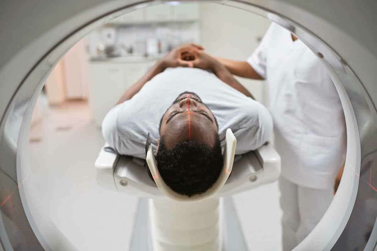
The Patient Experience: What to Expect During Your PET Scan
A PET scan is a key tool for doctors. Knowing what to expect can make it easier. We’ll help you understand the steps and make sure you’re comfortable.
Preparation Guidelines and Restrictions
Getting ready right is key to good results. Patients must follow diet rules and skip some meds before the scan. Here’s what to do:
- Fast for 4-6 hours before, drinking only water.
- Don’t do hard exercise for 24 hours before.
- Tell your doctor about all meds, including diabetes ones.
- Wear comfy, loose clothes and no metal jewelry.
Also, arrive 15 minutes early to fill out forms and get ready.
The Scanning Process and Comfort Considerations
During the scan, you’ll lie on a table in a big machine. The scan is painless and lasts 30-60 minutes.
To keep you comfy:
- We’ll give you a blanket to stay warm.
- If you’re nervous, we might give you a mild sedative.
- Our team will talk to you through an intercom.
After the scan, you can go back to normal unless your doctor says not to. The results will be reviewed and talked about with you later.
| Preparation Step | Description | Importance Level |
| Fasting | 4-6 hours before the scan, consume only water | High |
| Medication Adjustment | Inform your doctor about all medications | High |
| Comfortable Clothing | Wear loose, comfortable clothing without metal | Medium |
Knowing about the PET scan helps make it easier. Our team is here to support you every step of the way.
Advanced PET Technologies Revolutionizing Lymphoma Diagnosis
PET technology is changing how we diagnose and treat lymphoma. These new technologies are making a big difference in how we approach this disease.
PET/CT and PET/MRI Integrated Imaging
PET technology is getting better with the help of CT and MRI. PET/CT combines metabolic info from PET with CT’s detailed images. This gives a full picture of the disease.
PET/MRI adds MRI’s soft tissue details to PET’s metabolic activity. This is great for checking lymphoma in hard-to-reach places.
These new imaging methods help doctors stage lymphoma more accurately. They combine functional and anatomical info. This helps doctors plan better treatments.
Novel Radiotracers Beyond FDG
Even though FDG is common in PET scans, researchers are working on new tracers. These new tracers aim to be more specific and sensitive. They target different parts of tumor biology.
Researchers are exploring many new tracers. Some target how fast cells grow, how much oxygen they use, and more. These could lead to more accurate diagnoses and better care for patients.
Conclusion: Maximizing the Value of PET Scans in Lymphoma Care
PET scans play a key role in managing lymphoma, from finding the disease to checking how well treatments work. By looking at lymphoma PET scan results, doctors can make better choices. They can tailor treatments to fit each patient’s needs.
PET scans give important clues about the disease. They show where the lymphoma is active, helping to tell it apart from other issues like scars or inflammation. To get the most out of PET scans, it’s important to know their strengths and weaknesses. This ensures the results are understood correctly.
As we keep working to improve lymphoma treatment, PET scans will be even more important. Using PET scan results in full care plans can lead to better outcomes. This means a better life for those with lymphoma. Using PET scans well is key to better managing lymphoma.
FAQ
What is a PET scan, and how is it used in lymphoma diagnosis?
A PET scan uses a special sugar molecule to find cancer cells in the body. It helps doctors see active lymph nodes. This is key for diagnosing, staging, and planning treatment for lymphoma.
Why do lymph nodes light up on PET scans?
Lymph nodes light up because they take up more of a radioactive sugar. This can happen for many reasons, like cancer, infection, or inflammation. The scan shows this activity, making the nodes stand out.
How do PET scans differentiate between viable tumor tissue and scar tissue?
PET scans can tell the difference by looking at how active the tissue is. Tumors are usually very active, showing up bright on the scan. Scar tissue is less active, so it’s harder to see.
What is the significance of SUV values in PET scan results?
SUV values show how much sugar a body part takes in. High values mean high activity, which is often cancer. Doctors use this to see how serious the disease is and if treatment is working.
Can PET scans detect all types of lymphoma?
PET scans work well for many lymphomas, but not all. Some types might not show up because they’re not very active. It depends on the type and stage of lymphoma.
How do PET scan protocols differ between Hodgkin’s and non-Hodgkin’s lymphoma?
Protocols for PET scans vary because Hodgkin’s and non-Hodgkin’s lymphomas act differently. Knowing these differences helps doctors make accurate diagnoses and plans for treatment.
What is the Deauville five-point scoring system, and how is it used?
The Deauville system is a way to check how well treatment is working on PET scans. It looks at how much sugar uptake there is in lymph nodes and other areas. This helps doctors see if treatment is effective.
Are there non-malignant reasons for lymph nodes to light up on PET scans?
Yes, sometimes lymph nodes light up for non-cancer reasons like inflammation or infection. Doctors look at the whole picture to tell if it’s cancer or not.
What should I expect during a PET scan?
During a PET scan, you’ll get a small dose of radioactive sugar. Then, you’ll wait and then get scanned. The scan takes about 30-60 minutes. You’ll need to stay very quiet and follow any instructions given.
How are advances in PET technology impacting lymphoma diagnosis and management?
New PET technologies like PET/CT and PET/MRI are making diagnosis and treatment better. They help doctors find and understand lymphoma more clearly. This leads to more effective treatments.
Can PET scans predict outcomes in lymphoma patients?
Yes, PET scans can predict how well a patient will do. They show how the disease is responding to treatment. This helps doctors know if treatment is working and if the disease might come back.
Reference
- Zanoni, L. et al. (2023). PET/CT in non-Hodgkin lymphoma: An update. European Journal of Radiology, 157, 110575. https://www.sciencedirect.com/science/article/abs/pii/S0001299822000964


