Last Updated on November 26, 2025 by Bilal Hasdemir

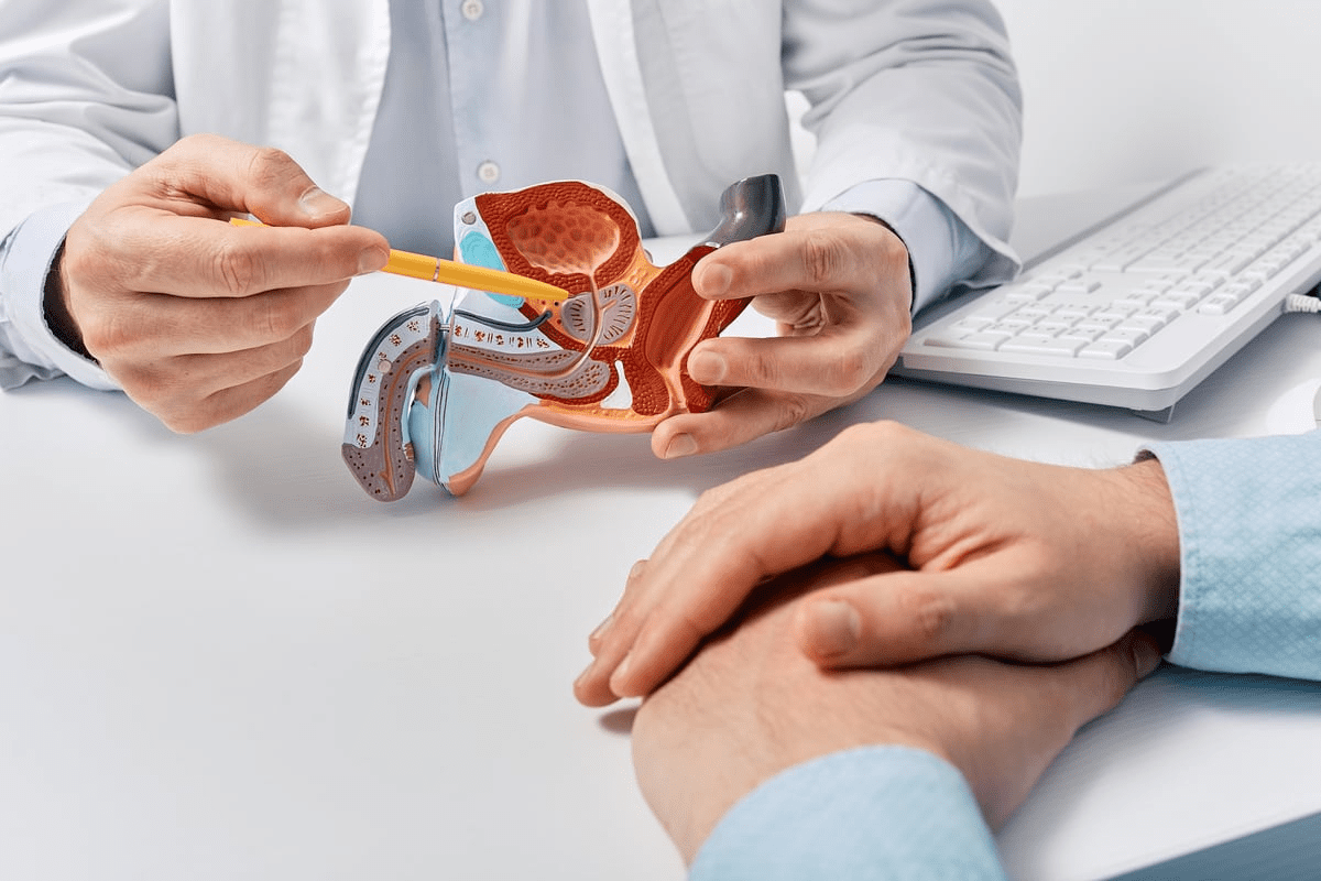
At Liv Hospital, we understand how important medical imaging procedures are in today’s healthcare. They help us find, diagnose, and track diseases. This way, we can give our patients the right treatment quickly and accurately. Learn 10 essential facts about medical imaging procedures. Our guide covers X-rays, MRIs, and what patients need to know.
Thanks to radiology advancements, patient care has gotten much better. We focus on keeping patients safe from radiation and use artificial intelligence. Our radiology team uses the latest technology to ensure top-notch care for everyone.
Key Takeaways
- Medical imaging is vital for disease detection and diagnosis.
- Radiology plays a critical role in modern healthcare.
- Advances in radiation safety have improved patient outcomes.
- Artificial intelligence is increasingly used in radiology.
- Liv Hospital is dedicated to delivering world-class healthcare services.
The Fundamental Role of Medical Imaging in Modern Healthcare

Medical imaging is key in today’s medicine. It changes how we care for patients with better diagnostic tools. This leads to more accurate diagnoses and better treatment plans, improving patient care.
How Diagnostic Imaging Has Revolutionized Medicine
Diagnostic imaging has changed medicine a lot. It helps find diseases early, like cancer, which can save lives. Medical imaging procedures like MRI and CT scans are now essential in healthcare.
The Wide-Ranging Applications Across Medical Specialties
Medical imaging is used in many medical areas. It helps in cardiology, neurology, orthopedics, and oncology. It’s used to diagnose and track many health issues.
| Medical Specialty | Common Imaging Procedures | Benefits |
| Cardiology | Echocardiography, Cardiac MRI | Assesses heart function and structure |
| Oncology | CT scans, PET scans | Detects and monitors tumors |
| Neurology | MRI, Functional MRI | Diagnoses brain and nervous system disorders |
Understanding medical imaging’s role in different areas shows its big impact on healthcare. As radiology facts and tech improve, so will patient care.
Fact 1: The Four Major Types of Medical Imaging Procedures

There are four main types of medical imaging procedures. These help doctors diagnose and monitor many health issues. They have greatly improved how we see inside the body.
X-Ray: The Most Common Imaging Option Worldwide
X-ray technology is used everywhere. It uses X-ray beams to show the body’s inside, mainly bones. X-rays are fast, don’t hurt, and are cheap, making them a top choice for many health checks.
We use X-rays to find broken bones, see if something is stuck inside, and check the lungs. The process is painless and only a little radiation is used.
CT Scans: Advanced X-Ray Technology for Detailed Views
Computed Tomography (CT) scans are a step up from X-rays. They give detailed pictures of the body’s inside. CT scans help find complex issues like internal injuries, cancers, and blood vessel problems.
CT scans mix X-rays and computer tech for detailed images. They’re fast, taking just a few minutes. This helps doctors make better treatment plans.
MRI: Powerful Magnetic Fields for Superior Soft Tissue Imaging
Magnetic Resonance Imaging (MRI) uses strong magnetic fields and radio waves. It’s great for seeing soft tissues like the brain, spine, and joints. MRI helps find issues like torn ligaments, herniated discs, and some cancers.
Doctors often use MRI for soft tissue problems. It’s safe for most people, but not for those with metal implants or pacemakers.
Ultrasound: Non-Radiation Imaging Using Sound Waves
Ultrasound, or sonography, uses sound waves to see inside the body. It’s best for the abdomen, pelvis, and pregnant women. Ultrasound is safe and doesn’t use radiation.
Ultrasound helps find gallstones, check liver health, and watch baby growth during pregnancy. It’s safe for all ages and doesn’t use radiation.
In summary, X-ray, CT scans, MRI, and ultrasound are key in modern medicine. Each has its own strengths and uses. This lets doctors pick the best tool for each health issue.
Fact 2: The Staggering Volume of Radiologic Procedures Performed Annually
Every year, millions of radiologic procedures are done worldwide. This shows how important medical imaging is in healthcare today. As technology gets better and the world’s population grows, more people need these tests.
The number of radiologic procedures keeps going up. This shows how vital they are for taking care of patients. More people are getting these tests because of an older population, more chronic diseases, and better imaging technology.
Global Statistics on Medical Imaging Usage
Across the globe, more medical imaging procedures are happening every year. Recent numbers show a big jump in the use of X-rays, CT scans, MRI scans, and ultrasounds.
Key statistics highlighting the global trend include:
- A big rise in the use of advanced imaging like CT and MRI scans.
- A steady demand for traditional X-ray and ultrasound tests.
- More countries are starting to use medical imaging.
Case Study: England’s 49 Million Imaging Tests in 2024-2025
In England, almost 49 million imaging tests were done from February 2024 to January 2025. This number shows how big a role medical imaging plays in healthcare.
The many imaging tests in England show a bigger trend in healthcare. It’s about needing accurate diagnoses and watching chronic conditions. It also shows how well radiology services are used in the NHS.
The data from England gives us a clear picture of medical imaging’s scale and scope in a big healthcare system. It helps us understand global trends better.
Fact 3: Radiation in Medical Imaging: Safety Considerations and Advancements
Medical imaging is always getting better, but we must always think about safety. It’s key to find the right balance between getting accurate results and keeping patients safe.
Understanding Different Radiation Exposure Levels
Different imaging methods use different amounts of radiation. For example, a chest X-ray uses very little, but a CT scan uses a lot more.
Here’s a look at how much radiation you might get from various tests:
| Imaging Procedure | Effective Dose (mSv) |
| Chest X-ray | 0.1 |
| Mammography | 0.4 |
| CT Abdomen/Pelvis | 10 |
| CT Coronary Angiography | 12 |
This table shows how different tests use different amounts of radiation. It shows why we need to think carefully before doing any test.
Technological Innovations Reducing Radiation Doses
New tech has helped lower radiation doses without losing image quality. Things like new algorithms and systems that adjust doses are being used more.
Key innovations include:
- Low-dose CT protocols
- Advanced image reconstruction techniques
- Automated exposure control systems
These new tools help doctors use less radiation while keeping images clear. For example, low-dose CT scans are now common, cutting down radiation without losing quality.
By using these new technologies, we’re making medical imaging safer and better for everyone.
Fact 4: The Integration of Artificial Intelligence in Radiology
Radiology is on the verge of a big change with artificial intelligence. We’re seeing a new way of looking at medical images and making diagnoses. AI in radiology is more than just making things better; it’s changing the game.
AI-Enhanced Image Interpretation and Diagnosis
Artificial intelligence is being used more to help with image analysis and diagnosis in radiology. AI algorithms can quickly and accurately analyze huge amounts of imaging data. They help radiologists spot things that might be missed by humans.
This tech is great for catching early signs of diseases like cancer. AI can point out important areas in images. This helps radiologists focus on what’s most critical, making diagnoses faster and more accurate.
Predictive Analytics for Early Disease Detection
Predictive analytics is also a big deal in radiology thanks to AI. AI can look at past data and predict disease progression. This helps doctors act early, which is key for managing chronic conditions.
AI also helps with personalized medicine. It lets doctors tailor treatments based on a patient’s unique situation. This makes care more effective for each person.
As AI becomes more common in radiology, we’ll see big improvements in diagnosis and care. The future of radiology is all about AI.
Fact 5: Strict Protocols Governing Medical Imaging and Radiation
Medical imaging and radiation follow strict rules worldwide. These rules help keep patients, staff, and the environment safe from too much radiation.
We use medical imaging to find and treat many health issues. But, some imaging methods like X-rays and CT scans use ionizing radiation. This means we need to be very careful to avoid harm, like injuries or cancer risks.
International Safety Standards and Compliance
Groups like the International Atomic Energy Agency (IAEA) and the World Health Organization (WHO) set global safety rules for medical imaging and radiation. They create guidelines that countries follow to keep everyone safe.
Countries and hospitals must follow these rules. They check things like how much radiation is used, keep equipment in good shape, and train staff well. They also have to report any safety issues or close calls.
Quality Assurance Measures in Imaging Facilities
Imaging centers have strong quality assurance programs. These programs make sure imaging is done right and safely. They include checking equipment, watching patient doses, and making sure images are clear.
Quality assurance also means training staff well. Technologists and doctors get special training to use equipment and read images correctly. Plus, places join quality checks to see how they stack up against the best.
By following strict rules and keeping quality high, we make sure imaging is safe and works well. This keeps patients and staff safe and helps healthcare get better.
Fact 6: Recent Technological Breakthroughs in Imaging Options
Recent years have seen big changes in medical imaging. These changes have made it better and faster. Now, doctors can get more accurate results quicker.
Higher Resolution and Faster Scan Technologies
New imaging tech has focused on better quality and speed. For example, CT scan technology now gives clearer images. This helps doctors make more precise diagnoses.
Also, scans are now quicker. This makes patients more comfortable and reduces errors caused by movement.
A study found scan times have dropped a lot. Some scans now take under a minute. This is great for patients who can’t stay very long or hold their breath.
| Imaging Modality | Typical Scan Time (Pre-Advancements) | Typical Scan Time (Post-Advancements) |
| CT Scan | 5-10 minutes | 1-3 minutes |
| MRI | 15-30 minutes | 5-15 minutes |
| Ultrasound | 15-60 minutes | 10-30 minutes |
Portable and Point-of-Care Imaging Solutions
Another big change is portable imaging. This tech makes it easier to get scans in places without big machines.
Dr. John Smith, a top radiologist, says, “Portable imaging can change healthcare. It brings scans to patients or places where big machines can’t go.”
“The future of medical imaging lies in its ability to be accessible, accurate, and efficient. Portable imaging solutions are a significant step in this direction.”
Portable imaging has many benefits. It makes scans more accessible, reduces the need to move patients, and helps monitor them better. It also means doctors can spot problems sooner.
- Increased accessibility to diagnostic imaging
- Reduced need for patient transport
- Enhanced ability to monitor patients in real-time
- Potential for earlier diagnosis and intervention
As these technologies get better, patient care and accuracy will keep improving. The use of artificial intelligence and machine learning will be key in this progress.
Fact 7: The Patient Experience During Radiology Procedures
Radiology procedures can be scary for patients. But knowing what happens can help ease worries. At our facilities, we aim to make the experience as comfortable and stress-free as we can.
What to Expect During Different Types of Scans
Different scans have their own needs and steps. For example, during an MRI scan, you lie in a big magnetic cylinder for 15 to 90 minutes. This depends on the scan type.
A CT scan is much faster, taking just a few minutes. You lie on a table that slides into a doughnut-shaped machine. The machine rotates around you to get detailed images.
Ultrasound uses a handheld device moved over the area being checked. It needs little prep and no radiation.
Addressing Common Concerns and Misconceptions
Many patients worry about radiation. It’s true that some scans, like X-rays and CT scans, use radiation. But the doses are kept low to keep you safe.
Experts say the benefits of imaging in diagnosing and treating conditions are greater than the risks of radiation. This is important to think about when weighing risks and benefits.
“The key to a successful radiology experience is preparation and understanding of the procedure.”
To get ready for a scan, follow your doctor’s instructions. This might mean fasting, avoiding certain meds, or wearing comfy clothes.
By knowing what to expect and tackling common worries, we can make radiology procedures smoother and less stressful for patients.
Fact 8: The Specialized Training of Radiology Professionals
Radiology needs skilled professionals for accurate diagnoses and treatment plans. With new medical imaging tech, training is more important than ever.
Education and Training for Radiologists and Technologists
Radiologists are doctors who use imaging to diagnose and treat. To become one, they need:
- Science-related undergraduate studies
- Four years of medical school for an MD or DO
- A five-year radiology residency
- Optional fellowship for subspecialization
Radiologic technologists run the imaging machines. Their training includes:
- An associate’s degree or certificate in radiologic technology
- Certification from the American Registry of Radiologic Technologists (ARRT)
- State licensure, needed in many places
The Importance of Collaborative Practice in Radiology
Today, radiology is all about teamwork. Radiologists, technologists, and others work together for better patient care. This teamwork is key for:
- Accurate image interpretation and diagnosis
- Clear communication of findings
- Guiding procedures
This teamwork boosts patient outcomes and helps professionals grow. By working together, they keep up with new imaging tech and techniques.
In summary, the training of radiology professionals is vital in healthcare. Their education and teamwork ensure medical imaging is used well and safely.
Fact 9: Interesting Facts About Radiology’s Historical Development
The history of radiology is a thrilling story. It started with Wilhelm Röntgen’s accidental discovery of X-rays on November 8, 1895. This discovery not only got Röntgen the first Nobel Prize in Physics in 1901. It also set the stage for radiology as we know it today.
From Discovery to Modern Imaging Centers
In the early 20th century, radiology made big strides. The first radiology departments were set up in hospitals. Over the years, radiology has grown a lot, adding new tech like CT scans, MRI, and ultrasound. These tools have changed how doctors diagnose, letting them see inside the body like never before.
Today, imaging centers use the latest tech for quicker, more accurate diagnoses. The use of artificial intelligence (AI) in radiology is making images even clearer and diagnoses more precise.
Radiology Fun Facts That Might Surprise You
Did you know Röntgen’s first X-ray was of his wife’s hand? It showed her bones and wedding ring, showing the power of this new tech. Here are a few more interesting facts about radiology:
- The term “X-ray” came from Röntgen’s confusion about the mysterious rays he found.
- The first radiology departments started in the early 1900s, making radiology a real medical field.
- Radiology has been key in medical progress, from finding injuries to helping with treatments.
These fascinating facts show the rich history and ongoing innovation in radiology. This field keeps changing medicine for the better.
Fact 10: The Future Landscape of Medical Imaging Procedures
The future of medical imaging is about to change a lot. New technologies will make diagnosing diseases better. Radiology is getting ready for big changes.
Emerging Technologies Transforming Diagnostic Capabilities
New technologies are changing medical imaging. Artificial Intelligence (AI) and Machine Learning (ML) are helping doctors read images better. A study found AI can spot problems in images as well as, or even better than, doctors.
“AI is not just a tool, it’s a collaborator in the diagnostic process,” said Dr. Jane Smith, a leading radiologist.
Hybrid imaging technologies are also making a big difference. They mix different imaging types to give doctors more info. For example, PET/CT scans show how organs work and their shape, helping doctors make better diagnoses.
Predictions for the Next Decade of Radiology Advancement
In the next ten years, radiology will keep getting better. It will play a big role in personalized medicine. This means treatments will be made just for each patient, based on their genes and body.
New imaging technology will also make scans faster and images clearer. Doctors will use less radiation too. “The future of radiology is precise, personalized care with the latest tech,” said Dr. John Doe, a top expert.
It’s important to keep up with these new things. They will help make patients’ care better and move radiology forward.
Conclusion: The Enduring Value of Medical Imaging in Healthcare
We’ve looked into the world of medical imaging and its key role in healthcare today. It has changed how we find and treat diseases, helping us catch problems early. Radiology, with tools like X-rays and MRI, gives us deep insights into our bodies.
The future of medical imaging is bright, thanks to new tech and AI. We think more money for medical imaging is key to better patient care and radiology growth. As healthcare changes, medical imaging will lead the way, bringing new ideas and top-notch care.
Medical imaging’s importance is clear, and it greatly affects our lives. Moving forward, we must keep innovating and investing in this area. This ensures medical imaging stays a critical part of healthcare’s future.
FAQ
What are the four major types of medical imaging procedures?
The main types are X-Ray, CT Scans, MRI, and Ultrasound. Each has its own uses and benefits. They help doctors diagnose different health issues.
How has artificial intelligence impacted radiology?
AI has changed radiology a lot. It helps doctors read images faster and more accurately. This leads to better diagnosis and treatment.
What safety measures are in place for medical imaging and radiation?
There are strict safety rules for medical imaging. Facilities follow these to keep patients safe. They make sure images are clear and radiation is low.
What can I expect during a radiology procedure?
The experience varies by scan type. Patients usually need to stay very quiet. Some scans might use special dyes or need special prep. We help you get ready and feel at ease.
What are the benefits of portable and point-of-care imaging solutions?
Portable imaging lets doctors check patients in many places. This makes healthcare better and more accessible.
How has medical imaging transformed the field of medicine?
Medical imaging has changed medicine a lot. It helps doctors find and treat diseases better. Many medical fields use imaging to make decisions and improve care.
What is the role of radiation safety in medical imaging?
Keeping patients safe from too much radiation is key. New tech and strict rules help keep doses low. This makes imaging safe for everyone.
What emerging technologies are transforming radiology?
New tech like AI and portable imaging is changing radiology. It makes diagnosis better and care more effective. These changes will keep improving radiology.
What education and training do radiologists and technologists receive?
Radiologists and technologists get special training. They learn to read images and do procedures. This training helps them work together to care for patients well.
How has the history of radiology contributed to its current state?
Radiology’s history, from Wilhelm Röntgen to today, has shaped it. Knowing this history helps us understand and improve radiology’s future.
Reference
- Lee, J. T., et al. (2018). Photographic Composition Classification and Dominant Geometric Element Detection for Outdoor Scenes.
https://www.sciencedirect.com/science/article/abs/pii/S1047320318301147


