Last Updated on December 1, 2025 by Bilal Hasdemir

Nearly 700,000 brain surgeries are done every year in the United States. Many of these are complex and need a lot of skill and care.
Brain surgery includes operations for tumors, blood clots, and more. These are done by skilled neurosurgeons. They work with anesthesiologists who know a lot about anesthesia and care after surgery.
The challenge in brain surgery is how delicate it is. Neurosurgeons have to carefully work around the brain’s complex parts. This makes it one of the riskiest and most complicated surgeries.
Key Takeaways
- Brain surgery is a complex and delicate procedure.
- It is used to treat various conditions such as tumors and aneurysms.
- Neurosurgeons and anesthesiologists play critical roles in these operations.
- The complexity of brain surgery requires exceptional skill and precision.
- Brain surgery is considered one of the riskiest medical procedures.
The Extraordinary Challenges of Neurosurgery

Working on the brain is very delicate. It needs precision and a deep understanding of how the brain works. Neurosurgery is complex, involving both the surgeon’s skill and knowledge of brain anatomy and function.
The Delicate Nature of Brain Tissue
Brain tissue is very sensitive. Damage during surgery can cause big problems. Neurosurgeons must be extremely precise to avoid harm.
Aspect | Challenge | Requirement |
Brain Tissue | Delicate and sensitive | Precision and care |
Neurological Functions | Critical and complex | Deep understanding and mapping |
Surgical Risk | High risk of complications | Expertise and preparedness |
Navigating Critical Neurological Functions
Handling critical brain functions during surgery is tough. Neurosurgeons must know the brain’s areas well to avoid damage. They use neuroimaging and mapping techniques to find these areas.
These tools help reduce the risk of brain damage. They ensure the best results for patients.
Factors That Make Brain Surgery Exceptionally Difficult
Neurosurgeons face many challenges during brain surgery. They must navigate complex brain structures and keep vital functions intact. The brain’s detailed layout and many important functions make surgery very hard.
Anatomical Complexity and Variations
The human brain varies a lot from person to person, making surgery tricky. Anatomical complexity requires neurosurgeons to know the brain’s structures well. They need to understand how these structures can differ.
Functional Preservation Requirements
Keeping brain functions safe is key during surgery. Surgeons must find and protect areas that control important things like movement and speech. Functional mapping techniques help them do this, reducing the chance of problems after surgery.
Vascular Complications and Bleeding Risks
Bleeding risks are big worries in brain surgery. The brain’s many blood vessels make it hard for surgeons to avoid bleeding. A surgical risk calculator helps them figure out these risks and plan better.
The mix of complex anatomy, the need to keep functions safe, and bleeding risks shows how tough brain surgery is. Knowing these challenges helps improve surgery results.
Skull Base Surgery: The Most Dangerous Brain Surgery

Skull base surgery is known for its complexity and risks. It involves removing tumors or lesions at the skull’s base. This area is filled with important blood vessels and nerves.
Accessing Tumors at the Cranial Base
Surgeons face a tough challenge when they operate on the skull base. They must carefully move through complex structures to reach tumors. They use advanced tools and images to guide them.
Proximity to Critical Neurovascular Structures
The skull base is close to vital blood vessels and nerves. This closeness raises the risk of problems during surgery. Surgeons must be very precise and plan carefully to avoid complications.
Mortality and Morbidity Statistics
Skull base surgery comes with big risks, shown in death and complication rates. The death rate can be between 1% and 5%. More people suffer from complications like infections and nerve damage.
Complication | Rate (%) |
Mortality | 1-5 |
Cerebrospinal Fluid Leakage | 5-10 |
Neurological Deficits | 10-20 |
It’s important for surgeons and patients to know these risks. This knowledge helps in making the right choice about skull base surgery.
Brain Stem Surgery: Operating in the Neural Control Center
The brain stem controls vital functions like breathing and heart rate. It connects the cerebrum to the spinal cord. This makes surgery here very delicate.
Anatomical Challenges of Brain Stem Access
Getting to the brain stem is tough. It’s surrounded by important nerves and blood vessels. Neurosurgeons have to be very careful to avoid damage.
Critical Functions at Risk
The brain stem handles essential functions for life. Damage during surgery can cause serious problems. It’s key to understand its anatomy for surgery success.
Case Studies and Outcomes
Many case studies show the challenges of brain stem surgery. They highlight the need for careful planning and techniques. Thanks to new methods, more people are surviving and recovering better.
In summary, brain stem surgery is very complex. It requires advanced techniques and technology. These advancements are vital for better results.
Arteriovenous Malformation (AVM) Resection
Removing arteriovenous malformations (AVMs) is a tough job. This is because of the complex blood vessel structures involved. AVMs are abnormal blood vessel tangles in the brain. They can cause serious symptoms and even life-threatening problems.
Complex Vascular Tangles
It’s key to understand the complex blood vessel tangles in AVMs for a successful surgery. These malformations have many feeding arteries and veins. This makes surgery planning and execution very complex. Doctors use advanced imaging like angiography to see the AVM’s structure.
Surgical Approaches and Techniques
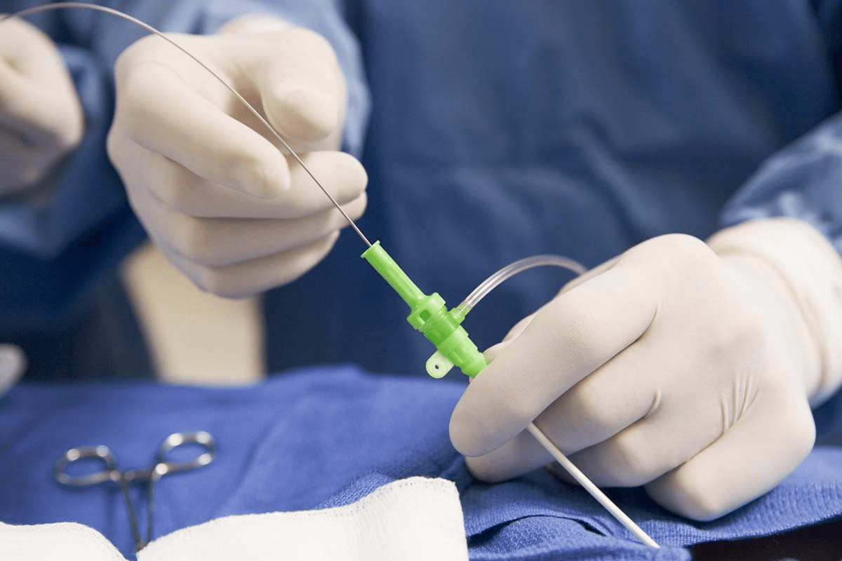
The way to remove AVMs depends on their size, location, and depth. Neurosurgeons use microsurgery. They use high-powered microscopes and special tools to remove the AVM carefully. Sometimes, they do embolization before surgery to lower bleeding risks.
Intraoperative Hemorrhage Management
Controlling bleeding during surgery is very important. Surgeons must act fast to stop bleeding and protect the brain. They use special agents and techniques. They also keep a close eye on the patient’s health and brain function during surgery.
Surgical Technique | Description | Advantages |
Microsurgery | Use of high-powered microscopes and specialized instruments | Precise dissection and removal of AVM |
Preoperative Embolization | Blocking blood flow to the AVM before surgery | Reduced risk of bleeding during surgery |
Intraoperative Monitoring | Continuous monitoring of vital signs and neurological status | Quick response to complications |
Awake Craniotomy: Operating on a Conscious Brain
Neurosurgeons face a big challenge when they operate on a patient who is awake. They must make sure the surgery is a success and the patient feels comfortable. This complex procedure lets the patient give feedback in real-time, which is key to keeping brain functions intact.
The Procedure Explained
The awake craniotomy starts with local anesthesia to numb the area. Then, the neurosurgeon makes an incision and removes part of the skull to reach the brain. The patient is kept awake during some parts to check brain function through tests.
Challenges for Surgeons
Surgeons have to manage the patient’s pain and anxiety. They also need to control the instruments precisely and understand the patient’s feedback. This requires a lot of skill and teamwork from the surgical team.
Patient Experience and Psychological Aspects
The experience for the patient can be very intense, with feelings of anxiety and vulnerability. It’s important to manage pain well and communicate clearly. The psychological impact of being awake during surgery is significant, and the team must be ready to support the patient.
Aspect | Description | Importance |
Real-time Feedback | Patient’s ability to respond during surgery | High |
Pain Management | Effective use of anesthesia and pain relief | High |
Patient Comfort | Ensuring the patient remains comfortable during the procedure | High |
Deep Brain Stimulation: Precision in Neural Pathways
Deep brain stimulation is a cutting-edge neurosurgical method. It involves placing electrodes in specific brain areas to treat diseases like Parkinson’s. This method needs great accuracy to hit the right neural paths.
Targeting Microscopic Structures
Success in deep brain stimulation depends on hitting tiny brain spots. Advanced scans like MRI and CT help find these spots. Doctors need to know the brain’s layout and the neural paths linked to the patient’s symptoms. Microelectrode recording during surgery adds to the precision by checking neural activity in real-time.
- Advanced imaging techniques for target identification
- Microelectrode recording for real-time neural activity feedback
- Detailed knowledge of brain anatomy and neural pathways
Technical Challenges of Electrode Placement
Placing electrodes in deep brain stimulation is tricky. The electrodes need to be placed with millimeter accuracy for the best results and to avoid harm. The process uses advanced equipment and live imaging to guide the electrodes. Also, the patient’s input during the surgery is key to perfecting the placement.
- Sophisticated stereotactic equipment for precise electrode guidance
- Real-time imaging to verify electrode placement
- Patient feedback for adjusting electrode positioning
In summary, deep brain stimulation is a complex surgery that needs precise targeting of neural paths. The challenges in placing electrodes are big, but new imaging and equipment have made it better for patients with neurological issues.
Intraventricular and Deep-Seated Tumor Removal
Surgery for tumors in the brain’s ventricles or deep areas is very tricky. These tumors are hard to get to because of their location.
Navigating the Ventricular System
The brain’s ventricles are filled with fluid and are complex. Intraventricular tumor removal needs surgeons to carefully move through this system. They must plan well and be precise to not harm other brain parts.
- Knowing the ventricular system’s anatomy is key.
- Advanced imaging helps plan the surgery.
- Microsurgery is used to protect surrounding tissue.
Challenges of Accessing Central Brain Regions
Deep-seated tumors are hard to reach without harming the brain. The main challenge is to get to these tumors without losing brain function. Surgeons use new methods and tools to balance this.
- They try not to damage important brain parts.
- They manage risks like bleeding or infection.
- They aim to remove the tumor completely to stop it from coming back.
By facing these challenges and using advanced surgery, doctors can help patients with these tumors more.
Cerebrovascular Bypass Procedures
Neurosurgeons use cerebrovascular bypass procedures to fix blood flow to the brain. They do this by going around blocked or damaged vessels. These surgeries need a deep knowledge of brain blood vessel anatomy and fine microsurgical skills.
These procedures make a detour around a blocked or damaged brain blood vessel. This is done through microsurgical vessel anastomosis. The surgeon connects a donor vessel to the affected area, making a new path for blood flow.
Microsurgical Vessel Anastomosis
Microsurgical vessel anastomosis is key in these procedures. It needs technical precision and a steady hand. The vessels involved are very small, about 1 mm in diameter. The success of the bypass relies on connecting these tiny vessels correctly.
The process includes several important steps:
- Identifying suitable donor and recipient vessels
- Preparing the vessels for anastomosis
- Performing the anastomosis under microscopic visualization
- Verifying the patency of the bypass
Technical Precision Requirements
The technical skill needed for cerebrovascular bypass procedures is very high. Surgeons must have advanced microsurgical abilities. They need to handle delicate tissues and understand complex brain blood vessel anatomy.
Technical Skill | Description | Importance Level |
Microsurgical Techniques | Ability to perform surgery under microscopic visualization | High |
Vascular Anatomy Knowledge | Understanding of cerebral vascular structure | High |
Tissue Handling | Delicate handling of brain and vascular tissues | High |
In conclusion, cerebrovascular bypass procedures are complex neurosurgical operations. They require a high level of technical precision and skill. The success of these procedures depends on the surgeon’s ability to accurately perform microsurgical vessel anastomosis and navigate the brain’s complex vascular anatomy.
Pediatric Brain Surgery: Unique Challenges
Pediatric brain surgery is very challenging because of a child’s developing brain. Neurosurgeons need to understand how the brain grows and changes. This knowledge is key to success in these surgeries.
Developing Brain Considerations
The young brain is more fragile and heals differently than an adult’s. Surgeons must think about these differences when they plan and do surgeries. The brain’s plasticity in kids can help them recover but also risks long-term problems if not handled right.
Congenital Malformation Correction
Fixing birth defects in the brain is a big part of pediatric surgery. These defects can be structural or vascular. Surgeons need to know exactly how these defects affect a child’s brain growth.
Long-Term Developmental Impacts
The effects of brain surgery on a child’s future must be thought about carefully. Things like thinking skills, movement, and emotional health are important. A good follow-up plan is key to help a child after surgery.
Consideration | Importance in Pediatric Brain Surgery |
Developing Brain | High |
Congenital Malformations | High |
Long-Term Follow-Up | Critical |
Epilepsy Surgery: Mapping and Removing Seizure Foci
Epilepsy surgery is a detailed process. It aims to map and remove the parts of the brain causing seizures. This surgery is for those whose seizures don’t respond to medication, affecting their life quality.
Functional mapping is a key challenge in this surgery. It’s about finding the brain areas that control important functions like speech and movement. This is to prevent damage during the surgery.
Functional Mapping Challenges
Functional mapping uses advanced techniques like EEG, MEG, and fMRI. These tools help doctors locate seizure zones and their relation to important brain areas.
- EEG records brain electrical activity to spot seizure patterns.
- MEG measures magnetic fields from brain activity, creating detailed maps.
- fMRI shows blood flow changes to map brain functions.
Balancing Seizure Control and Function Preservation
It’s important to remove enough brain tissue to control seizures but not harm important functions. Advanced imaging and mapping help surgeons plan with precision.
The surgery’s goal is to reduce or stop seizures and improve life quality. With careful planning and execution, neurosurgeons can greatly improve outcomes for those with drug-resistant epilepsy.
Surgical Outcomes and Recovery Considerations
Complex brain surgeries need careful thought about outcomes and recovery. These surgeries are complex, so it’s important to know the risks. They can affect a patient’s quality of life.
Mortality Rates Across Complex Procedures
Mortality rates for complex brain surgeries vary a lot. This depends on the surgery and the patient’s health. Knowing these risks helps patients make informed choices.
Quality of Life After High-Risk Surgery
The quality of life after high-risk brain surgery can change a lot. The surgery type, patient’s age, and health matter a lot. Some patients see big improvements, while others face challenges.
Rehabilitation Requirements
Rehabilitation is key for patients after complex brain surgery. The needs for rehab can differ a lot. Some might need a lot of physical, occupational, and speech therapy. A good rehab plan is vital for the best recovery.
Important things to think about for recovery include:
- Understanding the risks of the surgery
- The possible effects on quality of life
- The need for a detailed rehab plan
Training Requirements for Complex Brain Surgery
To master complex brain surgery, neurosurgeons must commit to a rigorous training program. This program covers both technical skills and psychological preparation. It’s essential for gaining the expertise needed for safe and effective surgeries.
Educational Pathway for Neurosurgeons
The path to becoming a neurosurgeon is long and challenging. It starts with a solid foundation in medical school. Then, there are years of residency training in neurosurgery.
During this time, future neurosurgeons get hands-on experience. They learn various surgical techniques and patient care.
Developing Technical Expertise
Building technical skills for complex brain surgery takes time. Neurosurgeons start with simpler surgeries and move to more complex ones. They practice and improve their skills through many surgeries.
They also keep up with the latest techniques and technologies through ongoing education.
Psychological Demands on Surgeons
Surgeons face big psychological challenges in complex brain surgery. They must handle high stress and make important decisions quickly. They also need to stay focused and calm during long surgeries.
Developing mental strength for these tasks comes from experience and training.
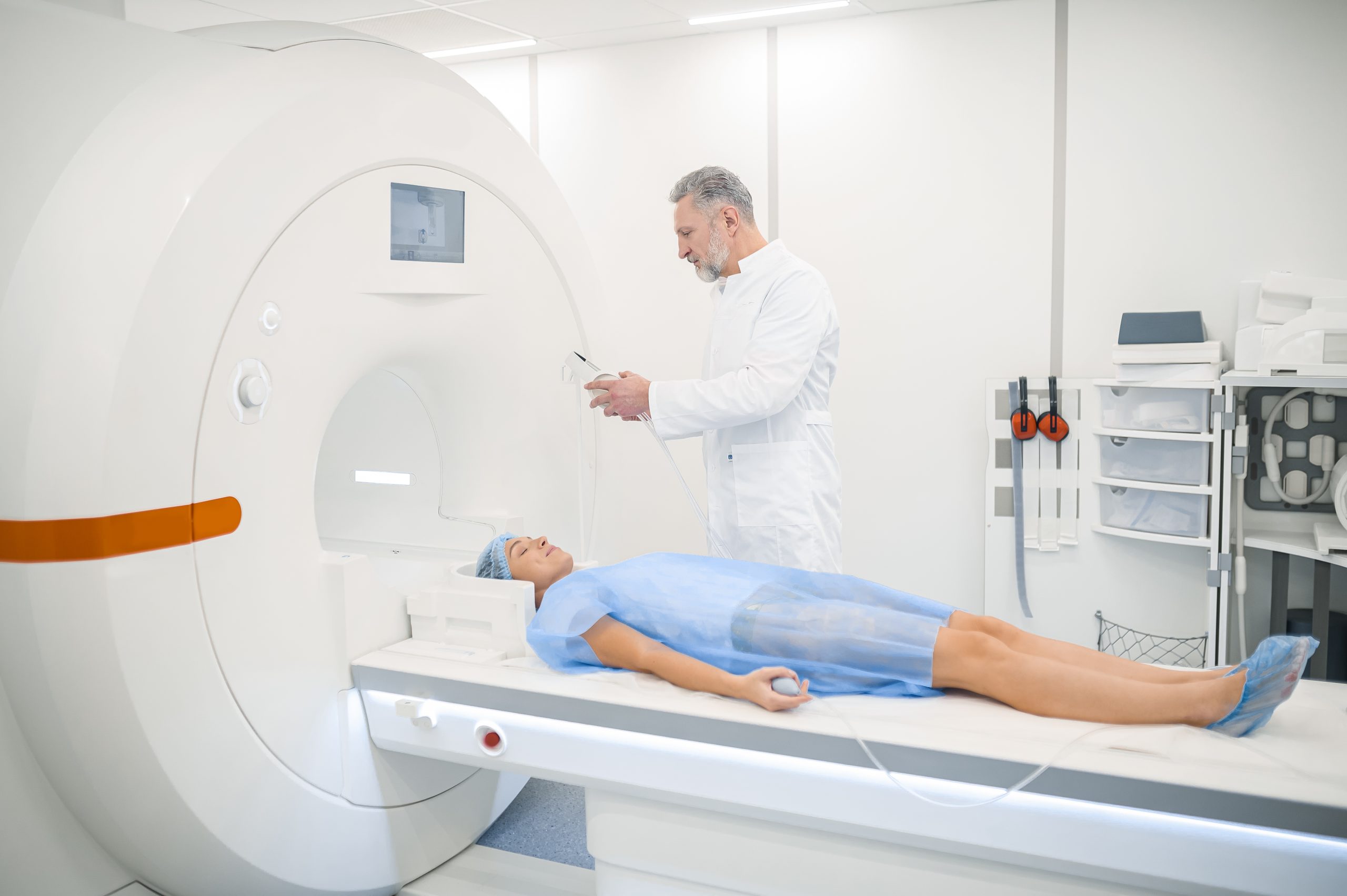
Technological Advancements in Complex Neurosurgery
Technological innovations are changing complex neurosurgery for the better. They make procedures more precise and successful. These new technologies help surgeons do detailed work with better results.
Intraoperative Imaging and Navigation
Intraoperative imaging and navigation are key in neurosurgery today. They give surgeons real-time feedback. This lets them work on complex areas with more accuracy.
Intraoperative MRI and CT scans are examples of these advancements. They give detailed views during surgery.
Robotic Assistance and Automation
Robotic systems are becoming more common in neurosurgery. They help surgeons do more precise work and reduce shaking. Robotic-assisted surgery has shown to improve results and shorten recovery times.
Augmented Reality Applications
Augmented reality (AR) is becoming a valuable tool in neurosurgery. AR gives surgeons real-time data and 3D views. This helps them understand complex anatomy better.
This technology could make planning and doing surgeries better.
Technological Advancement | Description | Benefits |
Intraoperative Imaging | Real-time imaging during surgery | Enhanced precision, better outcomes |
Robotic Assistance | Robotics aiding in surgical procedures | Reduced tremors, precise dissections |
Augmented Reality | 3D visualization and real-time data | Improved planning, execution |
Conclusion: The Future of Complex Brain Surgery
The future of complex brain surgery looks bright, thanks to new tech and better understanding of the brain. Patients will see better results as we explore new ways to operate. This is a big step forward.
New tools like intraoperative imaging and robotic help are changing how we do surgery. These tools make neurosurgeons more precise and accurate. They are key to the future of brain surgery.
As we keep researching and innovating, patient care will get even better. This is because we’re always finding new ways to help those with brain problems. Our goal is to give patients the best care possible.
The future of brain surgery is full of hope. We’re on the verge of big discoveries that will help many people. These advancements will make a real difference in people’s lives all over the world.
FAQ
What is the hardest brain surgery to do?
The hardest brain surgeries are those in complex and delicate brain areas. This includes brain stem surgery and skull base surgery. They are risky because they control critical functions.
What makes brain surgery so challenging?
Brain surgery is tough because brain tissue is very delicate. Surgeons must navigate through important functions and complex anatomy. There’s also a risk of bleeding and vascular complications.
What is skull base surgery?
Skull base surgery is a challenging procedure. It accesses tumors or lesions at the skull’s base. This area is rich in critical structures, making it risky.
Why is brain stem surgery considered high-risk?
Brain stem surgery is risky because it affects vital functions like breathing and heart rate. Any surgery here can have serious consequences.
What is an AVM resection?
An AVM resection is a surgery to remove a complex brain blood vessel tangle. It’s challenging because of the risk of bleeding during the operation.
What is an awake craniotomy?
An awake craniotomy is a surgery where the patient stays awake. This lets the surgeon get feedback and protect brain functions.
What is deep brain stimulation?
Deep brain stimulation targets tiny brain structures with electrodes. It’s used to treat neurological conditions and requires precision.
How long does it take to schedule a surgery?
Scheduling surgery time varies. It depends on the surgery type, the surgeon’s schedule, and the facility’s availability.
What are the risks of plastic surgery?
Plastic surgery risks include infection, scarring, and reactions to anesthesia. These risks vary by procedure.
What is the most complicated surgery?
The most complicated surgeries are those in delicate areas, like brain surgery. They require a lot of skill and precision.
What is the riskiest surgery?
The riskiest surgeries are those in critical areas, like the brain stem. Procedures like AVM resection are also very risky.
How do neurosurgeons develop their technical expertise?
Neurosurgeons gain expertise through a long education. This includes medical school, residency, and specialized training.
What technological advancements are being used in neurosurgery?
Neurosurgery uses new tech like intraoperative imaging and navigation. Robotic assistance and augmented reality also help improve precision and visualization.
References
Li Y., Suki D., Hess K., Sawaya R. “The influence of maximum safe resection of glioblastoma on survival in 1229 patients: Can we do better than gross-total resection?”https://pubmed.ncbi.nlm.nih.gov/26495941/

