Last Updated on December 1, 2025 by Bilal Hasdemir

Discover how MRI brain tumor detection works and its role in confirming diagnosis.
Did you know millions of people worldwide get diagnosed with brain tumors every year? Finding out if you have a brain tumor quickly and accurately is key. The MRI scan is a top tool for this.
We’ll look at how brain tumor MRI scans are essential for finding tumors. They give doctors clear pictures of tumors’ size, location, and more. This info is vital for choosing the right treatment.
Key Takeaways
- MRI scans are highly effective for diagnosing brain tumors.
- The accuracy of brain tumor diagnosis MRI helps in planning treatment.
- MRI brain tumor accuracy is key for knowing the tumor’s details.
- Understanding MRI’s role in diagnosing brain tumors helps patients make better choices.
- New MRI tech keeps getting better at finding tumors.
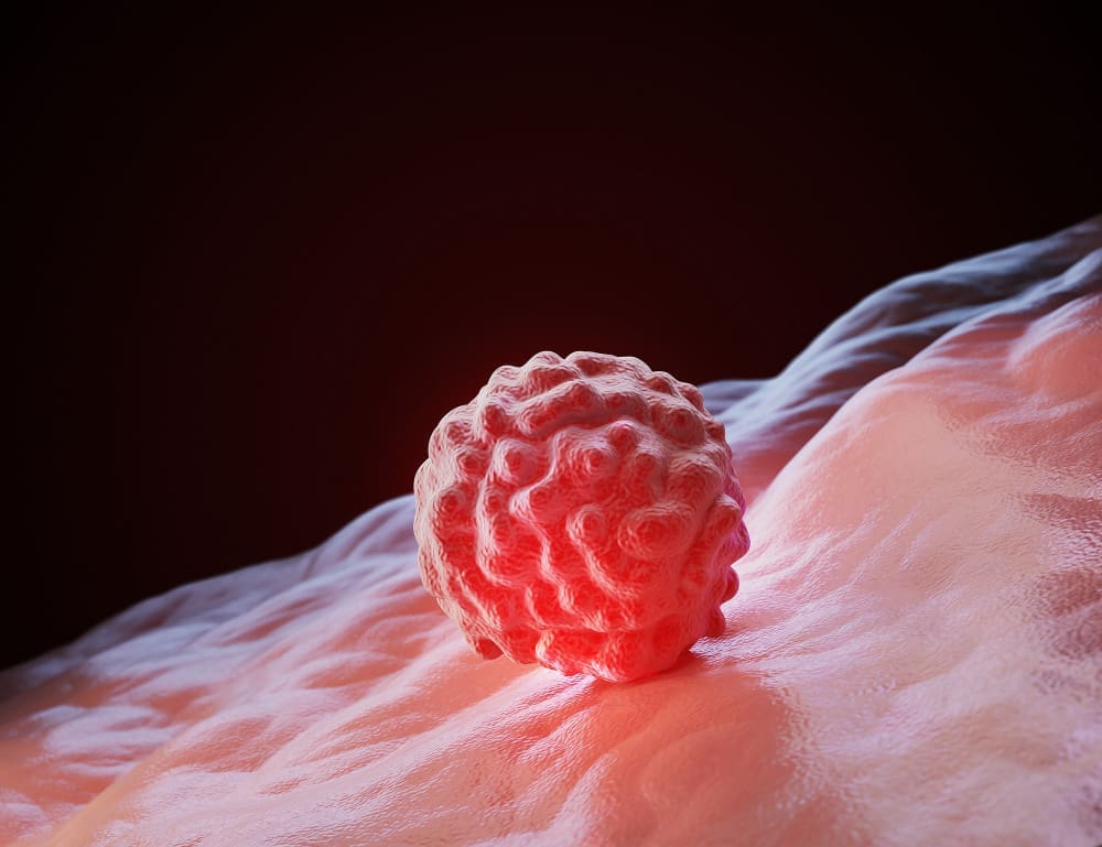
Understanding Brain Tumors and Their Detection
It’s key to know about brain tumors to find them early and right. There are two main types: primary and metastatic. Primary tumors start in the brain. Metastatic tumors come from other parts of the body.
Types of Brain Tumors
Brain tumors are sorted by their cell origin and how fast they grow. The main types are:
- Gliomas: These start in the brain’s glial cells, which support neurons.
- Meningiomas: These grow in the meninges, the brain and spinal cord’s covering layers.
- Pituitary Tumors: These happen in the pituitary gland, affecting hormone making.
- Medulloblastomas: These are very aggressive tumors mainly found in kids.
Each tumor type needs a special way to find and treat it.
Common Symptoms That Warrant Imaging

Symptoms of brain tumors vary based on their location, size, and type. Common signs that might need an MRI include:
- Severe Headaches: Often worse in the morning and with nausea or vomiting.
- Seizures: New seizures can mean a brain tumor.
- Cognitive Changes: Trouble with memory, focus, or other thinking skills.
- Motor Symptoms: Weakness, numbness, or paralysis in body parts.
When these symptoms show up, MRI is often used to find and diagnose brain tumors.
MRI is vital for finding and understanding brain tumors. It gives detailed images for treatment planning.
How MRI Technology Works for Brain Imaging

MRI technology uses magnetic resonance to create detailed brain images. It helps doctors diagnose many neurological conditions. MRI technology is key in medical diagnostics because it shows soft tissue without using harmful radiation.
The Science Behind Magnetic Resonance Imaging
MRI works by using nuclear magnetic resonance. When a patient is in the MRI machine, a strong magnetic field aligns hydrogen nuclei in the body. Then, radio waves disturb this alignment, creating signals for detailed images.
This process is complex. It uses advanced technology to make high-quality images. These images can show small changes in brain tissue.
Advantages of MRI for Brain Tissue Visualization
MRI’s main advantage is its ability to show brain tissue clearly. This is very important for diagnosing brain tumors. It helps doctors see the tumor’s size, location, and how it might affect nearby areas.
MRI’s sensitivity to soft tissue variations makes it a valuable tool. It offers insights that other imaging methods can’t. MRI is also safe because it doesn’t use harmful radiation.
It’s also non-invasive, which is good for patients. MRI can show how blood flows and tissues are perfused. This is important for understanding brain tumor activity.
MRI Brain Tumor Detection: Capabilities and Limitations
MRI is great at finding brain tumors, but it has its limits. It’s a key tool in neuro-oncology, showing detailed images of the brain and its problems.
What MRI Can Reveal About Brain Tumors
MRI gives a lot of info about brain tumors. It shows where they are, how big they are, and what they’re like. Advanced MRI can also look at how tumors work and their blood supply, helping doctors plan treatment.
MRI can tell if a tumor is harmless or cancerous by looking at its edges and how it affects the brain. This info is vital for doctors to decide the best treatment.
Size Limitations and Detection Thresholds
Even though MRI is very good, it can’t find all tumors, mainly the tiny ones. How well MRI can spot a tumor depends on the scanner’s quality and where the tumor is.
| Tumor Size | Detection Rate | Comments |
| >1 cm | High | Most tumors larger than 1 cm are detectable with MRI. |
| 0.5-1 cm | Moderate | Tumors in this range can be detected, but it may require advanced MRI techniques. |
| Low | Very small tumors may not be detectable with standard MRI protocols. |
It’s important for doctors and radiologists to know these limits. This knowledge helps them decide if more tests or closer checks are needed.
Types of MRI Scans Used for Brain Tumor Diagnosis
Diagnosing brain tumors needs different MRI scans. Each scan gives unique insights into the tumor. The choice depends on the tumor type, location, and the patient’s health.
Standard MRI vs. Functional MRI
Standard MRI shows the brain’s structure. It helps doctors spot abnormalities. Functional MRI (fMRI) looks at brain activity by tracking blood flow changes.
fMRI is key for planning surgery. It shows how tumors affect brain function. This info is vital for treatment planning.
Contrast-Enhanced MRI Techniques
Contrast-enhanced MRI uses a contrast agent to highlight brain areas. It’s great for spotting different tumors and their boundaries.
The contrast agent shows up in areas with blood-brain barrier issues. This makes tumors stand out on MRI images. It boosts diagnostic accuracy and helps plan treatment.
Advanced MRI Protocols for Tumor Characterization
Advanced MRI includes diffusion-weighted imaging (DWI) and perfusion-weighted imaging (PWI). DWI looks at tumor cell density. PWI checks the tumor’s blood supply.
| Advanced MRI Protocol | Information Provided | Clinical Utility |
| Diffusion-Weighted Imaging (DWI) | Cellular density of the tumor | Helps in assessing tumor aggressiveness |
| Perfusion-Weighted Imaging (PWI) | Tumor’s blood supply | Aids in evaluating tumor grade and planning treatment |
| MR Spectroscopy (MRS) | Metabolic information about the tumor | Assists in distinguishing between tumor types and grades |
Advanced MRI protocols, along with standard and functional MRI, give a full picture of brain tumors. This detailed approach is key for accurate diagnosis and effective treatment.
MRI vs. CT Scans for Brain Tumor Detection
Both MRI and CT scans are key tools for finding brain tumors. They work well in different situations.
Comparative Strengths and Weaknesses
MRI is great at showing soft tissues, which helps spot small or tricky-to-find tumors. CT scans, on the other hand, are fast and easy to get. They’re often used when quick results are needed.
MRI Strengths:
- Excellent soft tissue contrast
- No radiation exposure
- Ability to provide detailed images of brain structures
CT Scan Strengths:
- Quick and widely available
- Less sensitive to patient motion
- Good for detecting calcifications and bone abnormalities
When CT Might Be Preferred Over MRI
Even though MRI is usually the top pick for brain tumor checks, there are times when CT scans are better. For example, in emergency trauma cases or when fast bleeding detection is needed, CT scans are the go-to. They’re fast and good at spotting bleeding.
| Diagnostic Feature | MRI | CT Scan |
| Soft Tissue Contrast | Excellent | Good |
| Radiation Exposure | No | Yes |
| Speed of Procedure | Generally longer | Quick |
| Sensitivity to Calcifications | Limited | High |
In summary, picking between MRI and CT scans for brain tumor detection depends on the situation. It also depends on the patient’s health and what the doctors need to plan treatment.
Accuracy of MRI in Brain Tumor Diagnosis
Understanding how well MRI works in finding brain tumors is key. MRI is a top tool for spotting and studying brain tumors. It does this thanks to its clear images.
MRI is great at showing soft tissues, which helps find tumors. How well MRI works depends on the MRI type and the doctor’s skill.
Statistical Success Rates in Tumor Detection
Research shows MRI is very good at finding brain tumors. The success rate of MRI scans changes based on the tumor type and where it is. For example, a study showed MRI found brain tumors 95% of the time and correctly identified them 90% of the time.
| Tumor Type | MRI Sensitivity | MRI Specificity |
| Glioma | 92% | 88% |
| Meningioma | 95% | 92% |
| Metastatic Tumors | 90% | 85% |
The table shows MRI’s success in finding different brain tumors. It shows MRI is a reliable tool for doctors.
Factors Affecting Diagnostic Accuracy
Many things can change how well MRI works for finding brain tumors. The MRI machine’s quality, the use of contrast agents, and the doctor’s experience matter. Also, how the patient moves can affect the image quality.
We need to think about these factors when looking at MRI results. This helps make sure patients get the right treatment. Knowing MRI’s strengths and weaknesses helps doctors make better choices for their patients.
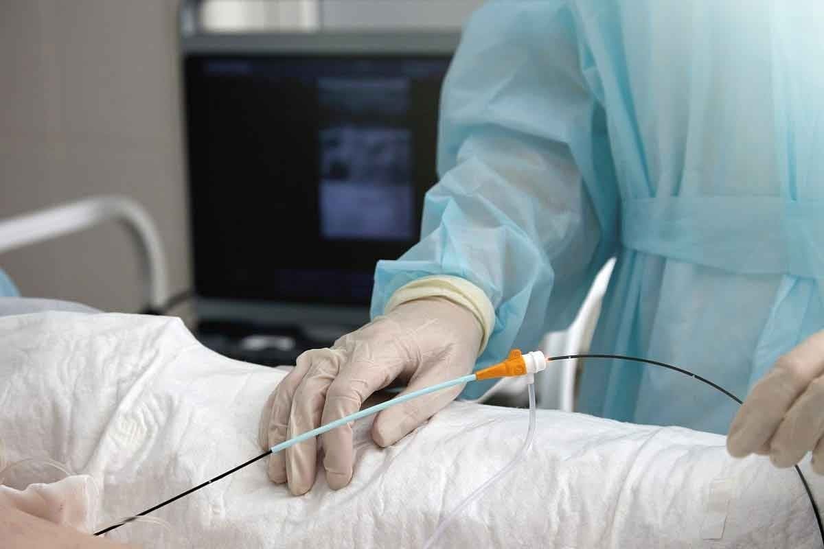
The MRI Brain Tumor Screening Process
Getting ready for an MRI to find brain tumors has several steps. We aim to make this process easy for you. We know tests can make you nervous, so we’re here to help.
Patient Preparation for Brain MRI
Before your MRI, you’ll get specific instructions. You’ll need to:
- Take off any metal items, like jewelry and glasses.
- Tell your doctor about any metal implants or pacemakers.
- Arrive early to fill out paperwork and wear a comfortable gown.
Telling your doctor about any fear of small spaces is also important. This helps us make sure you’re comfortable during the test.
What to Expect During the Procedure
During the MRI, you’ll lie on a table that slides into the machine. Our MRI technologist will help you get into the right spot. They’ll also give you a way to talk to them during the scan.
The MRI machine makes loud noises. We’ll give you earplugs or headphones to block out the sound. It’s important to stay very quiet during the scan to get clear pictures.
| Procedure Step | Description | Duration |
| Preparation | Getting into position and receiving instructions | 15 minutes |
| Scanning | The actual MRI scanning process | 15-30 minutes |
| Total Time | Including preparation and scanning | 30-45 minutes |
Duration and Comfort Considerations
The whole MRI process usually takes 30 to 45 minutes. We want to make it as comfortable as possible. If you’re scared of small spaces, we might offer an open MRI or sedation to help you relax.
Our team is committed to making your MRI as safe and precise as possible. If you have any worries or questions, please ask us.
Interpreting MRI Brain Tumor Results
Reading MRI results for brain tumors is a detailed job for skilled radiologists. They carefully look at the MRI images for any signs of tumors. This helps them understand what the images show.
How Radiologists Analyze Brain MRI Images
Radiologists follow a specific method to study brain MRI images. They check the size, shape, and where the abnormalities are. They also look at how these appear on different MRI sequences. This helps them figure out if the abnormalities are likely tumors.
They examine several things, including:
- Size and Location: Larger tumors or those in key areas are more concerning.
- Signal Intensity: Tumors can look different on MRI images based on their tissue.
- Contrast Enhancement: Contrast agents can make tumors more visible.
- Margins and Edema: The edges of a tumor and swelling around it give clues.
Common Findings That Indicate Tumors
Some patterns and features on MRI images suggest brain tumors. These include:
- Mass Effect: Tumors can push or distort normal brain structures.
- Heterogeneous Enhancement: Tumors may show different enhancement patterns after contrast.
- Necrosis: Necrotic areas in a tumor can indicate aggressive growth.
By studying these signs, radiologists can give important information. This helps in diagnosing and managing brain tumors.
We know MRI results can be worrying. Our team of experts is here to provide accurate and caring help. We make sure patients get the support they need during the diagnostic process.
MRI for Pediatric Brain Tumor Detection
Detecting brain tumors in children needs precise imaging. MRI is key in this process. It’s vital to consider the unique challenges and needs of children when diagnosing brain tumors.
Special Considerations for Children
Children’s brains are developing, and their size is smaller. This makes imaging harder. MRI is great because it gives clear images without harmful radiation. This makes it safer for kids.
Key considerations for pediatric MRI include:
- Minimizing radiation exposure
- Ensuring the child’s comfort and cooperation during the scan
- Using appropriate MRI protocols tailored for pediatric patients
Sedation Requirements and Protocols
One big challenge in MRI for kids is keeping them calm during the scan. Sedation helps, mainly for younger kids who can’t stay quiet or follow instructions.
| Sedation Method | Age Group | Monitoring Requirements |
| General Anesthesia | Typically under 6 years | Continuous monitoring by an anesthesiologist |
| Conscious Sedation | Often used for children over 6 years | Monitoring by a trained nurse or sedation specialist |
| No Sedation | Cooperative children, usually over 8 years | Minimal monitoring, preparation includes explaining the procedure |
It’s vital to tailor sedation to each child’s needs. This includes their age, medical history, and the MRI’s specific needs.
Understanding these special needs and sedation rules helps healthcare providers. They can use MRI better to find brain tumors in kids. This leads to better diagnoses and treatment plans.
Brain Tumor Staging Using MRI Technology
MRI has changed how we stage brain tumors, giving us detailed views of tumors. It’s key in diagnosing and staging brain tumors. This helps doctors decide the best treatment.
How MRI Helps Determine Tumor Grade
MRI is key in finding out how aggressive brain tumors are. It helps doctors see important details like:
- Tumor size and location
- How the tumor reacts to contrast agents
- Presence of dead tissue or cysts
- How much the tumor affects the brain
These details help doctors figure out the tumor’s grade. High-grade tumors need stronger treatments. Low-grade ones might get milder care.
Impact on Treatment Planning
What MRI scans show greatly affects how doctors plan treatment. They can:
- Make treatment plans that fit each patient
- Choose the best surgery approach
- Plan radiation therapy doses
- Check if treatments are working and adjust as needed
Knowing the tumor’s stage also helps predict how well a patient will do. This lets doctors give better advice and support to patients and their families.
In short, MRI is vital in brain tumor staging. It gives doctors the info they need to make treatment plans. By using MRI, we can help patients more and move neuro-oncology forward.
Advanced MRI Techniques for Complex Brain Tumors
Complex brain tumors need precise tools for diagnosis. Advanced MRI techniques are at the forefront. They give detailed insights into tumor characteristics, helping in diagnosis and treatment planning.
Diffusion and Perfusion Weighted Imaging
Diffusion-weighted imaging (DWI) and perfusion-weighted imaging (PWI) are key MRI methods. DWI shows how water moves in tissues, helping spot high-grade tumors. PWI looks at blood flow and volume, showing tumor vascularity and aggressiveness.
These methods are great for differentiating tumor types and checking treatment success. For example, DWI can tell the difference between tumor growth and radiation damage.
MR Spectroscopy Applications
Magnetic Resonance Spectroscopy (MRS) is a strong tool in MRI. It gives metabolic info about brain tumors. By analyzing data, doctors can spot specific metabolites linked to different tumors.
MRS also tracks how tumors change with treatment. This info is key for adjusting treatment plans and better patient outcomes.
Emerging Technologies in Tumor Imaging
The MRI field is always growing, with new techniques and technologies. Advanced methods like diffusion tensor imaging (DTI) and functional MRI (fMRI) are improving our understanding of tumors and brain structures.
New technologies like artificial intelligence (AI) and machine learning are being used in MRI. They promise to make diagnosis more accurate and efficient. These innovations will be key in future neuro-oncology, leading to more tailored treatments.
Limitations and Challenges in MRI Brain Tumor Detection
While MRI is a powerful tool for detecting brain tumors, it has its limits. The accuracy of MRI scans can be affected by various factors. It’s important to understand these challenges to improve diagnostic outcomes.
False Positives and Negatives
One big challenge in MRI brain tumor detection is false positives and negatives. False positives happen when a healthy tissue is mistaken for a tumor. This causes unnecessary stress and can lead to more testing. On the other hand, false negatives occur when a tumor is missed. This delays diagnosis and treatment.
To tackle these issues, radiologists must carefully analyze MRI images. They often use advanced techniques like contrast-enhanced MRI to improve visibility.
| Challenge | Description | Potential Solution |
| False Positives | Healthy tissue mistaken for tumor | Advanced MRI techniques like contrast-enhanced MRI |
| False Negatives | Tumor missed during diagnosis | Careful image analysis, potentially using multiple MRI sequences |
Distinguishing Tumors from Other Abnormalities
Distinguishing tumors from other abnormalities is another challenge. Conditions like inflammation, infection, or vascular lesions can look like tumors on MRI scans. This makes diagnosis harder.
Advanced MRI protocols, including diffusion-weighted imaging and MR spectroscopy, can help. They can better characterize these abnormalities and differentiate them from tumors.
Patient-Related Challenges
Patient-related factors can also affect MRI’s effectiveness in detecting brain tumors. For example, patient movement during the scan can lead to image artifacts. This reduces image quality. Certain patient conditions, like claustrophobia or MRI-incompatible implants, can also limit MRI use.
To address these challenges, healthcare providers may use sedation to help patients stay calm during the scan. They might also choose open MRI machines for claustrophobic patients.
When MRI May Miss a Brain Tumor
While MRI is a powerful tool, it’s not perfect. There are times when it can miss a brain tumor. It’s important for both doctors and patients to know about these limits.
Size and Location Factors
The size and where a brain tumor is located can affect if it’s seen on an MRI. Small tumors or those in hard-to-reach spots in the brain might be hard to spot. For example:
- Tumors smaller than 3 mm can be tricky to find because of MRI’s limits.
- Tumors near bone or air-filled spaces can be hidden because of the artifacts they cause.
Timing Considerations in Tumor Development
When the MRI is done compared to when the tumor starts growing is also key. Tumors that are just starting might not show up on an MRI. We need to remember that:
- Tumors can grow fast and become visible between scans.
- The time between MRI scans can affect how likely it is to see tumor growth or development.
Knowing these points helps us understand the challenges in finding brain tumors with MRI. It’s vital to use MRI results along with symptoms and other tests for a full picture.
Complementary Diagnostic Methods When MRI Is Inconclusive
When MRI results are unclear, more tests are needed to make a correct diagnosis. Doctors use other tests to get more details about brain tumors.
PET Scans and Other Nuclear Medicine Approaches
PET scans are very helpful when MRI results are not clear. They use a tiny bit of radioactive tracer that active cells absorb. Tumors, being active, show up well on PET scans.
PET scans give important info about tumor activity. They help tell if a growth is cancerous or not. They also show how cancer has spread and if treatments are working.
The Role of Biopsy in Definitive Diagnosis
Even with MRI and PET scans, a biopsy is the best way to confirm a diagnosis. A biopsy takes a piece of tumor tissue for a microscope check.
| Diagnostic Method | Advantages | Limitations |
| PET Scans | Provides metabolic information about tumors, helps in assessing cancer spread | Involves radiation exposure, may not be as detailed as MRI for certain tumor characteristics |
| Biopsy | Offers a definitive diagnosis by examining tumor tissue directly | Invasive procedure, carries risks such as infection or bleeding |
| MRI | Excellent for detailed visualization of tumor anatomy, non-invasive | May not always distinguish between tumor types or provide metabolic information |
In conclusion, MRI is a strong tool, but other tests like PET scans and biopsy are key. They help confirm brain tumor presence and type, even when MRI results are unclear.
Conclusion: The Value and Limitations of MRI in Brain Tumor Detection
We’ve looked at how MRI helps find brain tumors. It shows detailed images that help spot tumors accurately. MRI has changed how we diagnose brain tumors for the better.
MRI is great for seeing brain tissue clearly. This helps doctors find and understand tumors well. But, MRI has its limits, like size and detection issues. These need to be kept in mind for accurate diagnoses.
MRI is a key tool in finding brain tumors. It’s not perfect, but it’s getting better. MRI helps doctors plan treatments and care for brain tumor patients.
Knowing MRI’s good and bad points helps us see its importance in medicine. It helps us improve care for those with brain tumors.
FAQ
What is the role of MRI in detecting brain tumors?
MRI is key in finding brain tumors. It shows detailed images of brain tissue. This helps spot tumors and understand their size, location, and type.
How does MRI technology work for brain imaging?
MRI uses a strong magnetic field and radio waves. It creates images of the brain. This lets doctors see brain tissue and find tumors.
What are the advantages of MRI over CT scans for brain tumor detection?
MRI is better for brain imaging. It shows soft tissue details clearly. It can also find tumors that CT scans miss.
Can MRI detect all types of brain tumors?
MRI is very good at finding many brain tumors. But, it might miss some. This depends on the tumor’s size, location, and type, and the patient’s situation.
What are the different types of MRI scans used for brain tumor diagnosis?
There are several MRI scans for brain tumors. Standard, functional, and contrast-enhanced MRI scans give different information. Each helps understand the tumor’s behavior.
How accurate is MRI in diagnosing brain tumors?
MRI is very accurate for brain tumors. The success rate depends on the tumor’s type, size, and location. It also depends on the radiologist’s skill.
What can I expect during an MRI brain tumor screening?
During an MRI, you’ll lie on a table that slides into the machine. You must stay very quiet for 15 to 90 minutes.
How do radiologists analyze MRI images to diagnose brain tumors?
Radiologists look at MRI images closely. They check the tumor’s size, shape, and location. They also see how it affects the brain tissue around it.
Are there special considerations for pediatric brain tumor detection using MRI?
Yes, MRI for kids needs special care. They might need sedation to stay calm and safe during the test.
Can MRI be used to stage brain tumors?
Yes, MRI helps stage brain tumors. It shows how big the tumor is and if it has spread. This helps plan treatment and predict the outcome.
What are the limitations and challenges of MRI brain tumor detection?
MRI has its limits. It can sometimes give false results or miss tumors. Patient issues like claustrophobia can also be a problem.
When might MRI miss a brain tumor?
MRI might miss small tumors or those in hard-to-see areas. It can also miss tumors if they are not developed enough. This is why other tests are sometimes needed.
What complementary diagnostic methods are used when MRI is inconclusive?
When MRI is not clear, other tests like PET scans and biopsies are used. They help confirm the diagnosis and plan treatment.
References
- Palmieri, A., et al. (2021). Update on headache and brain tumors. Cephalalgia Reports, 4, 2514183X20968368. https://journals.sagepub.com/doi/10.1177/2514183X20968368
- Kleihues, P., Aguzzi, A., & Ohgaki, H. (1995). Genetic and environmental factors in the etiology of human brain tumors. Toxicology Letters, 82-83, 601–605. https://www.sciencedirect.com/science/article/abs/pii/0378427495035036


