Last Updated on December 1, 2025 by Bilal Hasdemir

Did you know millions of people worldwide get scans every year to check if their tumors are benign or cancerous? Medical imaging diagnosis is key in healthcare. It uses radiological assessment techniques to understand tumors.
MRI technology helps identify and understand tumors. It gives vital info for treatment plans. Knowing MRI’s strengths and weaknesses helps doctors and patients through diagnosis.
Exploring how MRI tumor benign identification works is important. We need to grasp MRI’s basics and its role in medical checks.
Key Takeaways
- Understanding the role of MRI in tumor characterization
- The importance of radiological assessment techniques in diagnosis
- How MRI technology aids in distinguishing between benign and malignant tumors
- The significance of accurate tumor diagnosis for treatment decisions
- Limitations and capabilities of MRI in tumor identification
Understanding Tumors: Benign vs. Malignant
It’s key to know the difference between benign and malignant tumors for good medical care. Tumors are abnormal tissue masses. Knowing if they are benign or malignant helps decide the right treatment.
Defining Tumors and Their Classifications
Tumors are sorted by how harmful they can be and their traits. Benign tumors are not cancerous and don’t spread. Malignant tumors are cancerous and can spread and grow in other parts.
Doctors classify tumors by their look, growth, and genetics. Getting the right classification is important for choosing the best treatment.
Key Differences Between Benign and Malignant Tumors

Benign and malignant tumors have some big differences. These include:
- Growth Patterns: Benign tumors grow slowly and stay in one place. Malignant tumors grow fast and spread.
- Metastasis: Benign tumors don’t spread. Malignant tumors can spread to other parts of the body.
- Cellular Characteristics: Malignant tumors have abnormal cells, like irregular shapes and more cell division.
Importance of Accurate Tumor Classification
Getting the right tumor classification is very important for treatment. A case report shows how complex managing diseases like malignant melanoma can be. MRI helps by giving detailed images to tell benign from malignant tumors.
Understanding tumors and using MRI helps doctors create treatment plans that fit each patient’s needs.
The Fundamentals of MRI Technology
Magnetic Resonance Imaging (MRI) is a key tool in diagnosing tumors. It has changed medical imaging by showing body structures in detail without harmful radiation.
Basic Principles of MRI
MRI uses a strong magnetic field to align hydrogen nuclei in the body. Radio waves then disturb these nuclei, creating signals for images. Different tissues react differently to these waves, helping to spot tumors.
Types of MRI Machines and Their Capabilities
There are many MRI machines, from standard to high-field systems. High-field systems, at 3 Tesla or more, give the best images for tumor checks. The right machine depends on the patient’s needs.
Advantages of MRI Over Other Imaging Techniques
MRI stands out for its clear images of soft tissues without harmful radiation. This is key for tumor diagnosis and tracking. It also shows how tissues function, like blood flow, which helps in tumor assessment.
| Imaging Technique | Soft Tissue Contrast | Use of Ionizing Radiation | Functional Information |
| MRI | Excellent | No | Yes |
| CT Scan | Good | Yes | Limited |
| Ultrasound | Fair | No | Limited |
Understanding MRI technology shows its importance in tumor diagnosis MRI and medical imaging diagnosis. MRI’s detailed images are vital for studying tumors. This helps doctors make accurate diagnoses and plan effective treatments.
MRI Tumor Benign Identification: Core Principles

MRI can tell the difference between benign and malignant tumors. It does this through several key principles. Knowing these is key for correct diagnosis and treatment.
How MRIs Visualize Tumor Tissue
MRI uses magnetic fields and radio waves to show internal structures. It’s great for soft tissue tumors. MRI’s high sensitivity to tissue differences lets it spot tumors other methods can’t.
Key Imaging Characteristics of Benign Tumors
Benign tumors show certain signs on MRI. These signs help tell them apart from cancerous ones. These include:
- Well-defined margins
- Homogeneous signal intensity
- Lack of invasive features
By looking at these signs, doctors can make more accurate diagnoses.
Contrast Enhancement in Tumor Identification
Contrast enhancement is key in MRI tumor analysis. Contrast agents, like gadolinium, make tumor features stand out. Dynamic contrast-enhanced MRI shows tumor blood flow. This helps tell if a tumor is benign or malignant.
| Tumor Type | Contrast Enhancement Pattern | Typical Characteristics |
| Benign | Gradual enhancement | Well-defined margins, homogeneous enhancement |
| Malignant | Rapid enhancement with washout | Irregular margins, heterogeneous enhancement |
Tumor Margin and Boundary Evaluation
Checking a tumor’s margin and boundary is vital. Benign tumors have clear, smooth edges. Malignant tumors have irregular or invasive edges. Accurate margin assessment is key for diagnosis and treatment.
Interpreting MRI Results for Tumor Assessment
Understanding MRI results for tumors involves a detailed look at different imaging signs. Advanced imaging helps us see the tumor’s details. These details are key for a correct diagnosis and treatment plan.
Visual Markers Radiologists Look For
Radiologists check MRI images for certain signs. They look at the tumor’s size, shape, and where it is. Visual assessmentis key to spotting possible cancers.
They search for irregular edges, mixed signal intensity, and dead spots in the tumor. These signs often point to cancer.
Signal Intensity Patterns in Benign vs. Malignant Tumors
Signal intensity on MRI differs between benign and malignant tumors. Benign tumors have uniform signal and clear edges. Malignant tumors show mixed intensity and irregular shapes.
The Role of Radiologists in Tumor Characterization
Radiologists are vital in MRI tumor interpretation. Their skill in spotting small details is key to telling benign from malignant tumors.
By mixing clinical info with imaging, radiologists give a precise diagnosis. This is vital for the right treatment plan.
Structured Reporting Systems for Tumor Classification
Structured reporting systems help standardize MRI findings for tumors. These systems categorize tumors based on imaging criteria. This makes communication among healthcare providers consistent.
With structured reporting, radiologists can classify tumors more accurately. This helps in making better treatment decisions and improves patient care.
Accuracy Rates of MRI in Benign Tumor Detection
MRI technology has changed how we diagnose tumors, showing high accuracy in finding benign ones. It’s important to know MRI’s strengths and limits in spotting benign tumors. This knowledge helps both patients and doctors.
Statistical Overview of MRI Diagnostic Accuracy
Research shows MRI is very accurate in finding benign tumors. Some studies say it’s right 85% to over 95% of the time. This is because MRI can show soft tissue details well, helping spot tumor traits.
Sensitivity and Specificity Measurements
Sensitivity and specificity are key in judging MRI’s performance in finding benign tumors. Sensitivity is about correctly identifying those with tumors. Specificity is about correctly identifying those without tumors. MRI does well in both, making it a key tool in diagnosing.
Factors Affecting Diagnostic Precision
Several things can affect MRI’s accuracy in finding benign tumors. These include the MRI machine type, the radiologist’s skill, and the tumor’s characteristics. Knowing these factors helps improve imaging strategies.
Comparing MRI Accuracy to Other Diagnostic Methods
Compared to other methods, MRI is often the most accurate for benign tumors. While CT scans and ultrasound have their benefits, MRI’s detailed soft tissue images without radiation are unique. This makes MRI very useful for some patients and tumors.
Limitations of MRI in Tumor Characterization
While MRI is a powerful tool, it has its limits in tumor characterization. Knowing these limits is key for radiologists and clinicians to make the best decisions for patients.
Technical Constraints of MRI Technology
One big limit of MRI is its technical constraints. The quality of MRI images can be affected by the magnetic field strength. Higher field strengths mean better images, but there are limits to how small tumors can be seen or how different tissues can be told apart.
Table: Comparison of MRI Field Strengths and Their Capabilities
| Field Strength | Typical Applications | Limitations |
| 1.5 Tesla | General MRI, soft tissue imaging | Lower resolution for small tumors |
| 3 Tesla | Advanced soft tissue imaging, functional MRI | Potential for increased artifacts |
Situations Where MRI May Be Inconclusive
In some cases, MRI may not be clear enough for tumor characterization. This can happen when tumors are near metal implants or in areas that move a lot. It can also be hard to tell apart certain types of tumors without more tools.
Common Misinterpretations and Their Causes
There are many reasons why MRI results might be misinterpreted. These include the experience of the radiologist, the quality of the images, and how complex tumors are. For example, it’s easy to mistake a benign lesion for a malignant one. Knowing these common mistakes is important for improving how well we diagnose tumors.
Patient-Related Factors Affecting Image Quality
Things about the patient can also affect MRI images. For example, how much the patient moves, their size, and if they have certain implants can all play a role. Larger patients might need special MRI settings to get good images.
By understanding these limits, healthcare providers can use MRI better in diagnosing tumors. Often, they use MRI with other tests to get a clearer picture of what’s going on.
Advanced MRI Techniques for Improved Tumor Differentiation
New MRI tech has made it easier to tell if a tumor is benign or malignant. These advanced methods give us more details about tumors. This helps us make more accurate diagnoses.
Diffusion-Weighted Imaging (DWI)
Diffusion-weighted imaging looks at how water moves in tissues. Tumors with lots of cells show up bright on DWI. This helps us tell malignant tumors from benign ones.
Key benefits of DWI include:
- Improved detection of malignant tumors
- Enhanced characterization of tumor cellularity
- Better differentiation between abscesses and tumors
Perfusion MRI and Dynamic Contrast Enhancement
Perfusion MRI checks how blood flows through tumors. Dynamic contrast enhancement (DCE) tracks a contrast agent through the tumor. This shows how vascular the tumor is. Malignant tumors often show quick enhancement and then washout.
The information gained from perfusion MRI and DCE can help in:
- Assessing tumor aggressiveness
- Monitoring response to treatment
- Differentiating between tumor types
Magnetic Resonance Spectroscopy (MRS)
Magnetic resonance spectroscopy looks at tumor metabolism. It analyzes spectral data to find specific metabolites. For example, high choline levels are often seen in malignant tumors.
MRS offers several advantages, including:
- Non-invasive assessment of tumor metabolism
- Improved characterization of tumor type and grade
- Potential for monitoring treatment response
Functional MRI Applications in Tumor Assessment
Functional MRI techniques, like blood oxygen level-dependent (BOLD) imaging, give insights into tumor oxygenation and function. These methods help assess tumor aggressiveness and monitor changes in tumor function over time.
By using these advanced MRI techniques, we can improve how accurately we differentiate tumors. Each method provides unique insights into tumor characteristics. This helps us make better decisions for patient care.
MRI Characteristics of Common Benign Tumors
Understanding MRI characteristics of benign tumors is key for accurate diagnosis and treatment planning. These non-cancerous growths can appear in many parts of the body. MRI gives valuable insights into their size, nature, and composition. This helps doctors make better decisions.
Lipomas and Their MRI Presentation
Lipomas are benign fatty tumors found under the skin. On MRI, they show up as well-defined, uniform masses. Their signal intensity is similar to subcutaneous fat on all sequences. They don’t enhance with contrast, making them easy to spot.
Fibroadenomas and Other Fibrous Tumors
Fibroadenomas are benign breast tumors common in young women. On MRI, they look like well-circumscribed, oval masses. They have low to intermediate signal intensity on T1-weighted images and high on T2-weighted images. They may show variable enhancement with contrast.
Hemangiomas and Vascular Lesions
Hemangiomas are benign vascular tumors found in various organs, like the liver and skin. On MRI, they appear as well-defined, lobulated masses. They have high signal intensity on T2-weighted images due to their vascular nature. They may show peripheral nodular enhancement with contrast.
Cystic Lesions and Their Identification
Cystic lesions are fluid-filled structures found in different parts of the body. On MRI, they look like well-defined, rounded masses. They have low signal intensity on T1-weighted images and high on T2-weighted images. They usually don’t enhance with contrast unless there’s inflammation or neoplasia.
| Tumor Type | Signal Intensity on T1 | Signal Intensity on T2 | Contrast Enhancement |
| Lipomas | Similar to fat | Similar to fat | No enhancement |
| Fibroadenomas | Low to intermediate | High | Variable enhancement |
| Hemangiomas | Low | High | Peripheral nodular enhancement |
| Cystic Lesions | Low | High | No enhancement unless inflamed or neoplastic |
Organ-Specific MRI Tumor Assessment
MRI is key in today’s medicine for checking tumors in different organs. Each organ has its own needs and challenges. MRI helps meet these needs.
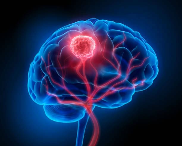
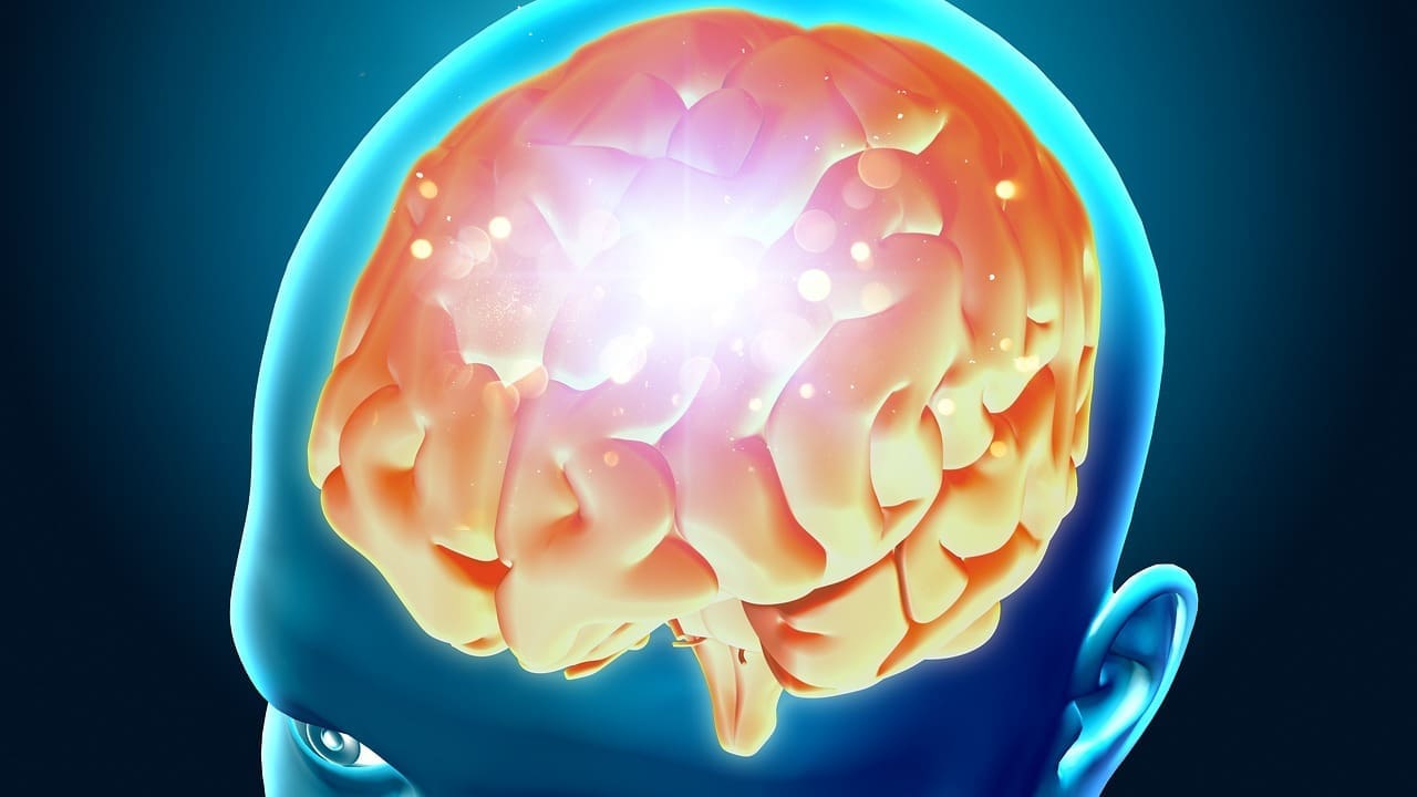
Brain and Neurological Tumors
MRI is great for looking at brain and neurological tumors. It shows soft tissue details clearly. It helps see where the tumor is, how big it is, and how it affects nearby areas.
Advanced MRI methods like diffusion-weighted imaging and perfusion MRI help us understand these tumors better. For example, diffusion-weighted imaging shows how dense the tumor cells are. This is important for figuring out what kind of tumor it is.
Breast Tumors and MRI Evaluation
MRI is very important for checking breast tumors. It’s great for seeing how big the tumor is and if there are more tumors that can’t be seen on other tests. It’s very good at finding breast cancer, which is why it’s so helpful for people at high risk.
Dynamic contrast-enhanced MRI is also very useful. It shows how the tumor looks with blood flow. This helps tell if a tumor is likely to be cancerous or not.
Abdominal and Pelvic Tumor Imaging
MRI is used a lot for looking at tumors in the abdomen and pelvis. It gives clear pictures of soft tissues. This is important for planning surgery and checking how well treatments are working.
Using T2-weighted imaging and diffusion-weighted imaging gives us more details about the tumor. It helps us spot any problems that might come up.
Musculoskeletal Tumor Characterization
MRI is also very helpful for looking at tumors in muscles and bones. It shows how big the tumor is and if it’s touching important structures. This is key for doctors planning surgery.
Using advanced MRI, like dynamic contrast-enhanced MRI, helps us see how the tumor reacts to treatment. This helps doctors plan the best treatment.
When MRI Alone Isn’t Enough: Complementary Diagnostic Methods
MRI is a powerful tool, but sometimes it’s not enough. This means we need other tests to get a full picture. MRI is often the first step, but more tests are needed for a clear diagnosis.
The Role of Biopsy in Definitive Diagnosis
A biopsy is key for a clear diagnosis, when MRI results are not clear. It takes a tissue sample from the tumor. This lets us see if the tumor is cancerous or not.
We use special biopsy methods to get accurate results without harming the patient too much.
Combining CT Scans with MRI Results
CT scans give us more information than MRI. They show details MRI can’t, like bone or lung issues. Together, MRI and CT scans give us a full view of the tumor.
PET Scans and Nuclear Medicine Approaches
PET scans use tiny amounts of radioactive tracers. They show how active tumors are. This helps us tell if a tumor is cancerous and how it’s growing.
We use PET scans with MRI to make sure we have all the information we need for treatment.
Ultrasound as a Supplementary Tool
Ultrasound is also helpful, used with MRI. It’s great for guiding biopsies and checking blood flow. It can give us live information that MRI can’t, helping us understand tumors better.
The Patient Experience: What to Expect During a Diagnostic MRI
Getting an MRI for tumor assessment can seem scary. But knowing what to expect can make it less stressful. We’ll walk you through the steps to make your experience as easy as possible.
Preparation for an MRI Tumor Assessment
Before your MRI, you need to get ready. Take off any metal items like jewelry or clothes with metal parts. They can mess with the MRI machine. Also, tell your doctor about any metal implants, claustrophobia, or other health issues.
You might need to arrive early to fill out forms and change into a gown. Being prepared in advance can reduce your anxiety on the day of the MRI.
| Preparation Step | Description |
| Remove Metal Objects | Take off jewelry, clothing with metal parts, and other items that could interfere with the MRI. |
| Inform Healthcare Provider | Disclose any metal implants, claustrophobia, or other relevant medical conditions. |
| Arrive Early | Come early to complete paperwork and change into a hospital gown. |
The Procedure: Duration and Process
The MRI process starts with lying on a table that moves into the machine. Scans usually last between 15 to 90 minutes. The machine makes loud noises, but you’ll get earplugs or headphones to block it out.
“The MRI machine is like a large, cylindrical tube that is surrounded by a magnetic field. It’s important to stay very quiet during the scan to get clear images.”
Our medical team will be with you the whole time. They’ll check on your comfort and answer any questions you have.
Understanding and Receiving Your Results
After your MRI, the images are checked by a radiologist. They’ll then talk to your doctor about the findings. Your doctor will explain the diagnosis and what to do next.
Waiting for results can take a few days. But we promise to get your results to you as soon as we can. We’re here to support you during this time.
Coping with Uncertainty During the Diagnostic Process
Diagnostic tests can be stressful and uncertain. It’s okay to feel worried about the results. If you have questions or concerns, don’t hesitate to ask your doctor.
Also, talking to loved ones or joining a support group can help. Our team is here to provide both medical care and emotional support during this challenging time.
Future Developments in MRI Tumor Characterization
The future of MRI tumor characterization looks promising. New technologies are on the horizon to boost diagnostic accuracy. Several areas are being explored to make MRI better at assessing tumors.
Artificial Intelligence and Machine Learning Applications
Artificial intelligence (AI) and machine learning (ML) are being used more in MRI. These tools can analyze complex data and find patterns. They can spot changes in tumors that might show they are cancerous.
Key Applications of AI and ML in MRI Tumor Characterization:
- Enhanced image analysis and interpretation
- Automated detection of tumors and their characteristics
- Predictive modeling for tumor behavior and treatment response
Higher Field Strength MRI Systems
New MRI systems, like 7 Tesla MRI, are being developed. They offer better detail and contrast, helping see tumors more clearly. This could make tumor diagnosis more accurate by showing tumor boundaries and internal details.
Molecular and Cellular Imaging Advances
New imaging techniques focus on tumor biology. They can show specific molecular markers or cellular processes. This helps choose the best treatment options.
| Imaging Technique | Application in Tumor Characterization |
| Magnetic Resonance Spectroscopy (MRS) | Analysis of metabolic changes within tumors |
| Diffusion-Weighted Imaging (DWI) | Assessment of tumor cellularity and water diffusion |
| Dynamic Contrast-Enhanced MRI (DCE-MRI) | Evaluation of tumor vascularity and perfusion |
Personalized MRI Protocols for Tumor Assessment
The future also includes personalized MRI protocols. Tailoring MRI to each patient can give more accurate information. This approach can lead to better diagnosis and treatment plans.
As we progress, using AI, high-field MRI, molecular imaging, and personalized protocols will be key. These advancements will help us better understand tumors and improve patient care.
Conclusion
MRI is key in finding out if tumors are benign or malignant. It’s a top-notch tool for medical imaging. MRI helps doctors tell the difference between harmless and harmful tumors.
This helps patients get better treatment plans. MRI’s advanced technology makes it easier to diagnose tumors accurately.
New MRI tech is important for better tumor checks. Things like stronger MRI machines and new imaging methods help a lot. These advancements make MRI even better for diagnosing tumors.
They also help doctors give more tailored care to patients. MRI’s role in finding out about tumors is growing. This shows how vital it is to keep improving MRI technology.
FAQ
Can MRI accurately distinguish between benign and malignant tumors?
MRI is very good at telling the difference between benign and malignant tumors. But, it depends on the tumor type, where it is, and the MRI methods used. New MRI methods like diffusion-weighted imaging and magnetic resonance spectroscopy have made it even better.
What are the key imaging characteristics of benign tumors on MRI?
Benign tumors show clear signs on MRI. They have well-defined edges, uniform signal, and don’t invade nearby tissues. The exact signs can change based on the tumor type and location.
How does contrast enhancement help in tumor identification on MRI?
Contrast enhancement is key in MRI tumor checks. It shows the tumor’s blood flow and how it’s fed. Benign tumors enhance slowly, while malignant ones enhance quickly and strongly.
What are the limitations of MRI in tumor characterization?
MRI is a strong tool, but it has limits. Technical issues like artifacts and resolution problems exist. So do patient issues like claustrophobia and movement. Some tumors look similar, making it hard to tell them apart.
How do radiologists interpret MRI results for tumor assessment?
Radiologists look at visual signs, signal patterns, and how the tumor enhances. They also consider the patient’s overall health and other tests to give a full picture.
What is the role of advanced MRI techniques in tumor differentiation?
New MRI methods like diffusion-weighted imaging and magnetic resonance spectroscopy help a lot. They give more info on the tumor’s biology and how it works. This helps figure out how aggressive the tumor is.
Can MRI be used to evaluate tumors in different organs and systems?
Yes, MRI can check tumors in many places like the brain, breast, and muscles. The MRI methods used depend on the tumor’s location and type.
How does MRI compare to other diagnostic methods in tumor assessment?
MRI is a great tool that works well with other methods like CT and PET. The best method depends on the tumor and the patient’s situation.
What can patients expect during a diagnostic MRI for tumor assessment?
Patients get ready by fasting, wearing the right clothes, and removing metal. The MRI is done while lying down in a comfortable table. They might get a contrast agent during the scan.
How will future developments in MRI technology impact tumor characterization?
New MRI tech like AI and higher field strength machines will make tumor checks even better. Advances in molecular imaging will also help a lot.
References:
- National Cancer Institute. (2011). Definition of solid tumor. NCI Dictionary of Cancer Terms.https://www.cancer.gov/publications/dictionaries/cancer-terms/def/solid-tumor


