Last Updated on November 27, 2025 by Bilal Hasdemir

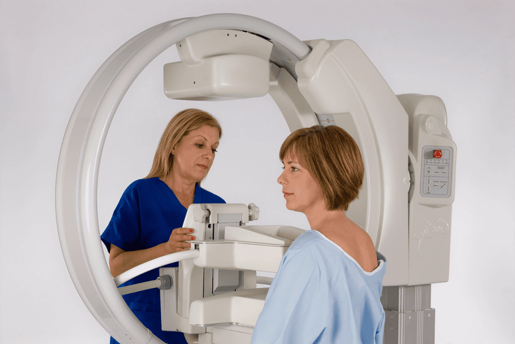
At Liv Hospital, we use the latest technology to care for our patients. A key tool is the gamma camera. It’s a special device used in nuclear medicine diagnostics.
A gamma camera, or scintillation camera, finds the radiation from special drugs. These drugs are used in nuclear medicine to help diagnose and treat patients. It turns this radiation into digital signals, making images that help us see what’s going on inside the body.
Key Takeaways
- Gamma cameras are key for diagnosing and tracking diseases in nuclear medicine.
- They find the radiation from special drugs used in medical tests.
- The tech helps us see things like tumors and big organs.
- Gamma cameras make digital signals that turn into images on a computer.
- Liv Hospital is dedicated to patient-centered, innovative, and respected healthcare.
The Role of Nuclear Medicine Cameras in Modern Diagnostics

The nuclear medicine camera, also known as a gamma camera, is key in modern diagnostics. It detects gamma radiation from the body. This technology is vital in medical imaging, helping doctors see and understand the body’s functions.
Definition and Basic Principles
A nuclear medicine camera, or gamma camera, detects gamma rays from radioactive tracers in a patient’s body. It has one to three camera heads, each flat, to get close to the patient for clear images. The camera works by catching gamma radiation to create images for diagnosing medical conditions.
According to the National Center for Biotechnology Information, gamma cameras track radiopharmaceuticals in the body. This gives doctors important diagnostic information. The process uses a scintillation crystal to turn gamma rays into light, which photomultiplier tubes detect.
Historical Development of Gamma Cameras
The history of gamma cameras started in the mid-20th century. The first one was made by Hal O. Anger in 1958. This was a big step in nuclear medicine. Over time, gamma cameras have gotten better, with better images and new materials.
Gamma cameras have changed a lot, with different types for different needs. They’ve also become part of hybrid systems with CT and MRI. This shows how gamma cameras have grown in importance.
| Year | Development | Significance |
| 1958 | The first gamma camera was invented by Hal O. Anger | Marked the beginning of gamma camera technology |
| 1970s | Introduction of microprocessor technology | Improved image processing and analysis capabilities |
| 1990s | Development of dual-headed gamma cameras | Enhanced sensitivity and reduced imaging time |
| 2000s | Advancements in detector technology and hybrid imaging | Improved diagnostic accuracy and expanded clinical applications |
As a renowned expert said, “The gamma camera has changed nuclear medicine. It lets us see the body’s functions in new ways.” The ongoing improvements in gamma camera tech make it more important in diagnostics today.
The Science Behind Gamma Radiation in Medical Imaging

Gamma radiation has changed how we diagnose diseases. It lets us see inside the body, like the heart and kidneys. This gives us important info about how they work.
Properties of Gamma Rays
Gamma rays are high-energy waves from radioactive stuff. They’re great for medical images because they can go through tissues and show us what’s inside. They also tell us how different parts of the body are working.
- High Energy: Gamma rays have enough energy to be seen outside the body.
- Penetration Power: They can go through tissues, letting us see inside organs.
- Detectability: Special detectors can catch gamma rays, making images possible.
Radioactive Tracers and Their Function
Radioactive tracers are special substances that give off gamma rays. They help us see how the body works in nuclear medicine. When given to a patient, they light up areas of interest, and we can see them with a gamma camera.
We pick different tracers for different parts of the body. For example, technetium-99m is good for the thyroid, bones, and heart.
Crystal Detector Systems
Crystal detector systems are key in gamma cameras. They use a scintillation crystal, like sodium iodide, to turn gamma rays into light. This light is then boosted by photomultiplier tubes to make an image.
How well these systems work is key to clear images. New gamma cameras use better crystals and designs to catch more light and show more detail.
Collimators: Types and Functions
Collimators help focus gamma rays onto the detector. This makes images clearer and cuts down on background noise.
- Parallel-hole Collimators: Good for general images, these have parallel holes for clear views.
- Pinhole Collimators: With a single hole, they’re for detailed views of small areas, like the thyroid.
- Converging Collimators: They magnify images, perfect for seeing small organs.
Understanding gamma radiation and gamma cameras shows us how advanced medical tech is. It’s a big help in diagnosing diseases.
How Gamma Camera Imaging Works
Gamma camera imaging detects gamma rays from a radioactive tracer in the body. This is painless and doesn’t cause discomfort. It’s a key tool for diagnosing and monitoring diseases, like cancers, by showing details other methods miss.
Image Formation and Processing
The gamma camera captures gamma rays from the tracer to form images. It uses a collimator, crystal detector, and photomultiplier tubes. The collimator focuses the rays, and the crystal detector turns them into visible light. This light is then amplified to create an electrical signal for the final image.
For more details on how it works, visit https://rnmcenter.com/how-does-a-gamma-camera-scan-work/. This site offers more on the technical and clinical uses of gamma camera imaging.
Digital Output and Analysis
The gamma camera’s digital output is analyzed with special software. This improves image quality and gets important diagnostic info. The images help doctors diagnose and manage patient care.
We use advanced software to process these images. This makes gamma camera imaging essential in healthcare today. It helps in precise diagnosis and treatment planning.
Types of Nuclear Medicine Gamma Camera Systems
Gamma camera systems in nuclear medicine vary to meet different needs. They help us diagnose and treat many conditions. This variety is key to our success in medical imaging.
Planar Gamma Cameras
Planar gamma cameras are simple and early types. They take 2D images of radioactive tracers in the body. These cameras work well for static images and are used in thyroid studies and bone scans.
SPECT Gamma Cameras
SPECT cameras are a big step up from planar cameras. They rotate around the patient to capture images from many angles. This lets us create 3D images, giving us more detailed info. SPECT cameras are great for complex conditions like heart diseases and some cancers.
Hybrid Imaging Systems
Hybrid systems mix nuclear medicine with CT or MRI. This mix gives us detailed images of both function and anatomy. They’re super useful in fields like oncology, cardiology, and neurology for precise diagnosis.
Cardiac Scans
Cardiac scans focus on the heart. They check heart function, viability, and perfusion. These scans are vital for diagnosing heart disease and guiding treatments. Cardiac SPECT is a common method that offers deep insights into the heart.
| Type of Gamma Camera System | Primary Use | Key Features |
| Planar Gamma Cameras | Static imaging, thyroid uptake studies, and bone scans | 2D imaging, simple, cost-effective |
| SPECT Gamma Cameras | 3D imaging, cardiac diseases, and cancer diagnosis | Rotates around the patient, 3D reconstruction, detailed functional information |
| Hybrid Imaging Systems | Oncology, cardiology, and neurology | Combines functional and anatomical imaging, enhancing diagnostic accuracy |
| Cardiac Scans | Myocardial perfusion, viability, and function assessment | Specialized for heart imaging, critical for coronary artery disease diagnosis and management |
Interpreting Gamma Camera Images in Clinical Practice
Understanding gamma camera images is key in healthcare. It helps shape treatment plans and improves patient care. These images help spot diseases like cancer and heart issues.
Normal vs. Abnormal Findings
It’s important to tell normal from abnormal images. Normal ones show even radiotracer spread. Abnormal ones might show hot or cold spots, hinting at disease.
In cancer care, hot spots mean tumors are active. We look at these images closely. We consider patient history and symptoms to make accurate diagnoses.
Quantitative Analysis Techniques
Quantitative analysis boosts image accuracy by giving numbers on radiotracer levels. SUV calculations help measure disease severity and treatment success.
We use software to analyze these images. This lets us track changes and adjust treatments. It’s very useful for long-term conditions and checking therapy results.
| Technique | Description | Clinical Application |
| SUV Calculations | Quantifies radiotracer uptake in regions of interest | Oncology, assessing tumor activity |
| Time-Activity Curves | Monitors changes in radiotracer uptake over time | Cardiovascular and renal function assessment |
| Attenuation Correction | Compensates for signal loss due to tissue attenuation | Improves image accuracy in various studies |
Common Artifacts and Limitations
Gamma camera imaging is powerful but has its limits. Artifacts can come from patient movement, equipment issues, or tissue blocking signals.
Knowing these issues helps us get better images. We work to avoid artifacts and correct them when they happen. The radiation from these scans is very low and safe.
By combining technical skills with medical knowledge, we can better understand gamma camera images. This helps doctors see how treatments are working. It lets us make changes to treatments as needed.
Clinical Applications of Gamma Imaging Camera Technology
Gamma imaging camera technology is key in today’s diagnostics. It gives us functional info about the body’s inner workings. We use gamma cameras for scintigraphy scans, giving us detailed insights into organ functions.
Oncology Applications
In oncology, gamma camera tech is vital for tumor detection and tracking. We use radioactive tracers that build up in cancer cells, showing us how far cancer has spread. This info is key for cancer staging and treatment planning.
Gamma cameras also help in checking how tumors react to treatment. By comparing images, doctors can see if tumors are getting smaller or bigger. This helps them adjust treatment plans as needed.
Cardiovascular Diagnostics
In heart health, gamma cameras help check blood flow and heart function. This helps spot coronary artery disease and check the heart’s pumping power. These scans are essential for finding heart muscle areas with low blood flow.
Gamma imaging also helps diagnose heart attacks. It lets doctors see the heart’s structure and function. This helps them make better care decisions for patients.
Neurological Assessments
Gamma imaging is used in neurology to study the brain and diagnose brain disorders. We can use special tracers to see different brain activities, helping diagnose Alzheimer’s and some types of epilepsy.
The detailed images from gamma cameras help doctors understand brain damage or disease. This guides treatment and rehabilitation plans.
Endocrine System Evaluation
Gamma camera tech helps check the endocrine system, like thyroid disorders. Thyroid scintigraphy is common, using radioactive iodine to diagnose hyperthyroidism and find thyroid nodules.
By looking at the tracer’s spread, doctors can figure out thyroid problems. They can then plan the right treatment, like meds, radioactive iodine, or surgery.
Comparing Gamma Ray Camera Imaging with Other Modalities
Gamma ray cameras are key in diagnostic imaging. But how do they compare to PET scanning, CT, and MRI? It’s important to know the strengths and weaknesses of each technology.
Gamma Cameras vs. PET Scanning
Gamma cameras and PET scanning are both used in nuclear medicine. But they work differently. Gamma cameras detect single photons, while PET scans look for pairs of photons. PET scanning is often better at showing details, which is great for cancer and brain studies.
Yet, gamma cameras are popular because they can use many different types of radioactive materials. “The choice between gamma camera and PET scanning depends on what you need to know,” says a nuclear medicine expert.
Advantages Over CT and MRI
CT and MRI are good at showing body structures. But gamma cameras can show how the body works. They help find out how diseases affect the body, which is key for diagnosis and treatment.
- Gamma cameras offer unique insights not found in CT or MRI.
- They’re great for certain cancers and heart issues.
- They help doctors make better treatment plans.
Complementary Roles in Diagnostic Imaging
Gamma cameras, PET scans, CT, and MRI work together, not against each other. For example, combining gamma camera info with CT or MRI images can make diagnoses more accurate.
Using all these imaging methods together is becoming more common. This way, doctors can get a clearer picture of what’s going on in the body. It helps in making better treatment plans.
In summary, gamma-ray camera imaging has its own benefits. But it’s just one tool in a bigger toolkit that includes PET scanning, CT, and MRI. Knowing how each tool works is key to the best care for patients.
Patient Experience During a Gamma Camera Scan
A gamma camera scan is a tool that helps doctors understand many health issues. It uses a small amount of radioactive material to see inside the body. This helps doctors find problems in specific areas.
Preparation for the Procedure
Before the scan, patients get a small dose of a radioactive drug. This drug makes the body emit tiny amounts of radiation. The gamma camera then picks up this radiation.
The type of drug used depends on what the doctor wants to check, like bones or the heart.
To get ready for the scan, patients might need to:
- Fast for a certain period
- Avoid certain medications
- Remove jewelry or other metal objects
- Wear comfortable clothing
What to Expect During the Scan
The scan’s length varies based on the drug used. For example, a bone scan can last from 1 to 4 hours. Patients must stay very quiet while the camera takes pictures.
The scan is usually painless. But some might feel uncomfortable because they have to stay in one spot for a long time.
Post-Procedure Care and Radiation Precautions
After the scan, patients can usually go back to their normal activities. The radioactive material leaves the body in a few hours to days. To protect others from radiation, patients might be told to:
- Drink lots of water to help get rid of the radioactive material
- Avoid being close to pregnant women and young kids for a bit
- Practice good hygiene, like washing hands well after using the bathroom
Knowing what to expect and following precautions makes the gamma camera scan safe and easy. Our team is here to help and support you every step of the way.
Technological Advancements in Med Camera Systems
Med camera systems have seen big changes thanks to digital tech and AI. These updates have made gamma cameras better at taking images. Now, they can show more details and be more precise.
Digital Gamma Cameras
Digital gamma cameras are a big step up from old models. They are more sensitive and accurate. They help doctors see diseases early and track them better. For example, they can check heart health, find tumors, and see how cancer spreads.
Experts say digital gamma cameras have changed nuclear medicine a lot. They make diagnoses more accurate and quicker.
AI and Machine Learning Integration
AI and machine learning are changing nuclear medicine. AI helps make images clearer and analyzes data faster. This makes doctors better at diagnosing and saves them time.
- Enhanced image processing capabilities
- Automated detection of abnormalities
- Predictive analytics for patient outcomes
Future Directions in Gamma Imaging
Technology will keep getting better, and so will gamma imaging. We might see even smarter AI, better materials, and new designs. There’s also talk of combining gamma cameras with CT or MRI.
“The future of gamma imaging lies in its ability to integrate with other diagnostic modalities, providing a more complete understanding of patient health.”
These updates will make gamma cameras even more important in medicine. They will help doctors give better care to patients.
Conclusion: The Evolving Role of Gamma Cameras in Modern Medicine
Gamma cameras are key in modern medicine, thanks to new tech that boosts their ability to diagnose. They are the main way to create images in nuclear medicine. This makes them a game-changer, helping make nuclear medicine more personalized.
We see how important gamma cameras are in medical care and diagnostics. Their role is changing fast, showing how quickly medical tech is advancing. With new tech coming, gamma cameras will keep giving vital insights. These insights help doctors make better treatment plans and improve patient care.
The future of gamma cameras in medicine looks bright. We expect more innovations to make them even more useful. Gamma cameras will keep being a vital tool for diagnosing and treating many health issues. This shows how critical they are in healthcare.
FAQ
What is a gamma camera and how does it work?
A gamma camera is a key tool in nuclear medicine. It detects gamma radiation from radioactive tracers in the body. This helps create images for diagnosing and monitoring diseases.
What is the role of nuclear medicine cameras in modern diagnostics?
Nuclear medicine cameras, like gamma cameras, are essential in today’s diagnostics. They provide detailed information about the body’s internal workings. This helps doctors diagnose and treat diseases more effectively.
How have gamma cameras evolved over time?
Gamma cameras have seen a lot of changes over the years. From their early beginnings to today’s digital and hybrid systems, they’ve improved a lot. Now, they offer better image quality and diagnostic abilities.
What are the different types of nuclear medicine gamma camera systems?
There are many types of nuclear medicine gamma camera systems. These include planar, SPECT, hybrid imaging, and cardiac scans. Each type has its own benefits and uses.
How do you interpret gamma camera images in clinical practice?
Interpreting gamma camera images requires knowing what’s normal and what’s not. It also involves using quantitative analysis and being aware of common issues. This ensures accurate diagnoses.
What are the clinical applications of gamma imaging camera technology?
Gamma imaging camera technology has many uses in healthcare. It’s used in oncology, cardiovascular diagnostics, neurological assessments, and endocrine system evaluations. It helps doctors diagnose and treat diseases well.
How does gamma ray camera imaging compare to other imaging modalities?
Gamma ray camera imaging has its own strengths and uses in diagnostics. It differs from other imaging methods like PET scanning, CT, and MRI. It’s often used with these methods to get a full picture of what’s going on in the body.
What is the patient experience during a gamma camera scan?
Patients going through a gamma camera scan need to prepare and stay calm during the scan. They also have to follow instructions after the scan, including radiation safety tips. This ensures a safe and effective test.
What are the technological advancements in medical camera systems?
Med camera systems, including gamma cameras, have seen big improvements. These include digital cameras, AI, and machine learning. These advancements are making diagnostics better and improving patient care.
What is the correct spelling for the recording of radioactivity with gamma cameras?
The correct term for recording radioactivity with gamma cameras is gamma camera imaging or scintigraphy.
What is the significance of gamma cameras in medical diagnostics?
Gamma cameras are very important in medical diagnostics. They provide valuable information about the body’s internal workings. This helps doctors diagnose and treat diseases effectively.
How does a gamma camera produce images?
A gamma camera makes images by detecting gamma radiation from radioactive tracers in the body. It uses a crystal detector system and collimators to create and process the images.
References
- American Scientific Research Journal for Engineering, Technology, and Sciences. (2024). Gamma cameras: Exploring the technology, applications, and prospects. American Scientific Research Journal for Engineering, Technology, and Sciences, 98(1), 296-308. https://asrjetsjournal.org/American_Scientific_Journal/article/view/10875
- Alenezi, A. R., & Alswaidan, N. A. (2023). Advancements in nuclear medicine: A comprehensive review. Open Access Journal of Nuclear Medicine, 8(3), Article 16679. https://www.openaccessjournals.com/articles/advancements-in-nuclear-medicine-a-comprehensive-review-16679.html
- Han, D. H., & Lee, J. (2023). Development and evaluation of a compact gamma camera for nuclear medicine applications. Medical Physics, 50(7), 3500-3509. https://www.sciencedirect.com/science/article/pii/S1738573323002073


