Last Updated on November 27, 2025 by Bilal Hasdemir
Depression is a complex mental health disorder affecting millions worldwide. Advanced imaging techniques, such as PET scans, have changed how we understand it. At Liv Hospital, they use PET scans to uncover how depression changes brain activity.How are PET scan and depression linked? Get an essential comparison of metabolic activity between depressed and normal brains.
Studies show that depressed brains have less activity in areas like the prefrontal cortex. A study in Frontiers in Psychiatry found big differences in brain activity between depressed and healthy people.
Key Takeaways
- PET scans can reveal altered brain activity in individuals with depression.
- Depressed brains show reduced activity in areas like the prefrontal cortex.
- Advanced imaging techniques help compare depressed and normal brain activity.
- Liv Hospital utilizes cutting-edge PET scans for depression research.
- Research indicates significant differences in brain activity between depressed and healthy individuals.
The Science Behind Brain Imaging Technologies
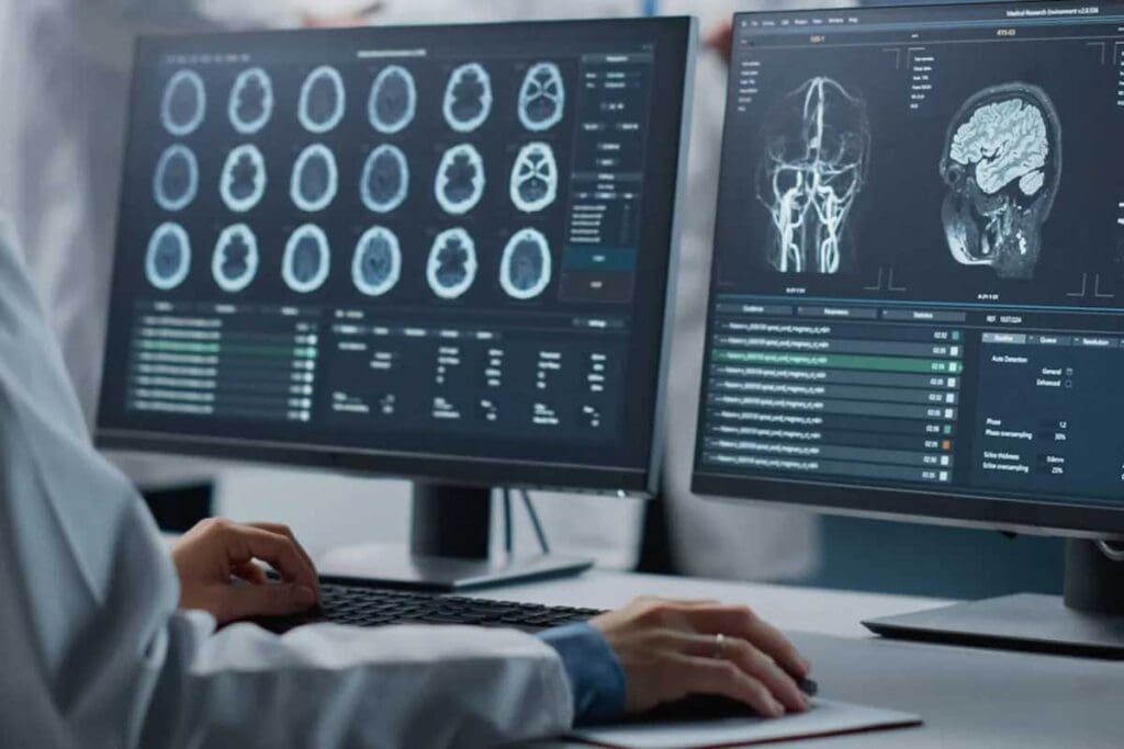
Brain imaging technologies have changed the game in neuroscience. They give us a peek into how depression affects the brain. These tools let us see the brain’s function and structure in amazing detail.
Principles of Neuroimaging
Techniques like positron emission tomography (PET) and functional magnetic resonance imaging (fMRI) show us the brain’s activity and structure. They work by linking brain activity to changes in blood flow and metabolism.
PET scans, for example, track the brain’s metabolic activity by using a radioactive tracer. This helps researchers spot brain areas affected by depression. To learn more about PET scans in depression research.
Evolution of Brain Scanning Techniques
Brain scanning has come a long way, from basic to advanced methods. The rise of PET scan psychology has been key in understanding depression’s neural roots.
| Technique | Description | Application in Depression Research |
| PET | Measures metabolic activity | Identifying areas affected in depression |
| fMRI | Measures changes in blood flow | Studying functional connectivity in depression |
| SPECT | Measures blood flow and perfusion | Assessing regional cerebral blood flow in depression |
How Functional Brain Imaging Works
Techniques like PET and fMRI detect changes in brain activity. These changes show up in blood flow and metabolism, signs of neural activity.
Understanding these imaging methods helps researchers uncover depression’s neural roots. This knowledge aids in creating better treatments and improving diagnosis.
Understanding PET Scan and Depression Research
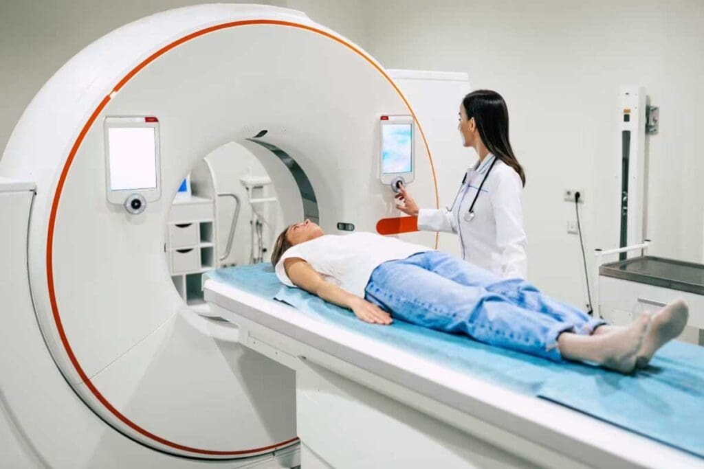
PET scans are key in studying depression. They show how the brain works in people with depression. This helps researchers understand the brain’s complex functions in depression.
What Positron Emission Tomography Measures
Positron Emission Tomography (PET) is a detailed brain imaging method. It looks at brain function, like how much glucose it uses and blood flow. A study in Nature shows PET scans find brain activity patterns linked to depression.
Glucose use shows how active brain cells are. Changes in this can point to depression. PET scans use special tracers to see and measure glucose use in the brain.
Radioactive Tracers Used in Depression Studies
Radioactive tracers are key for PET scans. They attach to brain targets, like receptors or glucose. In depression studies, FDG (fluorodeoxyglucose) is often used to check glucose use. Other tracers focus on neurotransmitters like serotonin or dopamine, which are important in depression.
The tracer choice depends on the study’s focus. For example, serotonin studies might use tracers that bind to serotonin receptors. This helps understand serotonin’s role in depression.
How PET Visualizes Brain Activity in Depressed Patients
PET scans show brain activity by tracking tracer signals. In depression studies, they find brain activity differences between depressed and healthy people. For instance, depressed people often have different glucose use in areas like the prefrontal cortex and limbic system.
By studying these differences, researchers learn about depression’s neural circuits. This knowledge is vital for better treatments and understanding the brain basis.
The Neurobiological Basis of Depression
Depression affects the brain in many ways. It changes how brain structures and neurotransmitters work. This complex condition impacts not just our minds but also our brain’s function and structure.
Key Brain Structures Involved in Mood Regulation
The brain has special areas that help control our mood. These include the prefrontal cortex, amygdala, and hippocampus. Together, they help us handle emotions and stay balanced.
The prefrontal cortex helps us make decisions and think clearly. The amygdala is key to feeling emotions. The hippocampus is important for memory and is hurt by stress and depression.
| Brain Region | Function | Impact iofDepression |
| Prefrontal Cortex | Executive Function, Decision Making | Impaired cognitive function, reduced decision-making ability |
| Amygdala | Emotion Processing | Hyperactivity, increased emotional response |
| Hippocampus | Memory Formation | Reduced volume, impaired memory |
Neurotransmitter Systems Implicated in Depression
Neurotransmitters like serotonin, dopamine, and norepinephrine are vital for mood. In depression, these systems often get out of balance.
Serotonin is very important for mood, hunger, and sleep. Medications like SSRIs help by making more serotonin available in the brain.
Neural Circuit Dysfunction in Depressive Disorders
Depression messes with the brain’s mood and emotional circuits. It changes how different parts of the brain talk to each other. This leads to depression’s symptoms.
Knowing how depression changes the brain is key to better treatments. Tools like PET scans help us see these changes in people with depression compared to those who are healthy.
Depression Brain Scan vs Normal: Observable Differences
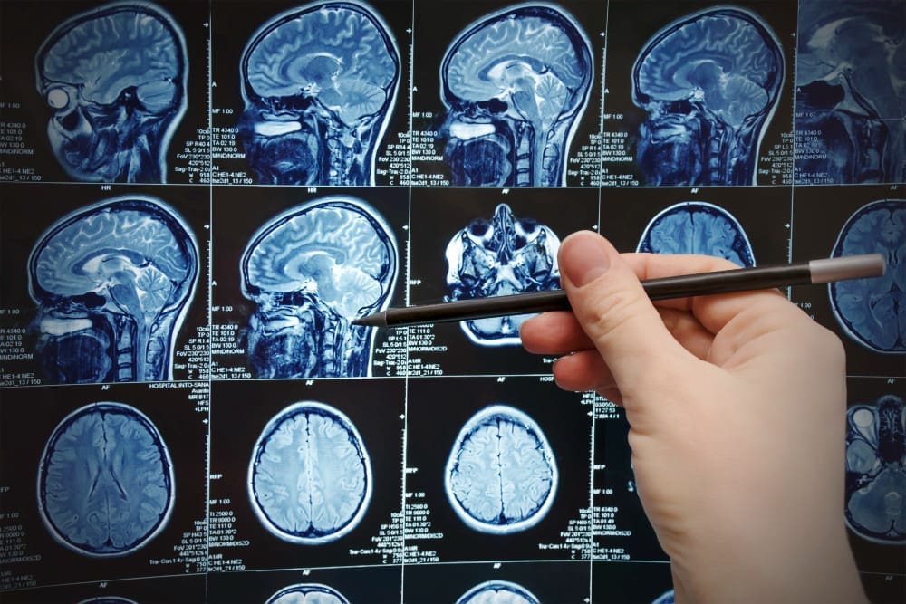
When we look at brain scans of people with depression and those without, we see clear differences. Advanced tools like Positron Emission Tomography (PET) help us see and measure these differences.
Regional Cerebral Blood Flow Alterations
Research shows that people with depression have changes in how blood flows to their brains. Some areas get less blood, like the prefrontal cortex. Others might get more blood. These changes can mess with how our brains handle mood.
Glucose Metabolism Patterns in Depressed vs. Healthy Brains
Glucose metabolism, as seen in PET scans, tells us about brain activity. Studies show that people with depression have different glucose patterns than healthy people. For example, some brain areas might use less glucose, while others use more. These changes could be linked to depression.
Interpreting Visual Differences in Brain Scans
Understanding the brain scan differences between depressed and healthy people requires special knowledge. Quantitative analysis of PET scans is key to spotting small differences. By using both visual checks and numbers, researchers can better understand depression’s effects on the brain.
These discoveries show how complex depression’s brain changes are. More research could help us find better ways to diagnose and treat depression.
Key Brain Regions Affected in Depressed Brain Scans
Studies using brain imaging have found key areas in the brain that differ in people with depression. These areas are important for understanding depression and how it changes brain function.
Prefrontal Cortex Abnormalities
The prefrontal cortex is key to mood, decision-making, and thinking. People with depression often have abnormalities in the prefrontal cortex. This can lead to problems with thinking and mood.
“The prefrontal cortex is less active in depressed individuals, which can lead to impaired cognitive function and mood regulation,” a study on depression and brain imaging found.
Anterior Cingulate Gyrus Findings
The anterior cingulate gyrus is important for catching mistakes, monitoring conflicts, and motivation. Research shows it’s affected in depression, with changes in activity. This can affect how emotions are processed.
As a researcher noted, “Alterations in the anterior cingulate gyrus can contribute to the emotional dysregulation observed in depression.”
Limbic System Alterations
The limbic system, including the amygdala and hippocampus, is key to emotions and memory. Depression is linked to alterations in the limbic system. This affects how emotions are processed and regulated.
The limbic system’s dysfunction can cause the intense emotional pain seen in depression.
Other Notable Regions Showing Functional Changes
Other brain areas, like the basal ganglia and thalamus, also change in depression. These changes can impact behavior and emotions. They add to the complex nature of depression.
Neurotransmitter Function Revealed Through PET Scan Psychology
PET scans have changed how we study depression. They use radioactive tracers to show how neurotransmitters work in the brain. This gives us a peek into the chemical changes that happen in depression.
Serotonin Receptor Binding Studies
Serotonin Receptor Binding Studies
PET scans have helped us understand serotonin in depression. Serotonin helps control our mood. Changes in how serotonin receptors work have been linked to depression.
Studies show people with depression have different serotonin receptor patterns. For example, some research found less binding to certain serotonin receptors in depressed brains. This suggests a connection between serotonin issues and depression.
Dopamine System Alterations
Dopamine is also key in depression studies with PET scans. It’s involved in feeling pleasure and motivation. People with depression often have problems with these areas.
Research shows depression changes how dopamine works in the brain. This includes how many dopamine receptors there are and how much dopamine is made. These changes help explain why people with depression might not feel pleasure or motivation.
Other Neurotransmitter Systems Implicated in Depression
While serotonin and dopamine get a lot of attention, other systems are important too. For example, the GABAergic and glutamatergic systems have been studied with PET scans. These systems help control how brain cells talk to each other.
By looking at these systems, PET scans help us understand depression better. This knowledge could lead to new treatments that target these systems.
Clinical Applications of Brain Imaging for Depression
Brain imaging has changed how we study depression. It uses advanced tools like Positron Emission Tomography (PET) to understand depression better. This has opened up new ways to diagnose and treat the condition.
Brain imaging helps in many ways, from better diagnosis to tracking treatment success. But there are also hurdles to cross.
Current Diagnostic Limitations
Today, doctors mainly use symptoms and patient history to diagnose depression. But brain imaging is starting to help, too. PET scans can spot brain activity patterns linked to depression, helping doctors make better diagnoses.
One big challenge is that depression is not just one thing. It’s a range of conditions with different brain effects. This makes it hard to find one imaging test that works for everyone.
| Diagnostic Challenge | Role of Brain Imaging |
| Heterogeneity of Depression | Identifying specific neurobiological subtypes |
| Lack of Clear Biomarkers | Developing imaging biomarkers for depression |
| Comorbidity with Other Conditions | Differentiating depression from other psychiatric conditions |
Treatment Response Prediction
Brain imaging is also used to guess how well treatments will work. PET scans can show brain activity patterns that match well with treatment success. This helps tailor treatments to each person, possibly leading to better results.
Studies have found that certain brain areas, like the anterior cingulate cortex, are key to how well treatments work. By looking at these areas, doctors might guess which treatments will help most.
Monitoring Depression Treatment Effects
Brain imaging also helps track how treatments are working. By using PET scans over time, doctors can see how brain activity changes. This gives them valuable insights into treatment success.
This is really useful in research, where it helps test new treatments. It also has a place in clinical practice, helping doctors adjust treatments based on brain activity changes.
By using brain imaging, doctors and researchers can understand depression better. There are challenges, but the benefits are huge.
Can You See Depression in a Brain Scan? Emerging Research
Scientists are working hard to detect pressure in brain scans. They’re looking for patterns in brain images to better diagnose depression.
Depression Biotypes and Their Neuroimaging Signatures
Studies show depression is made up of different types, each with its own brain scan pattern. A Stanford Medicine study found four main types based on brain scans. An expert says this could lead to better treatments for each type.
“Understanding these biotypes could lead to more personalized treatment approaches.”
This is important because it means different treatments might work better for different people.
Machine Learning Approaches to Depression Detection
Machine learning is helping to spot depression in brain scans. It looks at big datasets to find changes in brain activity that humans might miss.
Machine learning’s benefits include:
- It’s better at finding different types of depression
- It can quickly look through lots of data
- It might even guess how well treatments will work
Reliability and Reproducibility Challenges
Machine learning and brain scan patterns are promising, but there are big hurdles. Making sure these findings are reliable and can be repeated is key.
Researchers need to work on data quality, how many people are studied, and how to avoid bias. This will help create better, more tailored ways to diagnose depression.
Comparing PET to Other Brain Scan Technologies for Depression
Many brain imaging techniques are used to study depression. Positron Emission Tomography (PET) scans are valuable, but not the only option. It’s important to know how PET compares to other methods to better understand depression.
PET vs. fMRI in Depression Research
Functional Magnetic Resonance Imaging (fMRI) and PET scan brain activity. fMRI looks at blood oxygen levels, while PET scans measure glucose or neurotransmitters. PET gives direct measurements of some neurochemical processes, but fMRI has better spatial detail and no radiation.
Both methods show brain changes in mood regulation in depression. fMRI finds altered connections, and PET shows metabolism and neurotransmitter changes. Using both could give a deeper look into depression.
PET vs. SPECT for Neurotransmitter Studies
Single Photon Emission Computed Tomography (SPECT) is another imaging method. Like PET, it uses radiotracers but has longer tracers and lower resolution. PET is more sensitive and detailed, better for neurotransmitter studies.
PET and SPECT have shown neurotransmitter changes in depression. For example, serotonin receptor binding is altered. SPECT is cheaper and easier to get, but PET’s better resolution is more precise.
Advantages of Multimodal Imaging Approaches
Using many imaging methods in depression research is beneficial. Combining PET, fMRI, and others gives both functional and structural brain data. Multimodal imaging strengthens findings, making results more reliable.
- PET measures specific neurochemical processes.
- fMRI offers high spatial resolution and connectivity data.
- SPECT is an alternative for neurotransmitter studies with unique tracers.
This approach helps understand depression better, leading to better treatments.
Can an MRI Scan Detect Depression?
Magnetic Resonance Imaging (MRI) mainly looks at brain structure. While MRI can’t “detect” depression, advanced MRI techniques like fMRI or DTI can show brain function and connectivity. These might be linked to depression.
Studies have found differences in brain structure and function between depressed and healthy people. For example, altered connectivity in mood-regulating areas has been seen in depression. But MRI isn’t used to diagnose depression yet.
Combining MRI with other imaging, like PET, , T could improve our understanding of depression. It would combine structural, functional, and chemical brain data.
Ethical and Practical Considerations of Depression Brain Scans
Brain scans for diagnosing depression bring up ethical and practical concerns. As we use these technologies more, we must think about their impact on patients and healthcare. It’s important to consider the effects of these tools.
Cost and Accessibility Issues
One big worry is the cost of brain scans, like PET scans. They are pricey because of the tech and radioactive tracers needed. This high cost makes it hard for some to get these scans.
A study in the National Center for Biotechnology Information says we need to look more into whether these scans are worth the cost. Also, not everyone can get to these scans because they are mostly in cities. This leaves rural areas without access, making health care unfair.
Radiation Exposure Concerns
Radiation exposure is a big worry with PET scans. Even though the dose is safe, getting it too often could be bad. Researchers must think about the good and bad of PET scans, mainly for young people or those needing many scans.
Potential for Stigmatization and Misinterpretation
Using brain scans for depression could lead to stigmatization and misinterpretation. Seeing brain changes might make people with depression feel worse. Also, only experts can really understand these scans, and mistakes can happen.
Patient Privacy and Data Security
Keeping patient privacy and data security is key. Brain scan data is very personal and must be protected. Healthcare and research teams must make sure this data is safe from hackers and unauthorized access.
In short, brain scans help us understand depression, but we must think about the downsides. By doing this, we can use these tools in a way that helps patients.
Conclusion: The Future of Brain Imaging in Depression Diagnosis and Treatment
Brain imaging is getting better, which is great news for treating depression. Tools like PET scans are helping us understand depression better. They show us which parts of the brain and chemicals are involved.
Things are looking up for diagnosing depression with brain imaging. New studies on depression types and how to spot them are underway. Also, using computers to find depression early is becoming more common.
Using many imaging methods together will help us learn more about depression. This will lead to better treatments for patients. Doctors will be able to give treatments that really work for each person.
Brain imaging for depression is on the verge of a big change. It could bring new hope to those struggling with this serious condition.
FAQ
Can PET scans diagnose depression?
PET scans can show brain activity differences in depressed and normal brains. But they are not used alone to diagnose depression.
How do PET scans work in depression research?
PET scans measure brain activity by looking at glucose or blood flow. They use radioactive tracers to see brain activity in depressed patients.
What brain regions are affected in depression?
Depression affects areas like the prefrontal cortex and limbic system. These areas help regulate mood.
Can brain scans predict treatment response in depression?
Research shows brain imaging, like PET scans, might predict treatment success. But more studies are needed to confirm this.
How do PET scans compare to other brain imaging techniques like fMRI and SPECT?
PET scans are great for studying neurotransmitters. fMRI looks at blood oxygen changes, and SPECT checks blood flow. Using all these methods together gives a better view of the brain.
Are brain scans reliable for diagnosing depression?
Brain scans can show differences in depressed brains. But, they’re not reliable for diagnosing depression yet. This is because of individual differences and other factors.
Can an MRI scan detect depression?
MRI scans can spot some structural changes linked to depression. But, they’re not used for diagnosing depression. Functional MRI (fMRI) can look at brain activity changes.
What are the limitations of using PET scans in depression research?
PET scans use radiation, are expensive, and have limited detail. These limitations make them less useful in depression research.
How is machine learning being used in depression detection through brain scans?
Machine learning is being used to find patterns in brain imaging data. This could help improve the ddiagnosis andtreatment of depression.
What are the ethical considerations surrounding the use of brain scans for depression?
Ethical issues include patient privacy and data security. There’s also the risk of stigma and misinterpreting results. These concerns highlight the need for careful use.
Can brain scans be used to monitor treatment effects in depression?
Yes, brain imaging, including PET scans, can track changes in brain activity and structure. This helps assess how well treatments are working.
References
- National Institute of Mental Health. (2022). Depression. https://www.nimh.nih.gov/health/topics/depression


