Last Updated on November 27, 2025 by Bilal Hasdemir
Early cancer detection is key to saving lives. Many cancer cases worldwide show the need for good diagnostic tools. Positron Emission Tomography (PET) is a powerful tool in this fight. It helps doctors find and track cancer cells with great accuracy.
PET scans spot areas in the body with high glucose use, a sign of cancer cells. This makes PET scans, like PET/CT scans, very useful for finding and planning treatment for different cancers. They help doctors see how cancer is growing in the body.

Key Takeaways
- PET scans are a key tool in cancer detection and tracking.
- They find cancer cells by showing areas with high glucose use.
- PET scans are important for medical diagnosis and treatment planning.
- The technology combines functional info with anatomical details in PET/CT scans.
- Early detection with PET scans can greatly improve cancer treatment results.
Understanding PET Scan Technology
PET scans are a big step forward in medical imaging. They use Positron Emission Tomography to see how cells work. This tech is key in finding and treating many cancers. Let’s dive into how PET scans work, from their basics to the science behind them.
What is Positron Emission Tomography?
Positron Emission Tomography (PET) is a top-notch imaging method. It shows how tissues and organs work, not just their shape. It uses a special tracer, like fluorodeoxyglucose (FDG), to light up active areas.
The Science Behind PET Imaging
PET imaging works by catching positrons from the tracer. FDG goes to cells that use a lot of glucose, like cancer cells. When it decays, it sends out gamma rays. The PET scanner picks up these rays, making clear images of where the tracer is.
| Component | Description | Role in PET Imaging |
| Radioactive Tracer (FDG) | A glucose molecule attached to a radioactive atom | Accumulates in areas of high glucose metabolism, such as cancer cells |
| PET Scanner | A device that detects gamma rays emitted by the tracer | Creates images of metabolic activity based on gamma ray detection |
| Image Reconstruction Software | Software that processes data from the PET scanner | Generates detailed, cross-sectional images of the body’s metabolic activity |
Knowing how PET scans work helps us see their importance in fighting cancer. They let us see how cells are working, which is vital for finding and treating cancer. This tech gives doctors the info they need to plan and check on treatments.
How PET Scans Work in Cancer Detection
PET scans are a key tool for finding cancer. They use a special radioactive tracer, like fluorodeoxyglucose (FDG). This tracer is a sugar molecule with a radioactive tag. Cancer cells eat more sugar than normal cells, making them show up on scans.
Radioactive Tracers and Glucose Metabolism
First, a radioactive tracer is injected into the patient. Cancer cells eat more sugar, so they grab more of the tracer. This tracer sends out positrons, which meet electrons and create gamma rays. The PET scanner picks up these rays to make detailed images.
Key aspects of radioactive tracers include:
- They are absorbed by cells based on their metabolic rate.
- Cancer cells, having higher metabolic rates, absorb more tracer.
- The emitted gamma rays are used to create detailed images of the body’s metabolic activity.
Why Cancer Cells Appear Different on PET Scans
Cancer cells show up on PET scans because they use more sugar. This means they take in more of the radioactive tracer. They appear as bright spots on the scan images.
The difference in glucose metabolism is a hallmark of cancer that PET scans exploit for detection.
| Cell Type | Glucose Metabolism Rate | Tracer Uptake | Appearance on PET Scan |
| Normal Cells | Low | Low | Less Active |
| Cancer Cells | High | High | More Active |
“The ability of PET scans to detect cancer based on metabolic activity has revolutionized cancer diagnosis and treatment planning.”
” Expert in Nuclear Medicine
The PET Scan for Cancer: Effectiveness and Applications
PET scans have changed how we diagnose and treat cancer. They give us vital info on tumor activity and spread. We use them in oncology for diagnosing, staging, and tracking treatment progress.
PET scans help us see how active tumors are. This tells us how aggressive the cancer is. Knowing this helps us choose the best treatment.
Primary Uses in Oncology
PET scans are key in oncology for several reasons:
- Diagnosis: They spot cancer by showing where cells are most active.
- Staging: They show how far cancer has spread, which is key for staging.
- Treatment Monitoring: They check if cancer is responding to treatment.
Cancer Staging and Treatment Planning
Knowing the cancer stage is vital for planning treatment. PET scans help by showing tumor location and activity.
They help doctors:
- See how far cancer has spread.
- Choose the best treatment, like surgery or chemo.
- Check if treatment is working and make changes if needed.
Using PET scans in treatment planning makes care more personal and effective for cancer patients.
Types of Cancers Most Effectively Detected by PET Scans
PET scans are key in finding and treating some cancers. They show how tumors work by looking at their activity. This makes PET scans great for certain cancers.
Lung Cancer Detection
PET scans are very useful for lung cancer. They can tell if lung nodules are cancerous, which is important for early treatment. PET scans work well with CT scans to find lung cancer.
They also help see if lung cancer has spread. This is important for planning treatment. Knowing how far cancer has spread helps doctors choose the best treatment.
Colorectal Cancer Imaging
PET scans are also great for colorectal cancer. They find cancer that has come back in patients with high tumor markers. They also check if treatment is working.
- PET scans find cancer that has spread in colorectal cancer patients.
- They check if treatment is working.
- PET scans help surgeons plan to remove cancer in other parts of the body.
Lymphoma and PET Scan Accuracy
PET scans are also good for lymphoma. They find lymphoma very well. This is important for starting treatment, checking how well it’s working, and finding cancer that comes back.
PET scans are great for lymphoma because they find active tumor sites. This helps doctors plan treatment better.
In summary, PET scans are very good at finding and treating lung cancer, colorectal cancer, and lymphoma. They give important information that helps doctors make better treatment plans.
Cancers That May Be Difficult to Detect With PET Scans
PET scans are great for finding many cancers. But, some tumors are hard to spot. Knowing these limits helps us understand PET scan results better.
Slow-Growing Tumors
Slow-growing tumors are tricky for PET scans. They don’t use as much glucose as fast-growing cancers. This makes them hard to see on scans.
For example, some prostate cancers or low-grade lymphomas might not show up well. This is because they don’t use much glucose.
We might need other tests to find these tumors. This could include other imaging or biopsies.
Brain Cancer Challenges
Brain cancers are hard to find with PET scans. The brain uses a lot of glucose, which can hide tumors. Small tumors or those that don’t use much glucose are even harder to spot.
New PET scan methods might help find brain tumors better. But, these are not common yet.
Small Lesions and Detection Limitations
Small tumors, under 8-10 mm, are hard to see with PET scans. This is because the scans can’t see them well. They might look less active or not be seen at all.
Detection Challenges and Possible Solutions
| Cancer Type | Detection Challenge | Potential Solution |
| Slow-Growing Tumors | Low metabolic rate | Alternative imaging modalities or biopsy |
| Brain Cancer | High background glucose metabolism | Advanced PET tracers or PET-MRI combination |
| Small Lesions | Limited PET scanner resolution | Improved scanner technology or complementary imaging |
It’s key for doctors and patients to know these challenges. This shows why we need to use many tests together. This way, we get a clearer picture of cancer.
PET-CT Combination: Enhancing Cancer Detection
PET-CT hybrid imaging is a big step forward in finding cancer. It gives us both how the body works and what it looks like. This mix of Positron Emission Tomography (PET) and Computed Tomography (CT) scans makes cancer diagnosis and staging more accurate.
We use PET-CT to combine the best of both worlds. PET scans show how cells work, while CT scans give us detailed pictures. Together, they help us understand tumors better.
Benefits of Hybrid Imaging
Using PET and CT scans together has many advantages:
- Improved Diagnostic Accuracy: PET-CT combines cell function and body pictures for better tumor detection.
- Enhanced Staging Capabilities: It helps see how far cancer has spread.
- Better Treatment Planning: With detailed info, doctors can plan treatments more effectively.
Anatomical and Functional Information Integration
PET-CT lets us get both body pictures and cell activity at the same time. This is key for knowing how tumors work and where they are in the body.
| Imaging Modality | Information Provided | Clinical Benefit |
| PET Scan | Metabolic Activity | Identifies tumor activity and spread |
| CT Scan | Anatomical Detail | Provides precise location and structure |
| PET-CT Combination | Both Metabolic and Anatomical Information | Enhances diagnostic accuracy and treatment planning |
By merging PET and CT scans, we greatly improve cancer detection and care. The PET-CT combo is a strong ally in the battle against cancer, giving us a deeper look into the disease.
Preparing for a PET Scan: Patient Guidelines
Getting ready for a PET scan is important. This means following dietary rules and managing your medications. We know it can feel overwhelming, but these steps help make sure your scan is accurate.
Dietary Restrictions Before the Scan
Before your PET scan, you need to eat a special diet. Stay away from sugary foods and drinks for at least 24 hours. These can mess with the scan’s results. Eat low-sugar, low-carb foods in the days before.
Also, fast for 4-6 hours before the scan. Your doctor might give you different instructions. You can usually drink water, but check with your doctor first.
Medication Considerations
Some medicines can change your PET scan results. Tell your doctor about all your medications, including prescriptions and supplements. They might ask you to stop some or adjust your doses.
Make a list of your medicines and talk to your doctor early. This helps make sure your scan is accurate.
The “Dinner Glow” Phenomenon and Its Impact
The “dinner glow” is when the tracer picks up in muscles after eating or exercise. This can confuse the scan results. Try to avoid hard workouts and foods that raise tracer levels.
By managing these factors, you help get the most accurate PET scan. This lets your healthcare team make better decisions for you.
The PET Scan Procedure: What to Expect
For those facing a PET scan, knowing what to expect can be very reassuring. The thought of a PET scan might seem scary, but knowing what to expect can help a lot.
Before, During, and After the Scan
Before the scan, you’ll get instructions on how to prepare. This might include what to eat and what medications to take. It’s very important to follow these instructions to get accurate results. Then, you’ll get a radioactive tracer, like FDG, which helps find cancer cells.
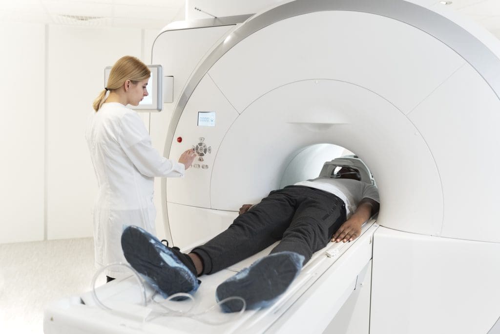
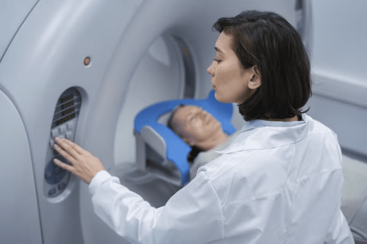
During the scan, you’ll lie on a table that slides into the PET scanner. The machine will pick up signals from the tracer. This helps create detailed images of your body’s inside. The whole process is painless and usually takes 30 to 60 minutes.
After the scan, you can go back to your usual activities unless your doctor says not to. A radiologist will look at the images, and your doctor will talk to you about the results.
Duration and Comfort Considerations
The pet scan duration is usually 30 to 60 minutes. To be comfortable, you’ll lie on a padded table in a room kept at a good temperature. Wear comfy clothes and avoid metal items.
To feel less anxious and more comfortable, try relaxation techniques. Deep breathing or listening to calming music can help, if allowed by the facility.
Knowing about the pet scan procedure can make you feel less anxious. If you have any worries or questions, talk to your healthcare provider.
Interpreting PET Scan Results in Cancer Diagnosis
Understanding PET scan results is key for accurate cancer diagnosis and treatment planning. PET scans show us how active our body’s tissues are. This information is vital for our health.
Understanding SUV Values
SUV stands for Standardized Uptake Value. It measures how much a radioactive tracer is taken up by our body’s tissues. Higher SUV values often mean more metabolic activity, which can be a cancer sign. But, SUV values can be affected by many things, like the cancer type, glucose levels, and when the scan is done.
Inflammation or infection can also raise SUV values. This can lead to wrong conclusions if not looked at with other health info.
False Positives and False Negatives
PET scans are not perfect. False positives happen when a scan shows cancer when there isn’t any. False negatives occur when cancer is there but the scan misses it. Size, location, cancer type, and scanning details can affect these results.
Knowing these limitations is key for correct PET scan results interpretation. Our healthcare team must weigh these factors to make the best decisions for us.
The Role of Radiologists in Interpretation
Radiologists are vital in reading PET scan results. They help tell the difference between harmless and harmful processes. They also check how far the disease has spread and spot any false positives or negatives. Their interpretation is a big part of the diagnosis process, helping doctors plan the best treatment.
By using their radiology skills and looking at all the health info, radiologists help make sure we get the right diagnosis and care.
PET Scans vs. Other Cancer Imaging Techniques
PET scans are just one tool used to find and treat cancer. They work alongside CT scans, MRI, and other methods. Each has its own benefits and drawbacks.
Comparison with CT Scans
CT scans show the body’s structure, like tumor size and location. PET scans, on the other hand, reveal how cells work. PET scans are great for spotting cancer spread and checking treatment success.
CT scans are good for seeing structural changes. PET scans catch changes at the cell level. Often, PET-CT scans are used together for a full picture.
Comparison with MRI
MRI is a strong tool for soft tissue imaging. It’s best for cancers in the brain and spine. MRI doesn’t use radiation, making it safer for some patients.
But MRI might not show cancer spread or tumor activity as well as PET scans. The choice between MRI and PET scans depends on the situation. For example, MRI is key for brain and spinal cord imaging, while PET scans check for cancer spread.
When to Use Different Imaging Modalities
The right imaging tool depends on the cancer type, stage, and patient health. For example, PET scans are key in staging lymphoma and lung cancer. They show how active the cancer is and where it’s spread.
- PET scans are top for finding metastasis and checking treatment.
- CT scans are best for detailed anatomy and biopsies.
- MRI is chosen for soft tissue and specific areas.
Knowing each tool’s strengths helps doctors choose the best imaging for each patient.
Limitations of PET Scans in Cancer Detection
PET scans are a key tool in finding cancer, but they have their limits. It’s important to know these limits to understand results and make good decisions for patients.
Technical Constraints
PET scans use advanced tech and radioactive tracers to spot cancer. But, technical constraints can make them less accurate. For example, they might miss small tumors because of their resolution.
- The sensitivity of PET scanners can vary between different models and manufacturers.
- Image reconstruction algorithms can influence the quality and accuracy of PET scan results.
- Technical issues during the scan, such as patient movement, can compromise image quality.
Patient-Related Factors
Patient-related factors also affect how well PET scans work. Blood sugar levels, for instance, can change how the tracer is taken up, impacting image quality.
Other factors include:
- Body size and composition, which can affect the distribution and uptake of the tracer.
- Certain medical conditions, such as diabetes, which can influence glucose metabolism and tracer uptake.
- Recent medical procedures or treatments that may alter the metabolic activity of tissues.
Also, access can be limited by:
- The availability of PET scan facilities in certain geographic regions.
- The need for specialized equipment and trained personnel to administer and interpret PET scans.
We need to think about these limits when using PET scans for cancer detection. Knowing the challenges helps healthcare providers use this tool better.
Radiation Exposure and Safety Concerns
PET scans use radiation, which raises safety concerns. We need to look closely at the risks and benefits, mainly for cancer patients. Understanding these is key.
Quantifying Radiation Dose
The dose from a PET scan is measured in millisieverts (mSv). The dose varies based on the tracer and scan protocol. For example, a F-FDG PET scan might give 7-10 mSv.
Background radiation is about 3 mSv a year. So, getting multiple scans can add up. It’s important to only do scans when needed to avoid too much radiation.
Risk-Benefit Analysis for Cancer Patients
PET scans are often very helpful for cancer patients. They help doctors plan treatments and check how well treatments are working. This can be very important for patients.
But, we must think about each patient’s situation carefully. We look at their age, the type of cancer, and if there are safer options. For younger patients or those with a good chance of recovery, we might choose safer scans.
Choosing to do a PET scan is a big decision. We weigh the benefits of knowing more about the cancer against the risks of radiation. By using the least amount of radiation needed, we aim to help patients while keeping them safe.
Advances in PET Scan Technology for Cancer Detection
PET scanning is getting better for finding cancer early and tailoring treatments. New tech is making PET scans more effective in fighting cancer.
New tracers are being developed to spot cancer cells better. For example, Fluorothymidine (FLT) checks how fast cells are growing. This helps doctors see how aggressive tumors are.
New Tracers Beyond FDG
Scientists are finding new tracers to make PET scans more accurate. Some examples include:
- Fluoromisonidazole (FMISO): Finds areas in tumors that don’t get enough oxygen, which can make them hard to treat.
- Fluoroestradiol (FES): Helps find estrogen receptors in breast cancer, which is important for treatment.
- PSMA-targeting tracers: Spot prostate cancer when other tests can’t.
These new tracers are making PET scans better at diagnosing and planning treatments.
Improved Resolution and Sensitivity
New PET scanners can see smaller tumors and learn more about how they work. This is thanks to better tech and hybrid scans like PET-CT and PET-MRI. They mix PET’s info on how cells work with CT or MRI’s body maps.
| Feature | Traditional PET | Advanced PET |
| Resolution | Lower | Higher |
| Sensitivity | Moderate | Higher |
| Tracer Variety | Limited (mainly FDG) | Diverse (multiple tracers available) |
AI and Machine Learning Applications
AI and Machine Learning are changing PET imaging. AI can spot things in scans that humans might miss. This means faster and more accurate diagnoses.
AI in PET scans is used in many ways, like:
- Image Segmentation: AI makes it easier to see where tumors start and end.
- Quantification: AI helps measure how much tracer is in cells, giving more precise numbers.
- Predictive Analytics: AI looks at scan data to guess how well a patient will do and how they’ll react to treatment.
As we keep improving PET scan tech, we’re getting closer to better cancer care. The future of cancer detection and treatment looks bright with these new tools.
When Doctors Recommend a PET Scan for Cancer
PET scans are key in cancer care. Doctors suggest them in important situations. Knowing when and why helps patients understand their cancer journey better.
Initial Diagnosis Scenarios
At first cancer diagnosis, a PET scan might be suggested. It’s often for cancers that spread quickly. PET scans show the cancer’s stage, helping plan treatment.
They can also find the cancer’s main source if it’s spread. This is key for choosing the right treatment.
Treatment Monitoring Applications
After starting treatment, PET scans check how the cancer responds. They compare before and after treatment scans. This helps doctors adjust treatment for the best results.
PET scans also watch for cancer coming back. This allows for quick action if needed.
Recurrence Detection
For those who’ve finished treatment, PET scans watch for cancer coming back. Early detection means quicker treatment, improving chances of success.
Some cancers need regular PET scans to catch recurrence early. This gives patients and their families peace of mind.
Understanding PET scans in cancer care shows their importance. They help manage the condition effectively.
Conclusion: The Role of PET Scans in Modern Cancer Care
PET scans are key in modern cancer care. They give important info for diagnosis, treatment planning, and tracking. We’ve learned how they show cancer metabolism, guiding the best treatment plans.
In today’s cancer diagnosis, PET scans are a must-have. They help doctors spot cancer early, check how treatments work, and find signs of cancer coming back. Using PET scans with CT scans makes them even better at finding problems.
As PET scan tech gets better, so will cancer care. New tracers and imaging methods will make PET scans even more useful. This will lead to better care for patients everywhere.
In short, PET scans are essential in modern cancer care. Their growth will help shape the future of cancer treatment. By using PET scans, we can offer more effective and caring care for patients all over the world.
FAQ
What is a PET scan, and how does it work in cancer detection?
A PET (Positron Emission Tomography) scan uses a radioactive tracer to find active areas in the body. It looks for where cells are using a lot of glucose, which cancer cells do. This helps doctors spot cancer cells.
What types of cancers can be detected using PET scans?
PET scans are good for finding lung, colorectal, and lymphoma cancers. They work best on cancers that use a lot of energy.
How do PET scans compare to other imaging techniques like CT and MRI?
PET scans show how active cells are. CT scans give detailed body pictures. MRI shows both body structure and activity. The right choice depends on the situation.
What is the “dinner glow” phenomenon, and how does it affect PET scan results?
The “dinner glow” is when eating increases glucose in the stomach. This can make PET scans harder to read. Doctors often ask patients to fast before a scan.
How should I prepare for a PET scan?
Before a PET scan, you might need to fast and avoid exercise. Tell your doctor about any medicines you’re taking.
What can I expect during a PET scan procedure?
During a PET scan, you’ll get a tracer injection and lie on a table in a scanner. It’s usually painless but you must stay very quiet. The time needed varies by scan type.
How are PET scan results interpreted, and what do SUV values signify?
Radiologists look at how much tracer is taken up in tissues. SUV values show how much, with higher numbers often meaning aggressive cancer.
What are the risks associated with radiation exposure during a PET scan?
PET scans use low amounts of radiation, but it’s important to consider the risks. Your doctor will talk about the benefits and risks with you.
Are there any advancements in PET scan technology that improve cancer detection?
Yes, new tracers and better technology are being developed. These advancements help find cancer better and faster, thanks to AI and machine learning.
When do doctors recommend a PET scan for cancer diagnosis or treatment monitoring?
Doctors use PET scans for diagnosing cancer, checking treatment success, and finding cancer return. The choice depends on the cancer type, stage, and patient health.


