Last Updated on October 22, 2025 by mcelik

Diagnosing and staging cancer often involves PET scans. These scans help find areas with high metabolic activity, like cancer cells. Cancerous lymph nodes can be hard to spot, but PET scans are very helpful.
A PET scan uses a radioactive tracer to show areas with high activity. While PET scans are great for finding cancer, like non-Hodgkin’s lymphoma, there’s a worry about radiation exposure. It’s important for patients to know how to reduce this risk.

PET scans give us a peek into how cancer cells work. They help doctors figure out what treatment to use. To get why PET scans are key in finding cancer, we need to know how they work.
A PET scan is a special imaging test. It uses a radioactive tracer to see how active the body’s cells are. Unlike other tests, PET scans show how cells work, not just their shape.
The tracer used most often is fluorodeoxyglucose (FDG). It’s a sugar molecule with a radioactive tag. Cancer cells eat more of this sugar than normal cells, making them easy to spot on PET scans.
Getting a PET scan involves a few steps. First, you get a shot of FDG. This tracer goes through your body, focusing on active areas. Then, you wait for about an hour before being scanned.
The scanner picks up the gamma rays from the tracer. It uses this info to make detailed images of your body’s activity. A radiologist then looks at these images to find any signs of cancer.
While FDG-PET is the most used, other tracers are used for different cancers. Some scans use tracers that target specific cancer cells or check other body processes.
| Tracer Type | Application | Metabolic Process Assessed |
| FDG (Fluorodeoxyglucose) | General Cancer Detection | Glucose Metabolism |
| FLT (Fluorothymidine) | Cancer Cell Proliferation | DNA Synthesis |
| F-MISO (Fluoromisonidazole) | Hypoxia Imaging | Oxygen Metabolism |
Knowing how PET scans work is key to understanding their role in fighting cancer. They help doctors find cancerous tissues, like in lymph nodes. This is important for planning the right treatment.

PET scans work by using special tracers and how cells use energy. They are a key tool in finding cancer. This is because cancer cells use energy differently than healthy cells.
PET scans use a special fact about cancer cells. They use more glucose than normal cells. Radioactive tracers that act like glucose help show this.
The most used tracer is Fluorodeoxyglucose (FDG). It goes to cells that use a lot of glucose. This helps doctors see where cancer might be.
FDG is the top choice for PET scans. It shows where cells are using a lot of glucose. Cancer cells use more glucose, so FDG is great for finding them.
The PET scanner makes images by detecting gamma rays from the tracer. These images show where cells are using a lot of energy. Doctors then look at these images to find cancer.
Doctors use SUV (Standardized Uptake Value) to see how active tissues are. A high SUV means a tissue is very active, like cancer.
Understanding PET scans helps doctors fight cancer better. These scans use special tracers and technology to see how cells work. This is a powerful way to find and track cancer.
The lymphatic system is key to our body’s defense. It’s important to understand how it works for cancer detection. This system is a network of organs, tissues, and vessels that fight off infections and diseases.
The lymphatic system includes lymph nodes, lymph vessels, the spleen, and lymphoid tissues. It filters out harmful substances and cells, like cancer cells. Lymph nodes are vital, acting as filters that catch pathogens and abnormal cells.
Cancer cells can move from the primary tumor to nearby lymph nodes through the lymphatic system. This is called metastasis. Once in the lymph nodes, they can grow and form new tumors.
Checking lymph nodes is key to knowing how far cancer has spread. This helps doctors plan the best treatment and predict the outcome. Doctors use imaging tests, like PET scans, and biopsies to assess lymph nodes.
PET scans can spot cancerous lymph nodes because cancer cells eat more glucose than regular cells. They use a radioactive glucose tracer, like FDG, which builds up in areas with lots of activity, like cancer.
Cancerous lymph nodes show up on PET scans because they use more glucose. This is because cancer cells need more glucose to grow. So, these nodes take up more FDG, showing as “hot spots” on the scan.
SUV (Standardized Uptake Value) measures how much glucose lymph nodes take up. High SUV values mean more glucose uptake, which often means cancer.
| SUV Value Range | Interpretation |
| 0-2.5 | Typically considered normal or benign |
| 2.5-4.0 | May indicate inflammation or low-grade malignancy |
| >4.0 | Often associated with high-grade malignancy |
PET scans are great for finding cancerous lymph nodes, but they’re not perfect. Inflammation can also make lymph nodes show up as active, leading to false positives. Doctors need to look at the whole picture, including the patient’s history and other tests, to make sure.
It’s tricky to tell if a lymph node is cancerous or just inflamed. Clinical correlation and more tests are often needed to be sure about what the PET scan shows.
PET scans are key for cancer diagnosis but come with radiation risks. It’s important for patients to know about these risks and the steps to reduce them.
PET scans use a radioactive tracer to see how the body works. The dose of radiation depends on the tracer and the patient’s health. On average, an adult gets 4 to 7 millisieverts (mSv) of radiation.
Radiation Exposure Levels: The dose can change based on the tracer, the patient’s size, and the scan’s details.
Comparing PET scan radiation to other tests helps understand it better. The dose from a PET scan is similar to many other imaging methods.
| Imaging Procedure | Average Radiation Dose (mSv) |
| PET Scan | 4-7 |
| CT Scan (Abdomen and Pelvis) | 10-20 |
| X-ray (Chest) | 0.1 |
| Mammogram | 0.4 |
The table shows PET scans have radiation, but the dose is similar to other tests.
Risks from PET scan radiation differ for each patient. Age, health, and other conditions play a big role.
Patient-Specific Risk Factors: Kids, pregnant women, and people with certain health issues need extra care. They might be more sensitive to radiation.
Doctors look at each patient’s situation to make sure the scan’s benefits are worth the risks.
Getting ready for a PET scan is key to getting good results. It’s important to prepare well to get clear and useful information. This helps doctors diagnose and plan treatments better.
Before a PET scan, you need to follow certain diet rules. Fasting for 4 to 6 hours is usually needed. This helps make the scan images clearer.
Also, avoid sugary foods and drinks before the scan. They can change how your body uses glucose, affecting the scan results.
Drink plenty of water before the scan. But, always check with your doctor about how much water to drink.
Tell your doctor about all the medicines you’re taking. Some medicines might need to be changed or stopped before the scan. For example, diabetic medications that affect blood sugar need careful management.
It’s very important to follow your doctor’s advice on managing your medicines. This ensures the PET scan is safe and effective.
On the day of the scan, wear comfortable, loose-fitting clothes without metal parts. Metal can interfere with the scan. Also, avoid jewelry or clothes with metal fasteners.
Bring any important medical records, like previous scans and a list of medicines. You’ll also need to bring identification and insurance information.
The PET scan procedure is complex but essential for finding cancerous cells. It has several steps that patients need to know for a smooth scan.
When you arrive, you’ll fill out paperwork and change into a hospital gown. This is to remove any metal objects that could mess with the scan.
Initial preparation also includes getting an intravenous (IV) line. This line is for the radioactive tracer injection.
The next step is the injection of the radioactive tracer through the IV line. This tracer is a special glucose that cancer cells take up quickly.
After the injection, there’s a waiting period, called the uptake period. It can last from 30 minutes to an hour. During this time, the body absorbs the tracer.
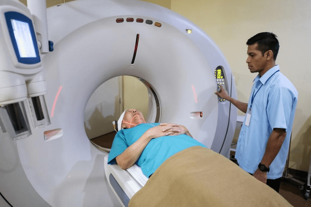
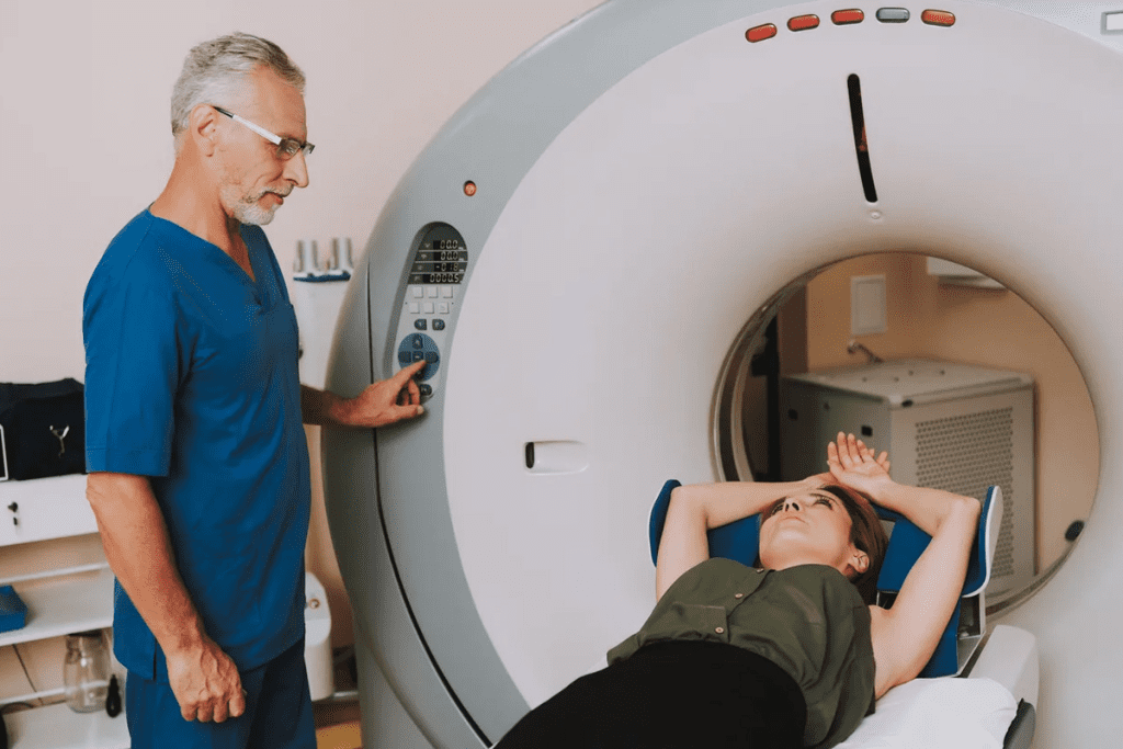
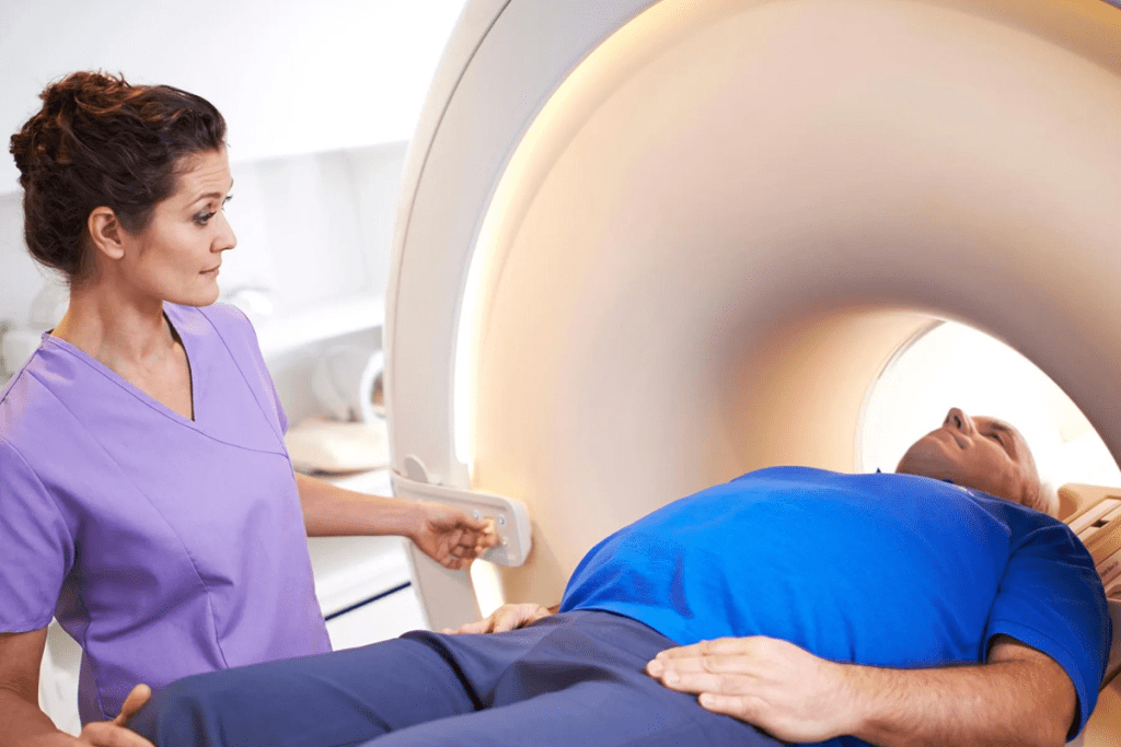
After the waiting period, you’ll go to the PET scanner. The scanning process has you lying on a table that moves through the scanner. It detects the radiation from the tracer.
The scan is painless and takes 30 to 60 minutes. It depends on the body area being scanned.
| Step | Description | Duration |
| Arrival and Preparation | Completing paperwork, changing into a hospital gown, and IV insertion | 15-30 minutes |
| Tracer Injection and Uptake | Injection of radioactive tracer and waiting for it to be absorbed | 30-60 minutes |
| Scanning Process | Lying on a table that moves through the PET scanner | 30-60 minutes |
Medical facilities take many steps to keep everyone safe during PET scans. They aim to reduce radiation exposure for patients, staff, and the environment.
Medical facilities stick to strict rules to lower radiation exposure. They use the least amount of radioactive tracer needed for each scan.
Key strategies include:
Protective gear is key to reducing radiation exposure. This includes:
Training staff is vital for radiation safety. Those working with PET scans learn a lot. They get trained on:
| Training Component | Description |
| Radiation Safety Principles | Understanding the risks associated with radiation and methods to minimize exposure. |
| Handling Radioactive Materials | Proper techniques for handling, storing, and disposing of radioactive tracers. |
| Emergency Procedures | Protocols to follow in case of accidental radiation exposure or spills. |
After a PET scan, there are important steps to follow for safety and comfort. It’s key to get rid of the radioactive tracer and get back to normal life.
Drink lots of water to get rid of the tracer. Also, remember to:
Hydration is key in getting rid of the radioactive material. Drinking lots of water helps remove the tracer, lowering body radiation. Aim for 6-8 glasses of water in 24 hours after the scan.
| Hydration Tips | Benefits |
| Drink plenty of water | Flushes out radioactive tracer |
| Avoid caffeinated beverages | Reduces side effects |
| Monitor urine output | Ensures tracer elimination |
Most can go back to normal activities right after the scan, unless told not to. But, it’s wise to:
By following these tips, patients can have a safe and easy recovery after their PET scan.
It’s important to know the rules for PET scans in special groups. These rules help keep everyone safe during the scan. Some people need extra care because of their health.
Pregnant or breastfeeding women should be careful with PET scans. The scans use radioactive tracers that might harm the baby. The risk depends on how far along the pregnancy is and the tracer used.
Women who are pregnant or breastfeeding should tell their doctor before a PET scan. Sometimes, other tests are suggested to lower the risk.
Children need extra care with PET scans because they are more sensitive to radiation. The tracer dose is adjusted for the child’s age, weight, and health issues.
PET scans in kids are only done when the benefits are clear. Steps are taken to reduce radiation exposure.
People with diabetes or kidney disease should be careful before a PET scan. Diabetic patients need to control their blood sugar. High sugar levels can mess up the scan results.
For those with kidney problems, the tracer or contrast might harm the kidneys. Drinking lots of water and being closely watched can help.
Following these guidelines helps doctors use PET scans safely for all patients.
Radioactive tracers in PET scans are key for cancer diagnosis. Yet, they’re filled with myths and misconceptions. It’s vital for patients to know the real risks and benefits of these tracers.
Many people, including some healthcare workers, think PET scans are very dangerous. They believe the radiation can harm the body a lot. But, the truth is, the radiation from PET scans is low and controlled.
Key facts to consider:
The risk of radiation from PET scans is a worry, but it’s important to understand it. Research shows PET scans help diagnose and manage cancer more than they harm.
It’s worth noting that:
Long-term safety is key when it comes to radioactive tracers. Studies show the risks from PET scan radiation are small when used right and with the right patients.
Long-term safety measures include:
PET scan results are key for checking lymph node involvement. It’s important to know what “lighting up” means, the chance of false positives and negatives, and the value of combining PET with other scans.
When lymph nodes “light up” on a PET scan, it means they are more active. This can be a sign of cancer. But, it’s not the only reason. Inflammation or infection can also make lymph nodes show up more.
Increased uptake of the radioactive tracer in lymph nodes is measured using Standardized Uptake Values (SUV). Higher SUV values often mean a higher chance of cancer. But, it’s not a sure sign. The doctor must look at the SUV values with the patient’s overall health in mind.
False positives happen when non-cancerous conditions make lymph nodes look active. False negatives occur when cancerous lymph nodes don’t show up because they’re too small or not very active.
Things like inflammation, infections, or recent surgery can lead to false positives. False negatives can happen because of small tumors or certain cancers that don’t show up well on PET scans.
To get a better look at lymph nodes, PET scans are often used with CT or MRI. This combination gives a clearer view of the lymph nodes and the area around them.
PET/CT fusion is very helpful. It mixes the metabolic info from PET with the detailed images from CT. This helps doctors spot and understand lymph node involvement better, leading to more accurate treatment plans.
Understanding PET scan results and using them with other tools helps doctors make better choices for patient care.
PET/CT fusion combines PET scans’ functional info with CT scans’ anatomical details. This gives a clearer view of lymph node involvement in cancer.
PET/CT fusion imaging has many benefits for checking lymph nodes. Its main advantage is combining metabolic and anatomical info. This makes it easier to spot and stage cancerous lymph nodes accurately.
By merging PET and CT scans, we can pinpoint active lymph nodes. This is key for planning surgeries or radiation therapy.
PET/CT fusion is great for diagnosis but involves radiation from both PET and CT. The total radiation dose is a big deal, mainly for those needing many scans.
Research shows PET/CT fusion’s radiation is similar to or a bit more than PET or CT alone. But the better accuracy makes the extra radiation worth it.
| Imaging Modality | Typical Radiation Dose (mSv) |
| PET Scan | 5-10 |
| CT Scan | 2-10 |
| PET/CT Fusion | 7-20 |
PET/CT fusion’s combined info boosts lymph node staging accuracy. This is vital for picking the right treatment and predicting how well a patient will do.
With a better grasp of lymph node involvement, PET/CT fusion helps tailor cancer treatment. This could lead to better results for patients.
Recent breakthroughs in PET scan technology are changing how we fight cancer. They focus on cutting down radiation exposure. The field of medical imaging has seen big changes, making patients safer while keeping diagnosis accurate.
The newest PET scanners use tech for lower radiation doses without losing image quality. They have advanced detector materials and smart algorithms for this purpose.
One big step is the high-sensitivity PET scanners. They can spot smaller amounts of radioactive tracer. This means patients get less radiation.
Scientists are looking for new tracers that need lower radiation doses. Some of these tracers are showing great results in tests. They could lead to less radiation for patients in the future.
Research on non-FDG tracers is also underway. These tracers might be used for certain cancers or conditions. They could mean less radiation for patients than traditional FDG PET scans.
AI and machine learning are key in making PET scans safer. They help figure out the least radiation needed for a good scan, based on the patient.
AI can also help make images better with lower doses. This mix of AI and PET tech is a big leap for patient care.
PET scans have changed how we diagnose and manage cancer. They offer big benefits but also come with risks from radiation. It’s important to weigh these carefully.
To use PET scans safely and effectively, we need to understand them well. Knowing how they work, how to prepare, and safety steps can help reduce radiation exposure.
Following certain rules before and after a PET scan can make it safer. This includes eating right, taking your medicine as told, and following after-care instructions. Hospitals also have a big role in keeping radiation low with the right equipment and rules.
New technology in PET scans is making them safer. Improvements in design and new tracers are cutting down on radiation risks. Using PET scans with other imaging and AI can make them even more accurate and safe.
In the end, PET scans are a key part of cancer care because of their benefits. Knowing the risks and how to lessen them helps both patients and doctors get the best results.
To reduce radiation, patients should follow dietary rules before the scan. They should also drink lots of water after to get rid of the tracer. It’s best to avoid being close to pregnant women, breastfeeding moms, and kids for a while after the scan.
A PET scan uses a small amount of radioactive tracer, like FDG, injected into the body. This tracer goes to areas with lots of activity, like cancer cells. The PET scanner then picks up the positrons to make detailed images.
Lymph nodes are key in finding cancer. They are where cancer often spreads first. Checking them helps figure out how far the cancer has gone and is important for treatment plans.
Cancerous lymph nodes show up on PET scans because they use more glucose. This means they take in more of the radioactive tracer FDG, making them stand out.
SUV values measure how much tracer a tissue takes in. They help doctors see how active a lesion is. Higher SUV values often mean more aggressive cancer.
To get ready for a PET scan, follow dietary rules and don’t exercise too much. Tell your doctor about any meds or health issues you have.
PET scans use ionizing radiation, which might slightly increase cancer risk. But, the scans are very helpful in finding and managing cancer, so the benefits usually outweigh the risks.
The tracer from PET scans leaves the body in a few hours to days. Drinking water helps get rid of it faster. This depends on how much you drink and your kidney health.
After a PET scan, avoid being close to pregnant women and kids for a bit. Also, drink lots of water to get rid of the tracer.
PET scans are not recommended for pregnant women because of radiation risks. For breastfeeding moms, it’s best to stop nursing for a while after the scan.
PET/CT fusion imaging combines PET’s function info with CT’s anatomy. This makes finding and staging lymph nodes more accurate by showing the body’s internal details.
New PET scanner tech, tracers with lower doses, and AI are being developed. These aim to cut down radiation exposure in PET scans.
Subscribe to our e-newsletter to stay informed about the latest innovations in the world of health and exclusive offers!