Last Updated on November 27, 2025 by Bilal Hasdemir

Understanding the risks and benefits of medical imaging starts with comparing the radiation dose panoramic dental X ray to other imaging procedures. At Liv Hospital, we prioritize patient safety and trust, providing clear and accurate information to help patients make informed decisions about their care.
A panoramic dental X ray delivers a relatively low radiation dose, roughly equivalent to a few days of natural background radiation. In contrast, a CT scan of the abdomen exposes patients to a much higher radiation dose, comparable to several years of background exposure.
By knowing the radiation dose panoramic dental X ray and comparing it with other scans, patients can better understand the safety and necessity of different imaging tests. Liv Hospital provides expert guidance to ensure that imaging is used safely and effectively.
Key Takeaways
- Panoramic dental X-rays have a low radiation dose, typically ranging from 4 to 30 microsieverts.
- Abdominal CT scans expose patients to a substantially higher radiation dose, often 8–10 millisieverts.
- The average annual radiation exposure from natural sources is about 3 millisieverts.
- A panoramic dental X-ray is equivalent to 2-3 days of natural background radiation.
- An abdominal CT scan is comparable to 2-3 years of background radiation exposure.
Understanding Radiation Measurement in Medical Imaging
Medical imaging uses radiation, which is important to know about. Techniques like X-rays and CT scans use ionizing radiation. This has benefits for diagnosis but also risks. Accurate dose measurement is key.
How Radiation Doses Are Quantified
Radiation doses are measured in specific units. The main unit is the millisievert (mSv). Other units include rad, rem, roentgen, sievert, and gray. The millisievert helps us understand the risk of different doses.
For example, a chest X-ray is like 10 days of natural background radiation. This helps patients see the risks of different scans.
The Significance of Millisieverts and Microsieverts
Millisieverts (mSv) and microsieverts (μSv) measure radiation doses. Millisieverts are for bigger doses, like CT scans. Microsieverts are for smaller doses, like dental X-rays. Knowing these units helps compare different scans.
A panoramic dental X-ray has 4-30 microsieverts. A CT abdomen scan has 8-10 millisieverts. This shows the big difference in radiation levels from different scans.

Measuring doses in millisieverts and microsieverts helps us understand risks. This knowledge helps us make better choices about medical imaging.
Radiation Dose Panoramic Dental X Ray: Surprisingly Low Exposure
When it comes to dental imaging, panoramic dental X-rays are key. They expose patients to a very low dose of radiation. Many wonder about X-ray safety, and it’s good to know panoramic dental X-rays are safe and useful.

Typical Dose Range: 4-30 Microsieverts
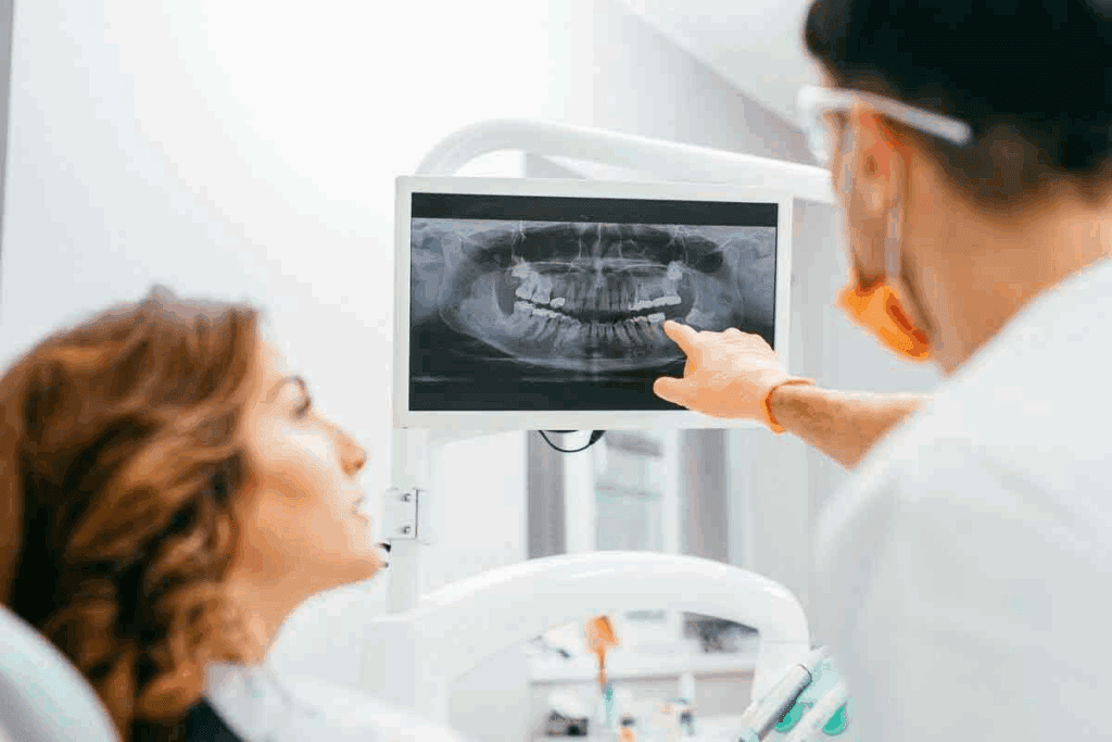
The radiation dose from a panoramic dental X-ray is between 4 to 30 microsieverts (µSv). For comparison, a single digital dental X-ray gives about 0.2 microsieverts (µSv) of radiation. Full-mouth X-rays are about 3.9 µSv, similar to a few days of natural background radiation.
- A panoramic dental X-ray dose is like a few days of natural radiation exposure.
- The dose range is generally considered safe for patients.
- Digital X-ray technology has significantly reduced radiation exposure compared to traditional film-based X-rays.
Equivalent to Just Days of Natural Background Radiation
To understand the low dose of radiation from panoramic dental X-rays, think about background radiation. The average person gets about 2.4 millisieverts (mSv) of background radiation yearly, or roughly 6.6 µSv daily. So, a panoramic dental X-ray with a dose of 4-30 µSv is like just a few days to a couple of weeks of natural background radiation.
So, the radiation dose from panoramic dental X-rays is quite low. This makes them a safe and valuable tool in dentistry.
CT Abdomen Scans: Substantially Higher Radiation Levels
CT scans of the abdomen expose patients to a lot of radiation. This is important to think about in medical imaging. The high dose is needed to see the inside of the abdomen clearly.
8 to 10 Millisieverts: Understanding the Standard Dose Range
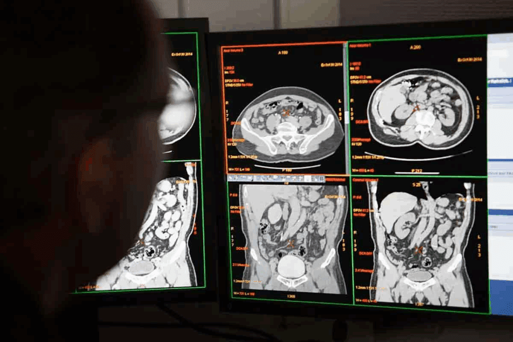
A CT abdomen scan usually has a radiation dose of 8 to 10 millisieverts (mSv). This is like getting 2.6 years of natural background radiation at once. If you have more scans, the dose can go up even more.
Knowing the radiation dose is key, as radiology guidelines tell us. It helps us understand the risks of CT scans.
The Necessity of Higher Radiation Doses in CT Scans
So, why do CT scans of the abdomen need so much radiation? It’s because they need to show a lot of detail. They have to see all the organs and any problems inside the abdomen. This requires a lot of radiation to get clear images.
Even though the dose is high, CT scans are very helpful. They are important in emergencies or for tracking complex conditions. The goal is to find the right balance between getting good images and keeping radiation low.
Comprehensive X-Ray Radiation Dose Chart and Comparison
It’s important to know how different medical imaging procedures affect radiation exposure. We’ll look at the doses for panoramic dental X-rays, CT abdomen scans, chest X-rays, and extremity X-rays.
Panoramic Dental X-Ray vs. CT Abdomen: Side-by-Side Analysis
A panoramic dental X-ray gives a dose of 4-30 microsieverts. A CT abdomen scan, on the other hand, has a dose of 8-10 millisieverts. This means a panoramic dental X-ray is like 2-3 days of natural background radiation. A CT abdomen scan is like 2-3 years of background radiation.
Choosing the right diagnostic tool is key because of these big differences in radiation exposure.
How These Compare to Chest X-Rays (CXR) and Extremity X-Rays
Chest X-rays have a low dose of about 0.1 millisieverts, similar to 10 days of natural background radiation. Extremity X-rays have even lower doses, around 0.001 millisieverts per image. These comparisons help us understand the risks of different imaging procedures.
Visualizing Radiation Exposure in Context of Daily Life
It’s easier to grasp these doses when we compare them to daily life. For example, a panoramic dental X-ray’s radiation is like what we naturally get in a few days. A CT abdomen scan’s dose is like several years of natural background radiation.
By comparing these doses and seeing them in terms of natural background radiation, we can make better choices about X-ray and CT imaging.
Modern Technology and Radiation Safety Advancements
Modern tech has changed how we handle radiation in medical imaging. We’ve made big steps in digital dental imaging and CT scans. These changes help lower the amount of radiation patients get. It’s all about finding the right balance between getting the info we need and keeping patients safe.
Digital Dental Imaging: Reducing Exposure Further
Digital dental imaging has moved us away from old film X-rays. Now, we use digital X-rays that use much less radiation. This is thanks to better sensors and tech that make images clearer with less exposure.
Some big pluses of digital dental imaging are:
- Lower radiation doses
- Instant image acquisition
- Enhanced image quality
- Easier image storage and transfer
CT Dose Reduction Techniques and Their Impact
CT scans are key in medical diagnosis but use more radiation than X-rays. To fix this, makers have come up with ways to cut down on radiation. These include:
- Automatic Exposure Control (AEC)
- Iterative Reconstruction (IR)
- Tube Current Modulation
These tools have greatly cut down the radiation from CT scans. For example, Iterative Reconstruction lets us use lower doses without losing image quality.
| Imaging Modality | Typical Radiation Dose |
| Panoramic Dental X-Ray | 4-30 microsieverts |
| CT Abdomen | 8-10 millisieverts |
| Digital Dental X-Ray | <1-5 microsieverts |
| Low-Dose CT Abdomen | 4-6 millisieverts |
Conclusion: The ALARA Principle and Balancing Diagnostic Benefits
The ALARA principle is key in medical imaging. It helps balance the need for clear images with the goal of reducing radiation. Medical experts aim to keep doses low while getting the needed information.
Even though the risk of cancer from medical imaging is very low, it’s wise to follow the ALARA principle. This ensures safety. The benefits of imaging, like accurate diagnoses and treatment plans, are usually greater than the risks.
Following the ALARA principle helps us reduce radiation exposure while keeping diagnostic benefits high. This shows our dedication to safety and quality in medical imaging.
FAQ
How is radiation dose measured in medical imaging?
Radiation dose is measured in millisieverts (mSv) and microsieverts (μSv). 1 mSv is equal to 1,000 μSv. These units help us understand how much radiation we get from medical imaging.
What is the typical radiation dose from a panoramic dental X-ray?
A panoramic dental X-ray has a low radiation dose. It ranges from 4 to 30 μSv. This is as little as a few days of natural background radiation.
How much radiation is involved in a CT abdomen scan?
A CT abdomen scan has a much higher radiation dose. It’s usually between 8 to 10 mSv. This is hundreds of times more than a panoramic dental X-ray.
Why do CT scans require more radiation than X-rays?
CT scans need more radiation because they show detailed images of internal organs. This requires a higher dose to get the quality needed for diagnosis.
How does the radiation dose from a chest X-ray compare to other X-ray procedures?
A chest X-ray has a low radiation dose, about 0.1 mSv. This is much lower than a CT abdomen scan but higher than a panoramic dental X-ray.
What advancements have been made to reduce radiation exposure in medical imaging?
New technologies like digital dental imaging and CT dose reduction have lowered radiation exposure. These advancements improve image quality while using less dose.
What is the ALARA principle in medical imaging?
The ALARA principle means “As Low As Reasonably Achievable.” It’s about keeping radiation exposure as low as possible while getting the needed diagnostic information. It helps doctors balance the benefits of imaging with the risks of radiation.
How can I understand the radiation dose from different medical imaging procedures?
To understand radiation dose, compare it to natural background radiation or other common procedures. For example, a panoramic dental X-ray is like a few days of natural background radiation. A CT abdomen scan is like several years.
References
- Ujike, C., Honda, E., & Shimoyama, T. (2014). Effective doses from panoramic radiography and cone-beam computed tomography. Oral Radiology, 30(1), 11–18. https://pmc.ncbi.nlm.nih.gov/articles/PMC4082268/


