Last Updated on November 26, 2025 by Bilal Hasdemir
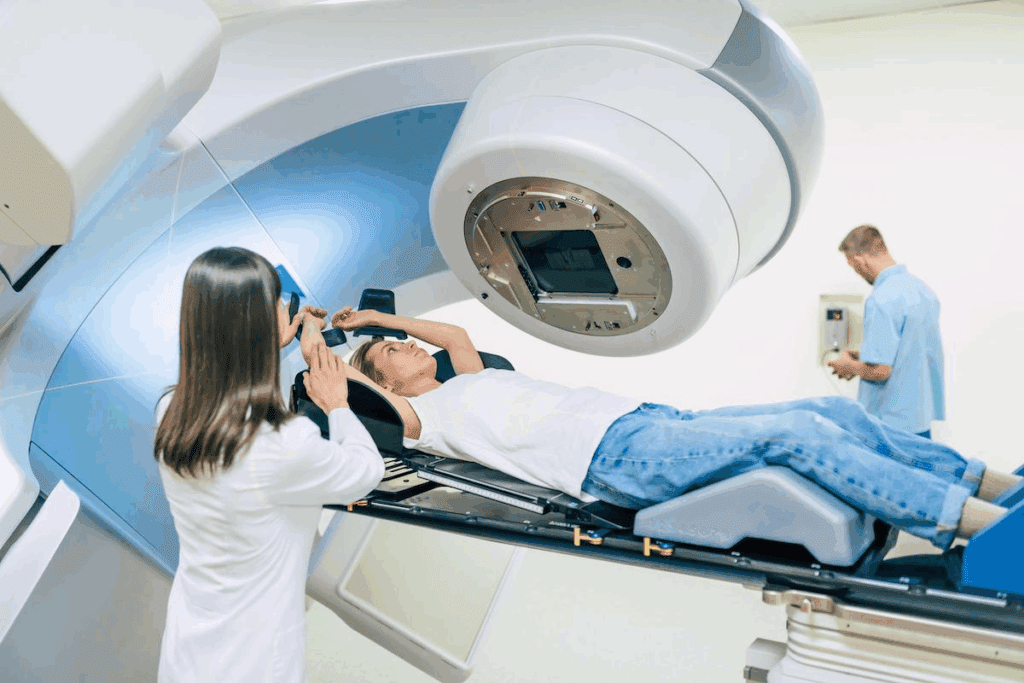
At Liv Hospital, we know how important radiation in medical imaging is for today’s health checks. Every year, over 80 million CT scans are done in the United States. This shows how big a role medical imaging radiation has in finding and treating health issues. How safe is radiation in medical imaging? Discover 7 key facts about the risks, benefits, and safety measures in healthcare.
We see that radiation and medicine go hand in hand. They are used in X-rays, mammography, CT, and nuclear imaging. As a trusted healthcare provider, we make sure patients are safe while using healthcare imaging.
Key Takeaways
- Understanding the role of radiation in medical imaging is key to safer healthcare.
- Over 80 million CT scans are done every year in the United States.
- Radiation is used in many medical imaging methods, like X-rays and nuclear imaging.
- Keeping patients safe is our main goal when using medical imaging radiation.
- Liv Hospital is dedicated to top-notch healthcare with full international patient support.
The Science Behind Radiation in Medical Imaging
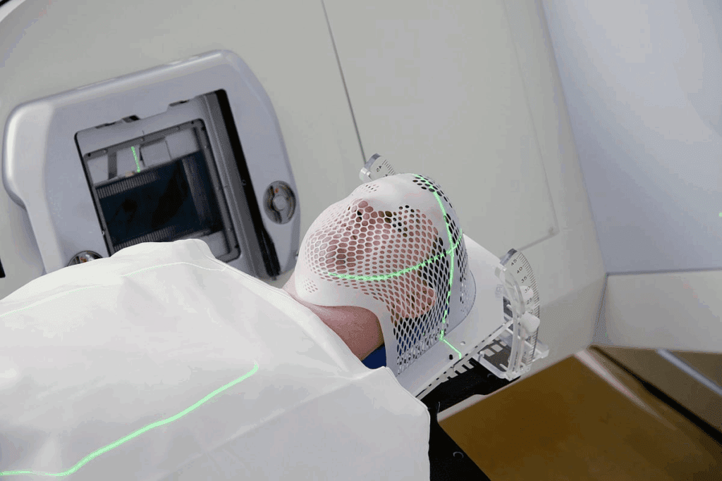
Medical imaging uses a lot of radiation. Knowing the science behind it helps us understand its good and bad sides. The history of using radiation in medicine is long and has changed a lot.
Historical Development of Medical Radiation Technology
Wilhelm Conrad Röntgen discovered X-rays in 1895. This was the start of using radiation in medical imaging.. It led to many new imaging technologies that changed how we diagnose diseases.
Medical radiation technology has grown a lot over time. The 1970s brought us CT scans, and MRI came later. These advancements have made medical imaging even better.
| Technology | Year Introduced | Primary Use |
| X-rays | 1895 | General imaging, bone fractures |
| CT Scans | 1970s | Detailed cross-sectional imaging |
| Mammography | 1960s | Breast cancer screening |
How Different Types of Medical Radiation Work
Medical radiation comes in different forms for different uses. X-rays help see bones and some soft tissues. CT scans give detailed pictures of the body’s inside.
X-rays: They pass through the body. Things like bone absorb more X-rays, making them visible inside.
CT Scans: Mix X-rays with computer tech for detailed body images. They’re great for detailed views.
Nuclear medicine uses tiny amounts of radioactive material to find and treat diseases. PET scans show how the body works by looking at its metabolic activity.
Knowing how these technologies work helps us see their benefits and risks. As medical imaging gets better, radiation will keep playing a big role.
Fact 1: The Prevalence and Importance of Medical Imaging
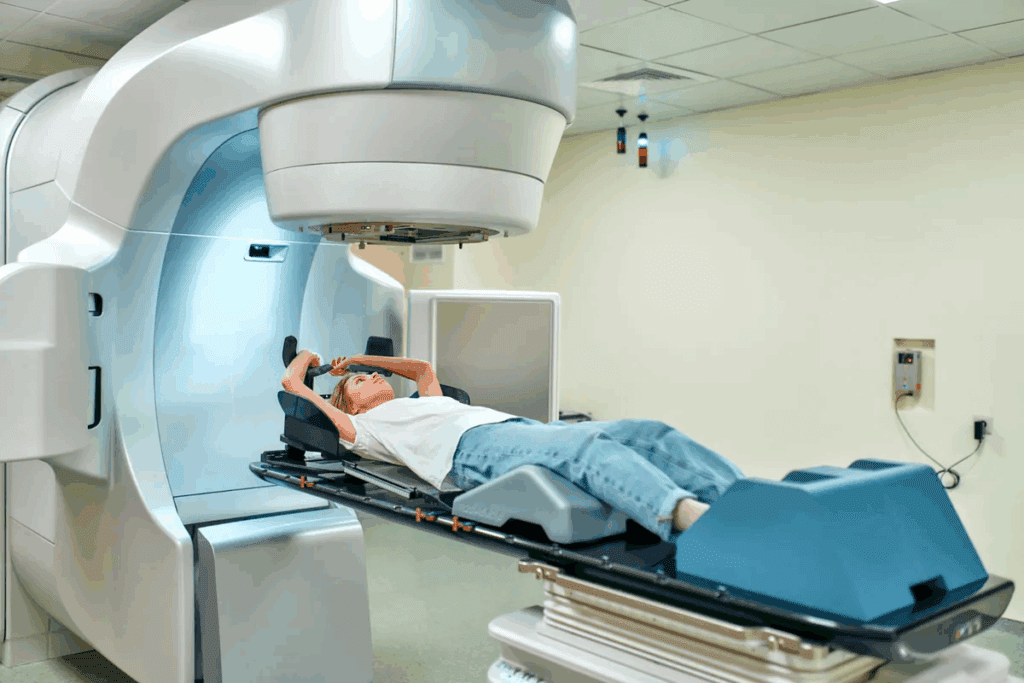
Medical imaging is now a key part of healthcare. It’s used more and more every year. This technology helps doctors diagnose and treat patients better.
Over 80 Million CT Scans Annually in the United States
Every year, over 80 million CT scans are done in the U.S. This shows how much we rely on imaging for health checks. It proves how important CT scans and other imaging are in medicine.
The number of CT scans keeps going up. This is because doctors need quick and accurate ways to diagnose. It shows how vital medical imaging is in healthcare.
The Growing Role of Radiological Testing in Diagnosis
Radiological testing has changed how we diagnose diseases. It lets doctors see inside the body. This helps find problems early and accurately.
“The integration of advanced imaging technologies into clinical practice has significantly enhanced our ability to diagnose and manage a wide range of medical conditions.”
Radiological testing is used in many areas of medicine. It helps find breaks and tumors. It also guides less invasive surgeries. Imaging radiation is key in many important uses.
Learning about radiation in medical imaging is important. It helps both doctors and patients understand its role. By knowing its value, we can see its benefits and challenges.
Fact 2: Applications of Radiation in Medicine
Radiation in medicine has many uses. It helps in several imaging methods that improve patient care. We use it to diagnose and treat many health issues.
X-rays and Digital Radiography
X-rays are key in medicine for seeing inside the body. Digital radiography has made X-rays better and faster.
New X-ray tech offers clearer images and less radiation. This is a big step forward.
Computed Tomography (CT) Scans
CT scans use X-rays to show detailed body images. They help find injuries and diseases.
CT scan tech has improved. Now, scans are quicker and use less radiation. This makes them safer and more useful.
Mammography and Breast Imaging
Mammography uses X-rays for breast imaging. It’s key to finding breast cancer early.
New mammography tech has better image quality. This helps doctors spot problems more easily.
Nuclear Medicine and Molecular Imaging
Nuclear medicine uses tiny amounts of radioactive materials. It helps diagnose and treat diseases by showing how the body works.
Molecular imaging, like PET scans, gives detailed body function. This helps manage complex conditions better.
| Imaging Modality | Primary Use | Radiation Level |
| X-rays | General diagnostic imaging | Low |
| CT Scans | Detailed cross-sectional imaging | Moderate to High |
| Mammography | Breast cancer screening | Low |
| Nuclear Medicine | Functional and metabolic imaging | Varies |
Fact 3: How Radiation in Medical Imaging Revolutionized Healthcare
The use of radiation in medical imaging has changed healthcare a lot. It has made diagnosing and treating diseases much better. Thanks to new radiation technology, we can now see inside the body without surgery.
Minimizing the Need for Invasive Procedures
One big plus of radiation in medical imaging is the reduced need for surgery. X-rays and CT scans let doctors see inside the body without surgery. This makes patients safer and helps them heal faster.
Enhanced Diagnostic Accuracy and Precision
Radiation-based imaging has made diagnosing better. Computed Tomography (CT) scans give clear pictures of the body. This helps doctors find problems early and plan better treatments.
Impact on Treatment Planning and Disease Management
Radiation in medical imaging has also changed how we plan treatments. It gives doctors clear images to make better plans. For example, in cancer treatment, radiation therapy uses these images to hit tumors right on target, protecting healthy tissue.
In short, radiation in medical imaging has greatly improved healthcare. It has reduced the need for surgery, made diagnoses more accurate, and helped in planning treatments. As technology gets better, we can expect even more progress in helping patients.
Fact 4: Comparing Radiation Levels in Different Radiographic Procedures
It’s important to know how much radiation different radiographic procedures use. This helps us see if they are safe and work well. Medical imaging is key for diagnosing and treating diseases, so we need to look at how much radiation each method uses.
We’ll look at how much radiation X-rays and CT scans use. We’ll also compare this to the background radiation we all get. We’ll talk about effective dose measurements and why they matter in medical imaging.
Radiation Dose Comparison: X-rays vs. CT Scans
X-rays and CT scans are two common imaging tools. X-rays are quick and show bone and some soft tissue issues. But CT scans give detailed images of cross-sections, helping diagnose many conditions.
CT scans use more radiation than X-rays. For example, a chest X-ray gives about 0.1 millisieverts (mSv) of radiation. But, chest CT scan can give up to 7 mSv. This shows we should think carefully about which imaging to use.
| Imaging Procedure | Typical Radiation Dose (mSv) |
| Chest X-ray | 0.1 |
| CT Scan of the Chest | 7 |
| Mammogram | 0.4 |
Background Radiation vs. Medical Imaging Radiation
It’s good to know how much radiation we get from medical imaging compared to background radiation. Background radiation is the natural radiation we all get. In the U.S., it’s about 3 mSv a year.
Medical imaging doses can be higher. For example, a CT scan of the abdomen and pelvis can give about 10 mSv. This is like getting 3-4 years of background radiation in one go.
Understanding Effective Dose Measurements
Effective dose helps us understand the risk of radiation harm. It looks at how much radiation different parts of the body get and how sensitive they are. This helps us compare the risks of different imaging methods.
Knowing about the effective dose is key for doctors and patients. It helps us make smart choices about imaging. We can balance the benefits of imaging with the risks.
By looking at radiation levels and understanding effective dose, we can make medical imaging safer. We can keep it useful for diagnosing and treating diseases.
Fact 5: Risk vs. Benefit Analysis of Medical Radiation
Medical radiation is key in healthcare. It’s important to know its risks and benefits. This helps make smart choices.
Scientific Evidence on Radiation Exposure Risks
Many studies have looked into radiation risks from medical imaging. High doses can raise cancer risk. But most medical imaging doses are low.
A study in the Journal of the National Cancer Institute found a small cancer risk from CT scans. This risk is small but real. It’s important to weigh this against the benefits of accurate diagnosis and treatment.
Why Benefits Often Outweigh Possible Risks
Medical radiation’s benefits often outweigh its risks. It helps find diseases early and track them. This leads to better treatments.
For example, mammograms help find breast cancer early. CT scans can spot serious problems quickly. This allows for fast action.
Special Considerations for Pediatric and Pregnant Patients
Children and pregnant women need extra care with medical radiation. Their bodies are more sensitive. Healthcare follows strict rules to reduce radiation for them.
For kids, doctors try to use less radiation and choose safer imaging. Pregnant women are told about risks and benefits. Imaging is done only when needed, with the lowest dose.
Healthcare providers aim to find a balance. They want clear diagnoses but also pto rotect vulnerable groups. This is very important for kids and pregnant women.
Fact 6: Technological Advances Reduce Medical Radiation Exposure
New technology has changed medical imaging, making it safer for patients. We’ve seen big improvements in how we test and diagnose diseases. This has made both safety and accuracy better.
Evolution of Low-Dose Imaging Protocols
Now, we use less radiation in imaging tests. This is thanks to new detector tech, better algorithms, and smarter scanning methods. These changes help doctors get the needed info without harming patients with too much radiation.
For example, iterative reconstruction techniques have cut down on image noise. This lets doctors use lower doses without losing quality.
Digital Technology and Dose Reduction
Digital tech has been key in cutting down radiation. Digital radiography has replaced old film methods. It brings many benefits, like:
- Better image processing
- Images available right away
- Fewer need for extra shots
AI and Machine Learning in Healthcare Imaging
AI and machine learning are now part of medical imaging. They help in many ways, like:
- More accurate image analysis
- Automated protocol tweaks
- Keeping patients safe with constant monitoring
AI can adjust imaging plans for each patient. This ensures the lowest dose needed while keeping image quality high. It’s a big leap in making imaging safer.
By using these new tech tools, we can keep making medical imaging safer. This improves care and safety for everyone.
Fact 7: Safety Guidelines and Radiation Protection in Healthcare
Radiation safety in medical imaging is very important. It affects how well patients are cared for and kept safe. It’s key that healthcare workers follow strict safety rules to lower radiation exposure.
Regulatory Standards for Radiation in Medical Imaging
Groups like the Nuclear Regulatory Commission (NRC) and the International Commission on Radiological Protection (ICRP) set rules. These rules help make sure radiation is used safely in medical imaging. They cover how safe the equipment is, who gets trained, and how to protect patients.
The ALARA Principle in Clinical Practice
The ALARA principle means keeping radiation exposure as low as possible. It’s a key idea in keeping people safe from radiation. It means planning carefully, using the right imaging methods, and always checking.
To follow ALARA well, healthcare places need to:
- Use the least amount of radiation needed for tests
- Make sure imaging plans work for all patients
- Keep imaging tools in top shape and check them often
Professional Training and Certification Requirements
Healthcare workers who deal with radiation need training. This includes doctors, techs, and physicists. Getting certified, like through the American Board of Radiology, ensures they know how to use equipment safely and read images right.
Following these safety rules and standards helps us get the best from medical imaging. It also lowers the risks from radiation.
How Healthcare Facilities Monitor and Control Radiation
Radiation safety is a top priority in healthcare facilities. At Liv Hospital, we actively support radiation safety measures. This ensures the well-being of our patients.
Quality Assurance Programs and Regular Testing
Quality assurance programs are key to maintaining high standards of radiation safety. These programs include regular testing and evaluation of imaging equipment. This ensures they operate within established safety parameters.
We implement detailed quality assurance programs. These include:
- Regular inspection and maintenance of imaging equipment
- Continuous training for radiologic technologists
- Periodic review of imaging protocols
Equipment Calibration and Maintenance Protocols
Proper calibration and maintenance of imaging equipment are critical. They ensure accurate and safe radiation doses. Our protocols include:
- Routine calibration checks on imaging devices
- Preventive maintenance schedules to avoid equipment downtime
- Documentation of all maintenance activities
By following these protocols, we minimize the risk of radiation exposure. This ensures the reliability of our imaging equipment.
Dose Tracking and Reporting Systems
Dose tracking and reporting systems are vital for monitoring patient radiation exposure. These systems track the dose administered during each imaging procedure. They ensure the dose is optimized for diagnostic quality while minimizing exposure.
Our dose tracking system includes:
| Feature | Description | Benefit |
| Automated Dose Recording | Captures radiation dose data for each procedure | Enhances accuracy in dose tracking |
| Real-time Monitoring | Allows for immediate review of radiation doses | Facilitates prompt adjustments to imaging protocols |
| Comprehensive Reporting | Generates detailed reports on radiation exposure | Supports compliance with regulatory standards |
By using these advanced systems, we can optimize radiation safety. This improves patient outcomes.
Effective radiation monitoring and control are essential. By implementing robust quality assurance programs, rigorous equipment calibration, and advanced dose tracking systems, healthcare facilities can significantly enhance patient safety and diagnostic accuracy.
How Can Radiation Be Controlled and Safely Used in Medicine
Healthcare providers work hard to use radiation safely in medical imaging. We aim to find the right balance between the benefits and risks of radiation.
To reach this balance, we use several important strategies. One key method is using the lowest effective dose of radiation needed for tests. This helps reduce radiation exposure while keeping image quality high.
Employing the Lowest Effective Dose
Using the lowest effective dose means making imaging protocols better. We do this by:
- Adjusting settings for each patient’s size and the imaging task
- Using new technologies that need less radiation
- Keeping imaging protocols up to date with the latest best practices
Patient-Specific Optimization Techniques
Another important step is patient-specific optimization techniques. We customize imaging for each patient, considering their age, size, and health condition.
For kids, we use special protocols because they are more sensitive to radiation. These protocols lower the dose but keep images clear.
Alternative Imaging Methods When Appropriate
When needed, we choose alternative imaging methods that don’t use radiation, like ultrasound or MRI. We decide if radiation-based imaging is really necessary. If not, we pick safer options.
By using these strategies—like the lowest dose, patient-specific plans, and choosing safer options when we can—we make sure radiation is used safely in medicine.
What Patients Should Know About Radiation Safety
As patients, knowing about radiation safety in medical imaging is key. It helps you make smart choices about your health. We aim to give you the info you need to stay safe during tests.
Important Questions to Ask Before Your Imaging Procedure
Before any imaging test with radiation, ask these questions. They ensure your safety:
- What is the purpose of the imaging procedure, and how will it help in my diagnosis or treatment?
- What type of radiation will be used, and what are the associated risks?
- Are there alternative imaging methods that do not involve radiation?
- What is the radiation dose for the procedure, and is it the lowest effective dose?
- Have you taken into account my previous radiation exposure history?
These questions help you grasp the procedure’s necessity and risks. This way, you can make informed choices about your care.
Tracking Your Personal Radiation Exposure History
Keeping track of your radiation exposure history is vital. It helps doctors choose the safest imaging for you. You should:
- Maintain a record of your previous imaging procedures that involve radiation.
- Inform your healthcare provider about any previous radiation exposure.
- Ask your provider to document your radiation exposure history in your medical records.
Being proactive about your radiation history can lower risks from cumulative doses.
Understanding Your Right to Informed Consent
Patients have the right to informed consent before any medical procedure, including those with radiation. Informed consent means your healthcare provider must:
- Explain the procedure and its associated risks and benefits.
- Discuss alternative procedures or treatments.
- Provide information about the radiation dose and possible risks.
- Answer any questions you may have about the procedure.
Knowing your right to informed consent lets you make informed decisions. This ensures you get the best care while avoiding risks.
Conclusion: Embracing the Future of Safer Medical Imaging
As we keep moving forward in radiological testing, it’s clear how vital safer medical imaging is. The future of medical imaging focuses on cutting down radiation exposure while keeping accuracy high. By using new tech and following strict safety rules, we can make healthcare imaging safer for everyone.
New imaging methods, digital tech, and AI are key in reducing radiation. These steps not only make patients safer but also help doctors make more accurate diagnoses. This leads to better treatments and results for patients.
Healthcare places must put a big focus on radiation safety. This includes regular checks, keeping equipment in top shape, and tracking doses. Doing this helps us keep providing top-notch care while lowering risks from medical radiation.
By embracing safer medical imaging, we can give patients the best care possible. We’ll use the latest in radiation safety and imaging to improve health outcomes and save lives.
FAQ
What is the role of radiation in medical imaging?
Radiation is key in medical imaging. It helps doctors diagnose and treat many health issues. It’s used in X-rays, CT scans, mammography, and nuclear medicine.
How does radiation in medical imaging work?
Radiation in medical imaging uses different types to show body parts inside. X-rays, for example, use high-energy photons to show bones and soft tissues. CT scans combine X-rays and computer tech for detailed images.
What are the benefits of radiation in medical imaging?
Radiation in medical imaging boosts accuracy and reduces the need for invasive tests. It helps in planning treatments and managing diseases. This has greatly improved healthcare by giving doctors valuable information.
How can radiation exposure be minimized in medical imaging?
To lower radiation exposure, use the least amount needed and tailor imaging to each patient. Choose other methods when possible. Healthcare places also focus on safety with quality checks and tracking systems.
What are the risks associated with radiation exposure in medical imaging?
Radiation can harm patients, but the benefits often outweigh the risks. Doctors aim to keep exposure low, which is safer for everyone.
How can patients ensure their safety during radiation-based imaging procedures?
Patients should ask questions before tests and keep track of their radiation history. Knowing their rights and sharing past exposure or conditions helps too.
What is the ALARA principle in radiation safety?
The ALARA principle means using the least radiation needed. It’s about keeping doses low while getting good images. This keeps everyone safe.
How do healthcare facilities monitor and control radiation?
Facilities use quality checks, calibrate equipment, and track doses. These steps ensure radiation is used safely and effectively.
What are some alternative imaging methods that do not use radiation?
Ultrasound and MRI are examples of non-radiation imaging. They use different tech to show body structures without radiation.
How is radiation safety regulated in healthcare?
Safety is regulated through standards, training, and certifications. Facilities must follow these to ensure radiation is used safely.
References
- United Nations Scientific Committee on the Effects of Atomic Radiation. (2022). *Sources, effects and risks of ionizing radiation: UNSCEAR 2020/2021 report*. United Nations. https://www.unscear.org/unscear/en/publications/2020_2021_2.html


