Last Updated on November 27, 2025 by Bilal Hasdemir
We’re about to dive into the world of brain surgery. We’ll see 9 amazing brain surgery videos and images. They show the complexity and new ideas in this field.
These videos and images give us a peek into the advanced methods used. They help treat different brain issues at places like Liv Hospital. This hospital is known for caring for patients and being a leader in medicine worldwide.
We aim to make you understand the details of neurosurgery. We want to mix medical knowledge with feeling and care.
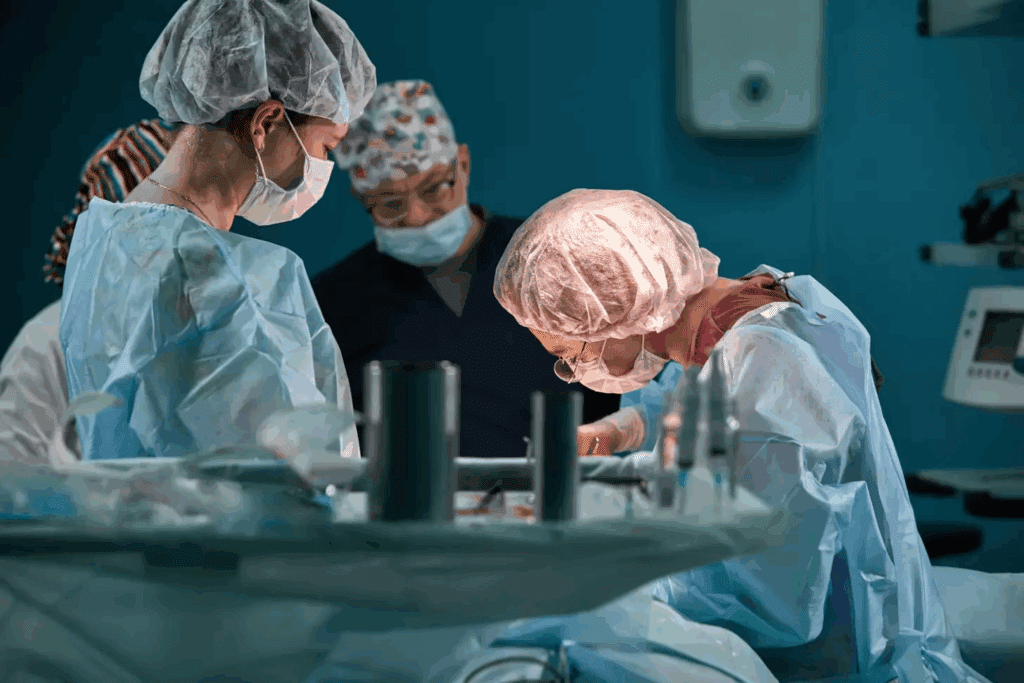
Real neurosurgery is a field that has made huge strides in recent years. It has caught the eye of doctors all over the world. The detailed and delicate nature of these surgeries is what draws people in.
“The intricacy of neurosurgery lies in its ability to balance precision with the need for swift decision-making,” says a renowned neurosurgeon. This quote captures the essence of why neurosurgery is both challenging and fascinating.
Neurosurgical procedures are complex and need a lot of skill and precision. Doctors find these operations both challenging and captivating. The use of advanced technology, like intraoperative MRI and neuro-navigation systems, has greatly improved these surgeries.
Also, neurosurgery is always changing, with new methods and tools being developed. For example, awake craniotomy lets surgeons work on patients who are awake. This makes it easier to remove tumors and reduces the chance of harming important brain areas.
The evolution of neurosurgical techniques has been amazing. It has turned a once-risky field into a highly advanced discipline. In the past, neurosurgery was risky and often didn’t have good results. But, with better technology and understanding of the brain, these techniques have improved a lot.
Today, neurosurgery uses many new methods, like minimally invasive surgery and stereotactic radiosurgery. These new approaches have made surgeries safer and faster, with better results for patients.
As we keep exploring what’s possible in neurosurgery, we see that “the future of neurosurgery lies in its ability to integrate technology with surgical expertise.” This mix is what keeps the field moving forward, giving hope to patients everywhere.
Brain surgery has changed a lot, moving from old open surgeries to new, less invasive methods. This change has made treatments better and helped more people get help.
Today, brain surgery comes in many forms. Each has its own good points and risks. Knowing about these options helps patients choose the best treatment for them.
Open brain surgery, or craniotomy, means taking part of the skull off to see the brain. It’s used for big surgeries like:
Open surgery lets doctors see and work on the brain directly. This is key for complex or critical cases.
Minimally invasive brain surgery is becoming more common. It aims to heal faster and cause less damage. These include:
Minimally invasive procedures mean smaller cuts and less brain harm. They also lead to shorter hospital stays. But, they work best for certain conditions and places.
Knowing the difference between open and minimally invasive surgeries helps patients. It lets them understand their options better and make choices that fit their needs.
Advanced medical technology is key in making high-quality neuro operation videos. It helps make surgeries more precise and safer for patients.
We use several important technologies for these videos. The main ones are Advanced Imaging Systems and Intraoperative Monitoring Tools.
Advanced imaging systems are essential in neurosurgery. They give surgeons clear views of the brain’s inside. MRI (Magnetic Resonance Imaging) and CT (Computed Tomography) scans help guide surgeries.
These tools help find and avoid important brain areas. This makes surgeries safer and more effective.
Intraoperative monitoring tools let surgeons watch the patient’s brain in real-time. This helps them adjust during surgery to avoid risks. electroencephalography (EEG) and somatosensory evoked potentials (SSEP) check brain activity.
Together, advanced imaging and monitoring tools improve patient care. They help make detailed videos of neurosurgery, showing the complexity of these procedures.
The awake craniotomy is a big step forward in brain surgery. It lets surgeons work on the brain while the patient is awake. This way, they can watch how the brain works in real time.
Awake craniotomy uses special brain surgery methods and careful use of anesthesia. First, the scalp is numbed with local anesthesia. The patient stays awake and can talk or move as needed.
Key steps in awake craniotomy include:
Patients who have this surgery often have a unique experience. Some might feel scared, but many stay calm. They might even play guitar or speak as the doctors ask.
The results of awake craniotomy are very good. It helps doctors remove tumors more accurately and safely. Here are some key benefits:
| Outcome | Description | Benefit |
| Precision in Tumor Resection | Real-time monitoring allows for more accurate removal of tumors | Reduced risk of tumor recurrence |
| Preservation of Brain Function | Functional mapping helps avoid critical brain areas | Improved post-operative neurological function |
| Patient Participation | Patients can assist during the procedure by performing tasks | Enhanced safety and precision |
Awake craniotomy is a big leap in brain surgery. It makes surgery safer and more precise. As technology gets better, we’ll see even more improvements in this field.
Complex brain tumors need a special surgical method. Brain tumor surgery is very delicate and requires a skilled team. Advanced imaging and monitoring are used to remove the tumor carefully and avoid harming the brain.
Neurosurgeons plan carefully for complex brain tumors. They use advanced imaging to understand the tumor’s shape and its location. Intraoperative MRI and neuro-navigation systems help make real-time changes during surgery.
The surgery method depends on the tumor’s size, location, and type. Tumors in important brain areas need extra care to avoid damage. Techniques like awake craniotomy might be used to check the patient’s brain functions during surgery.
The recovery after brain tumor surgery is very important. Patients are watched closely in the ICU for any signs of problems. The post-operative care team works hard to manage pain, prevent infections, and help the patient recover well.
Patients have follow-up appointments and imaging studies to check on the tumor removal and watch for any return. Rehabilitation might be needed to help the patient regain lost abilities. The table below shows the typical care plan for brain tumor patients after surgery.
| Post-Operative Day | Care Plan | Monitoring Parameters |
| 0-1 | ICU care, pain management | Vital signs, neurological status |
| 2-3 | Transfer to ward, mobilization | Neurological examination, wound check |
| 4-7 | Rehabilitation planning, discharge planning | Imaging studies (MRI/CT) |
“The advancements in neurosurgical techniques and post-operative care have significantly improved outcomes for brain tumor patients. Our team’s dedication to providing complete care has greatly helped patients recover and improve their quality of life.” -A Neurosurgeon
We know that brain tumor surgery and recovery are tough. Our team is dedicated to giving top-notch care and support to our patients at every step.
Now, we can control devices with just our thoughts, thanks to brain-computer interface technology. This breakthrough could change lives for those with paralysis, ALS, and other motor disorders.
Mind surgery videos show how neural implants are put in. The process is very detailed. It involves placing electrodes that can read brain signals, letting patients control devices outside their bodies.
Key components of brain-computer interfaces include:
People who got brain-computer implants have seen big improvements. Some can now control robotic arms, talk through computers, and even move again.
| Patient Condition | BCI Application | Outcome |
| Paralysis | Control of robotic arm | Regained ability to perform daily tasks |
| ALS | Communication through computer | Improved ability to interact with family |
| Motor disorder | Regaining motor functions | Enhanced independence |
These stories show how brain-computer interfaces can change lives. As the tech gets better, we’ll see even more amazing uses in neurosurgery.
Cerebrovascular surgery shows the detailed work of surgeons in live brain operations. They carefully handle complex blood vessels in the brain. This surgery treats conditions like aneurysms and malformations to avoid serious problems like stroke or death.
Aneurysms and vascular malformations need expert surgical care. Aneurysms can burst and cause bleeding in the brain. Vascular malformations can cause various symptoms. Surgeons use advanced imaging to see these problems and plan treatment.
Doctors from different fields work together to treat these conditions. Minimally invasive methods like coiling or embolization are used more often. This approach reduces the need for open surgery and helps patients recover faster.
Cerebrovascular surgery can have unexpected moments that require quick decisions. If an aneurysm bursts during surgery, doctors must act fast to stop bleeding. They use their experience and skill to handle these situations calmly and effectively.
Keeping the brain and its blood vessels safe is also key. Surgeons use intraoperative monitoring like EEG and SSEPs. This lets them adjust the surgery to protect the brain and avoid damage.
Neurosurgeons who work with kids face big challenges. They must carefully handle the growing brain to help their young patients. This field needs a lot of skill and knowledge about kids’ brains.
Brain surgery on kids is different from adults. Kids’ brains can heal in some ways but are also more fragile. We plan and do surgeries carefully to avoid risks and get the best results.
Kids’ brains change a lot as they grow. This means surgeons have to adjust their methods. Also, kids’ brains can sometimes make up for damaged areas, which is both good and tricky to plan for.
After surgery, we watch kids closely. Their growing brains can react differently to surgery. We help families give kids the right care and support while they recover.
Watching over kids after surgery is not just about immediate problems. We also keep an eye on how they develop over time. Surgery can affect how kids think, feel, and move, which is very important in pediatric neurosurgery.
| Aspect | Considerations in Pediatric Neurosurgery | Impact on Recovery |
| Brain Development | The surgery must be planned according to the child’s brain development stage. | Affects long-term cognitive and motor skills. |
| Surgical Technique | Surgeons must adapt techniques to the child’s anatomy and condition. | Influences the risk of complications and recovery speed. |
| Post-Operative Care | Close monitoring is required to address any immediate complications. | Critical for ensuring proper healing and minimizing long-term effects. |
We understand these factors and tailor our care to each child. This way, we can get the best results in pediatric neurosurgery. Our team is dedicated to giving top-notch care that helps kids grow and stay healthy.
Deep brain stimulation is a new hope for patients with movement disorders. It’s a surgery that implants a device called a “brain pacemaker.” This device sends electrical impulses to parts of the brain to help with symptoms.
The success of deep brain stimulation depends on where the electrodes are placed in the brain. We use MRI and CT scans to guide the surgery. This makes sure the electrodes are in the right spots to control brain activity.
Precision is key in this surgery. The spots for the electrodes can be very small and different for each patient. Our neurosurgeons work with a team to plan and do the surgery, considering each patient’s unique needs.
Deep brain stimulation can give patients quick relief from symptoms. Many people see a big drop in tremors, rigidity, and other motor symptoms.
The quick effects can change based on the condition and how the patient reacts. But, many patients see a big improvement in their daily life. They can do things more easily and on their own.
As we keep improving in neurosurgery, deep brain stimulation is a key treatment for movement disorders. Research and new tech are making it even better and safer for patients.
Traumatic head injuries need quick and skilled neurosurgery. Emergency trauma neurosurgery is a high-stakes field. It involves fast decision-making and precise surgery to save lives. We will look at the critical parts of this field, focusing on the urgent nature of these procedures and the chance for recovery.
In emergency trauma neurosurgery, the time for effective action is very short. Prompt action is key in preventing more brain damage and improving patient outcomes. Neurosurgeons must quickly assess the situation, make informed decisions, and perform complex procedures under intense pressure.
The process starts with quick imaging, like CT scans, to check the injury’s extent. This info helps the surgical team plan the best intervention. Time is of the essence in these cases, as delays can cause increased intracranial pressure, brain herniation, and other life-threatening issues.
Brain surgery images are vital in showing patients’ recovery after emergency trauma neurosurgery. These images provide visual proof of the surgical outcomes. They also help educate patients and their families about the recovery journey.
By looking at brain surgery images taken at different stages post-surgery, we can see the healing process and any complications. These images are key for tracking patient progress and making informed decisions about post-operative care.
Advanced imaging techniques are used throughout the recovery phase. They help ensure patients get the best care. As technology gets better, the quality and detail of these images improve. This gives us more insights into the recovery process and helps refine treatment plans.
Endoscopic neurosurgery uses advanced tools for precise brain surgery. It’s a safer and more effective way to treat brain conditions. This method helps patients recover faster and with less pain.
High-definition cameras and advanced lighting systems are key in endoscopic neurosurgery. They give surgeons a clear view of the brain. This helps them make precise cuts and avoid damaging nearby tissues.
Angled endoscopes also help surgeons reach hard-to-access areas. This is very useful in complex surgeries where accuracy is critical.
Endoscopic neurosurgery means less pain and shorter hospital stays for patients. The small incisions and less tissue disruption lead to faster recovery. Patients can get back to their daily lives sooner.
Endoscopic neurosurgery is a big step forward in neurosurgery. It offers a safer and more effective way to treat brain conditions.
Medical technology is getting better, and robotic neurosurgery is key in the future of brain surgery. We see big steps in robotic systems. They aim to make neurosurgical procedures more precise and accurate.
Robotic neurosurgery systems are made to offer unparalleled precision in brain operations. They use advanced navigation and control. This makes it possible to do complex procedures that were hard or impossible before.
This precision is great for complex brain surgeries. Minimally invasive techniques can cut down recovery time and improve patient results.
When we look at human and robotic surgical techniques, we see different strengths. Human surgeons have experience and intuition in the operating room. Robotic systems, on the other hand, offer enhanced precision and stability.
As we keep improving robotic neurosurgery, it’s clear that both human skill and robotic precision are vital. They will both play big roles in the future of brain operations.
Neurosurgery videos are key in teaching the next neurosurgeons. They let medical pros see and study complex surgeries up close. This is a unique chance to learn.
Neurosurgery videos are a big help in training neurosurgeons. They show clear and detailed views of how to do surgeries. You can watch them over and over to get it right.
They help understand the small details of tough surgeries, like brain tumors or aneurysms.
A study in a top medical journal found that videos improve skills and decision-making in residents.
“Video-based education has become an essential component of modern surgical training, providing a realistic and engaging learning experience.”
Neurosurgery videos are great for learning, but they raise big ethical questions. Keeping patient privacy and getting consent are top priorities. We must make sure all personal info is gone and patients agree to be recorded.
Thinking about how videos might affect patients and their families is also key. As teachers, we must weigh the benefits against the need to respect privacy and dignity.
In summary, neurosurgery videos are a powerful tool in teaching. By understanding their value and the ethics behind them, we can use them to train the next neurosurgeons well.
The field of neurosurgery is always moving forward. It’s thanks to new technologies and better techniques. We’ve looked at many complex surgeries, showing how advanced brain surgery is today.
New tools like advanced imaging and robotic systems have made surgeries better. We’ve seen how awake craniotomies and deep brain stimulation are changing care for patients.
We’re expecting even more progress in the future. Research and new tech will likely make surgeries safer and faster. This means better care for people with complex brain conditions.
As neurosurgery keeps getting better, we’re dedicated to top-notch healthcare. We support patients from around the world who need advanced medical treatments.
Neurosurgery is a medical field that deals with surgeries of the brain, spine, and nervous system. It treats conditions like brain tumors, aneurysms, and vascular malformations.
There are many brain surgery techniques. These include open brain surgery, minimally invasive methods, awake craniotomy, endoscopic neurosurgery, and robotic neurosurgery. Each has its own benefits and uses.
Awake craniotomy is a surgery where the patient stays awake. This lets surgeons watch brain function live. It helps them avoid harming important brain areas.
Deep brain stimulation is a surgery that implants electrodes in the brain. It treats movement disorders like Parkinson’s by changing brain activity.
Endoscopic neurosurgery has many advantages. It leads to quicker recovery, less scarring, and less invasive procedures. It’s a good option for some neurosurgical conditions.
Neurosurgery videos are a great learning tool for doctors. They show complex procedures and techniques. This helps in teaching and improving skills for future neurosurgeons.
Sharing neurosurgery videos raises ethical issues. These include patient privacy and consent. It’s important to handle these responsibly to ensure educational value.
Robotic neurosurgery uses robots to improve brain surgery precision. It could lead to better results and open up new possibilities for complex surgeries.
Advanced imaging and monitoring tools give surgeons real-time feedback. This helps them navigate complex anatomy and make accurate decisions. It improves patient outcomes.
Cerebrovascular surgery is key in treating aneurysms and vascular malformations. It offers surgical solutions to prevent rupture and improve symptoms. This leads to better patient outcomes.
Pediatric neurosurgery needs special care. It considers the unique anatomy and development of children’s brains. This requires precision and care for the best results.
