Last Updated on December 1, 2025 by Bilal Hasdemir

Rhabdomyosarcoma is a rare and aggressive cancer that starts in muscle cells. It’s tough to treat because it can spread quickly. Sadly, almost one-third of those diagnosed face this problem, making treatment harder.
Understanding the common metastasis sites of rhabdo sarcoma is crucial for effective treatment planning. It can go to the lungs, bones, and bone marrow. This means we need a full approach to help patients.
Key Takeaways
- Rhabdomyosarcoma is a rare and aggressive form of cancer.
- Nearly one-third of patients experience metastasis.
- Common sites for metastasis include the lungs, bones, and bone marrow.
- Understanding metastatic patterns is critical for effective treatment.
- Comprehensive care is essential for improving patient outcomes.
What Is Rhabdo Sarcoma: Definition and Classification

Rhabdomyosarcoma is a rare and complex cancer. It needs a deep understanding of its definition and types. We will look into its origins, cell characteristics, how common it is, and who it affects.
Origin and Cellular Characteristics
Rhabdomyosarcoma starts in skeletal muscle cells. The tumor cells look like developing muscle cells. This helps us understand the disease better.
The cells of rhabdomyosarcoma have rhabdomyoblasts, which are young muscle cells. These can be seen under a microscope and are key for diagnosing the disease.
Incidence and Demographics
Rhabdomyosarcoma is a rare cancer, making up a small part of all childhood cancers. Its occurrence changes with age, with some types more common in certain age groups.
To grasp the incidence and demographics of rhabdomyosarcoma, let’s look at the data in the table below:
| Age Group | Subtype | Incidence Rate |
| 0-9 years | Embryonal | Higher incidence |
| 10-19 years | Alveolar | Moderate incidence |
| 20+ years | Pleomorphic | Lower incidence |
The table shows the different incidence rates of rhabdomyosarcoma subtypes in various age groups. Knowing these demographics is vital for diagnosis and treatment planning.
Types and Subtypes of Rhabdomyosarcoma

Rhabdomyosarcoma is not just one cancer. It’s a group of cancers with different looks, genes, and symptoms. Each type needs its own treatment plan.
Embryonal Rhabdomyosarcoma
Embryonal rhabdomyosarcoma is the most common type. It mainly affects kids under 10. It looks like muscle tissue from an embryo. Key features include:
- Predominantly affects young children
- Often occurs in the head and neck region or genitourinary tract
- May have a more favorable prognosis compared to other subtypes
Alveolar Rhabdomyosarcoma
Alveolar rhabdomyosarcoma is aggressive and has unique genetic traits. It often involves chromosome translocations. Notable aspects include:
- More common in older children and adolescents
- Frequently occurs in the extremities
- Often associated with a poorer prognosis due to its aggressive nature and tendency to metastasize
Pleomorphic and Other Rare Subtypes
Pleomorphic rhabdomyosarcoma is rare and aggressive. It mainly affects adults. It has a mix of cell types. Other rare types include:
- Spindle cell/sclerosing rhabdomyosarcoma
- Epithelioid rhabdomyosarcoma
These rare types are hard to diagnose because of their unique features.
Knowing about these subtypes is key to better treatments. Doctors use a team approach to diagnose and treat rhabdomyosarcoma. They look at the cancer’s look, genes, and symptoms.
The Biological Mechanisms of Rhabdomyosarcoma Metastasis
It’s important to know how rhabdomyosarcoma metastasis works. This knowledge helps us find better treatments. Metastasis is a complex process. It starts with cancer cells moving into nearby tissues, then through the blood or lymph, and ends with them settling in other parts of the body.
Cellular Invasion and Migration Processes
Cellular invasion and migration are key steps in rhabdomyosarcoma spreading. Cancer cells can break through the tissue around them. They do this by breaking down the tissue and changing how they stick to each other. Cell migration happens because of changes in the cell’s structure and the genes it uses.
Genetic and Molecular Drivers of Metastasis
Genetic and molecular changes are big players in rhabdomyosarcoma metastasis. Certain genes and pathways help cancer cells spread. For example, changes in the TP53 and MYOD1 genes make the disease more aggressive.
| Genetic/Molecular Factor | Role in Metastasis |
| TP53 mutations | Impaired cell cycle regulation, contributing to increased cell proliferation and survival |
| MYOD1 mutations | Altered muscle differentiation, potentially enhances metastatic ability |
| PAX-FOXO1 fusion | Disrupts normal gene regulation, leading to more tumors and metastasis |
These genetic and molecular changes help start and grow rhabdomyosarcoma. They also affect how it spreads. Knowing about these factors is key to creating treatments that can stop metastasis.
Primary Sites of Rhabdomyosarcoma Before Metastasis
Knowing where Rhabdomyosarcoma first appears is key to treating it well. It can start in many parts of the body. Finding these spots is very important for doctors.
Head and Neck Region Tumors
Rhabdomyosarcoma in the head and neck is a big worry. It’s close to important parts of the body. Doctors group these tumors by where they are, like around the eye or in the throat.
Orbital Rhabdomyosarcoma is common in the head and neck. It can make the eye bulge or swell. Finding it early is vital to avoid serious problems.
Genitourinary Tract Involvement
The genitourinary tract is another common place for Rhabdomyosarcoma, mainly in kids. Tumors can show up in the bladder, prostate, vagina, or uterus. How they’re treated depends on where and how big they are.
Bladder and Prostate Rhabdomyosarcoma might cause trouble with urinating or block the flow. Quick action is needed to keep these organs working right.
| Primary Site | Common Symptoms | Treatment Considerations |
| Head and Neck | Swelling, pain, cranial nerve deficits | Surgery, radiation, chemotherapy |
| Genitourinary Tract | Urinary obstruction, hematuria | Organ-preserving surgery, chemotherapy |
| Extremities and Trunk | Swelling, pain, limited mobility | Surgery, chemotherapy, radiation |
Extremities and Trunk Presentations
Rhabdomyosarcoma can also show up in the arms, legs, or chest. These tumors can be hard to spot because their symptoms are not clear.
Dealing with Rhabdomyosarcoma in these areas needs a team effort. This includes surgery, chemo, and sometimes radiation. Each plan is made just for the patient.
Lymphatic System: First Station in Metastatic Spread
The lymphatic system is key in the spread of rhabdomyosarcoma. It defends the body against infections and diseases. But, it can also be a path for cancer cells to move from the original tumor to other parts of the body.
Patterns of Regional Lymph Node Involvement
Regional lymph nodes are where cancer cells from rhabdomyosarcoma often first go. The pattern of lymph node involvement depends on the tumor’s location. For example:
- Tumors in the head and neck region tend to metastasize to the cervical lymph nodes.
- Rhabdomyosarcomas in the extremities often spread to the axillary or inguinal lymph nodes, depending on whether the tumor is located in the upper or lower limb.
- Tumors in the genitourinary tract may metastasize to the pelvic or retroperitoneal lymph nodes.
Knowing these patterns is key for accurate staging and planning treatment.
Clinical Significance of Lymphatic Metastasis
Lymph node involvement in rhabdomyosarcoma metastasis is very important. It means a higher risk of further spread and a worse prognosis. So:
- Checking lymph node status is a big part of the diagnostic workup.
- Lymph node involvement affects treatment choices, like surgery, radiation, and chemotherapy.
- Having lymph node metastasis might mean more aggressive treatment is needed.
We understand that managing rhabdomyosarcoma needs a deep look at the lymphatic system’s role in metastasis. By correctly checking and dealing with lymph node involvement, we can make better treatment plans for patients with this tough disease.
Pulmonary Metastasis in Rhabdomyosarcoma
Understanding pulmonary metastasis in rhabdomyosarcoma is key to effective treatment. Cancer cells spread to the lungs, a common site for metastasis. This is true for many cancers, including rhabdomyosarcoma.
Frequency and Distribution
Pulmonary metastasis is a big concern in rhabdomyosarcoma management. Studies show the lungs are a common site for metastasis, mainly in advanced cases.
The location of metastatic deposits in the lung can vary. It often depends on the primary tumor’s characteristics and the patient’s overall health.
- Common patterns include:
- Multiple bilateral nodules
- Solitary nodules
- Diffuse lymphangitic spread
Detection Methods and Management Approaches
Spotting pulmonary metastasis early is vital for managing rhabdomyosarcoma. Several imaging methods are used:
- Computed Tomography (CT) scans: Great for finding small nodules and seeing how far the disease has spread.
- Positron Emission Tomography (PET)-CT: Helps check the activity of metastatic lesions.
- Chest X-rays: Less sensitive but useful for initial checks or follow-ups.
Managing pulmonary metastasis in rhabdomyosarcoma requires a team effort. This includes:
- Chemotherapy: Treats both local and distant disease.
- Surgical resection: Removes metastatic lesions in some cases.
- Radiation therapy: Helps control symptoms and treat limited metastasis.
The treatment plan depends on several factors. These include how much of the lung is involved, if there’s disease elsewhere, and the patient’s health.
Bone Marrow Infiltration Patterns
Understanding bone marrow infiltration patterns is key for managing rhabdomyosarcoma. It affects staging and treatment choices. Rhabdomyosarcoma is a cancer that starts in muscle cells and can spread to the bone marrow.
Prevalence and Clinical Manifestations
Bone marrow infiltration by rhabdomyosarcoma cells is common, mainly in advanced stages. Many patients with metastatic rhabdomyosarcoma have bone marrow involvement. Symptoms can range from none to severe blood cell shortages.
The high rate of bone marrow metastasis in rhabdomyosarcoma patients highlights the need for detailed diagnostic tests. We will look at how often bone marrow is involved and its impact on patient outcomes.
| Study | Number of Patients | Bone Marrow Involvement (%) |
| Study A | 100 | 25% |
| Study B | 50 | 30% |
| Study C | 200 | 20% |
Diagnostic Approaches for Bone Marrow Assessment
Diagnosing bone marrow infiltration in rhabdomyosarcoma involves several methods. These include bone marrow aspiration and biopsy, imaging studies, and molecular diagnostics. These help determine the extent of bone marrow involvement.
Bone marrow aspiration and biopsy are the top choices for diagnosing bone marrow metastasis. They allow for a direct look at bone marrow cells and tissue.
Imaging studies, like PET-CT scans, are vital for spotting bone marrow involvement and disease extent.
Central Nervous System Metastatic Spread
The central nervous system is a key area for metastasis in rhabdomyosarcoma. It brings unique challenges for diagnosis and treatment.
Rhabdomyosarcoma spreading to the CNS is a serious issue. It affects the brain and spinal cord. We will look at how common it is, its patterns, and its impact on patients.
Brain Metastasis: Incidence and Patterns
Brain metastasis in rhabdomyosarcoma is rare but serious. The chance of it happening varies by rhabdomyosarcoma type. Alveolar rhabdomyosarcoma is more likely to spread to the CNS.
Leptomeningeal and Spinal Cord Involvement
Leptomeningeal involvement happens when cancer cells reach the meninges. These are the protective layers around the brain and spinal cord. It can cause a lot of problems and needs strong treatment. Spinal cord involvement is less common but also poses challenges.
| CNS Metastasis Site | Incidence | Clinical Features |
| Brain | Variable, higher in alveolar subtype | Headaches, seizures, neurological deficits |
| Leptomeninges | Less common | Meningeal signs, cranial nerve palsies |
| Spinal Cord | Rare | Back pain, neurological deficits, bowel/bladder dysfunction |
Managing CNS metastasis in rhabdomyosarcoma needs a team effort. This includes surgery, radiation, and chemotherapy. Early action is key to better outcomes.
Skeletal System Metastasis in Rhabdomyosarcoma
It’s key to understand skeletal metastasis in rhabdomyosarcoma for better treatment. Rhabdomyosarcoma is a muscle tumor that can spread to the skeletal system. This spread greatly affects patient outcomes and life quality.
Common Bone Sites for Metastatic Deposits
Rhabdomyosarcoma can spread to any bone, but some are more common. The long bones of the extremities, like the femur and humerus, and the pelvis are often affected. These areas have a lot of bone marrow, making them perfect for metastatic cells to grow.
Other bones like the vertebrae, ribs, and skull can also be involved. The type of bone affected can depend on the rhabdomyosarcoma subtype and the patient’s condition.
Clinical Presentation and Management Strategies
The symptoms of skeletal metastasis in rhabdomyosarcoma can vary. Patients might feel pain, swelling, or have limited mobility if a joint or weight-bearing bone is affected. Sometimes, skeletal metastasis can cause pathological fractures, which need quick medical care.
Managing skeletal metastasis requires a team effort. This includes pain management, radiation therapy to stop tumor growth and ease pain, and surgical intervention for fractures. Systemic treatments, like chemotherapy, are also key in controlling the disease.
We stress the need for a detailed treatment plan for skeletal metastasis in rhabdomyosarcoma. Understanding the nature of skeletal metastasis and using the right management strategies can enhance patient outcomes and life quality.
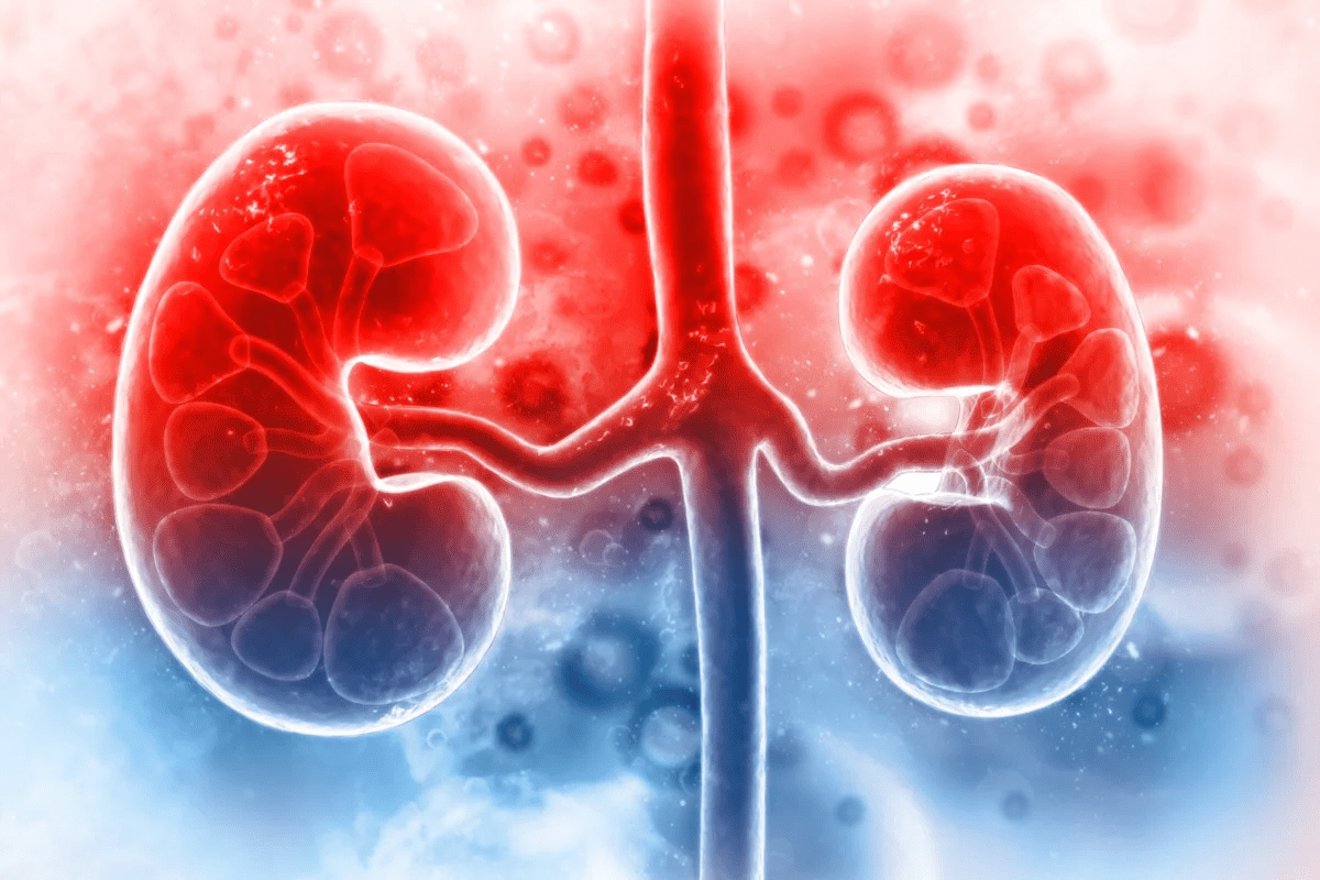
Liver and Abdominal Organ Metastases
Metastasis to the liver and other abdominal organs is a key part of rhabdomyosarcoma’s growth. It’s important to know how often this happens, where it happens, and what it means for patients.
Hepatic Metastasis: Frequency and Distribution
Hepatic metastasis in rhabdomyosarcoma is a serious issue. Research shows the liver is involved in about 10% to 20% of cases. Where these metastases appear in the liver can depend on the tumor’s location and type.
Key factors influencing hepatic metastasis include:
- The primary site of the tumor
- The histological subtype of rhabdomyosarcoma
- The presence of lymph node metastases
Other Visceral Organ Involvement
Other organs in the abdomen can also get involved. These include the spleen, pancreas, and adrenal glands. While less common, it means the disease has spread further, making treatment harder.
The symptoms of metastasis in these organs can be vague. Advanced imaging is often needed to find them. Early detection is key to managing the disease effectively.
Advanced imaging like CT, MRI, and PET-CT scans are essential. They help see how far the disease has spread.
In summary, metastasis to the liver and other abdominal organs is a big part of rhabdomyosarcoma’s growth. Knowing how often, where, and why this happens is vital for finding better treatments.
Differences in Metastatic Patterns Between Rhabdomyosarcoma Subtypes
Rhabdomyosarcoma, a muscle tumor, has different subtypes. Alveolar and embryonal are the most common. Knowing these differences helps doctors create better treatment plans.
Alveolar Rhabdomyosarcoma Metastasis Characteristics
Alveolar rhabdomyosarcoma grows fast and spreads early. It often goes to the lungs, bone marrow, and lymph nodes. Its genetic makeup, like the PAX-FOXO1 fusion, helps it spread.
This subtype has a worse outlook. So, finding it early and treating it aggressively is key.
Embryonal Rhabdomyosarcoma Spread Patterns
Embryonal rhabdomyosarcoma spreads in different ways. It starts in the head, neck, or genitourinary tract. It can go to nearby lymph nodes, lungs, and liver. Its spread is influenced by genetic changes, like RAS pathway mutations.
| Characteristics | Alveolar Rhabdomyosarcoma | Embryonal Rhabdomyosarcoma |
| Common Metastatic Sites | Lungs, bone marrow, lymph nodes | Regional lymph nodes, lungs, liver |
| Genetic Features | PAX-FOXO1 fusion | RAS pathway mutations |
| Prognosis | Generally poorer | More variable |
Understanding how alveolar and embryonal rhabdomyosarcoma spread is vital. It helps doctors tailor treatments for each subtype. This approach can lead to better patient care.
Advanced Imaging for Detecting Rhabdomyosarcoma Metastasis
Advanced imaging has changed how we find rhabdomyosarcoma metastasis. These new technologies help us see and track where cancer has spread. This is key for planning treatment.
Finding metastasis is very important in treating rhabdomyosarcoma. Advanced imaging helps us see how far the disease has spread.
CT, MRI, and PET-CT Applications
Computed Tomography (CT), Magnetic Resonance Imaging (MRI), and Positron Emission Tomography-Computed Tomography (PET-CT) are key in finding rhabdomyosarcoma metastasis.
- CT scans give us detailed pictures of the body, showing where the cancer has spread to organs like the lungs and liver.
- MRI shows soft tissues clearly, helping us find cancer in the brain, spine, and other soft tissues.
- PET-CT combines images of how the body works and its structure, helping us spot active tumor sites, which is key for finding metastasis.
Novel Molecular and Functional Imaging Techniques
New molecular and functional imaging methods are also helping us find rhabdomyosarcoma metastasis.
Some of these new ways include:
- Molecular imaging with special probes
- Diffusion-weighted MRI to see how dense tumor cells are
- Dynamic contrast-enhanced MRI to check how blood flows through tumors
These advanced imaging methods help us better find and understand metastasis. This leads to better care for patients.
Staging Systems and Risk Assessment Based on Metastatic Spread
Accurate staging and risk assessment are key in managing rhabdomyosarcoma, even with metastatic spread. Staging systems help doctors understand how far the disease has spread. This is vital for choosing the best treatment.
TNM and IRS Staging Classifications
The TNM staging system looks at the tumor size (T), lymph node involvement (N), and metastasis (M). It’s used for many cancers, including rhabdomyosarcoma. The IRS staging system, specific to rhabdomyosarcoma, groups patients based on disease extent and tumor resectability.
The IRS system categorizes patients by surgical outcome and disease extent. For example, Group I includes patients with fully removed tumors. Group IV includes those with distant metastasis. Knowing these systems is key for predicting outcomes and treatment planning.
Risk Stratification and Prognostic Groups
Risk stratification in rhabdomyosarcoma sorts patients into low, intermediate, and high-risk groups. Factors like age, tumor site, and metastasis play a role. This helps tailor treatment to each patient’s risk level.
Prognostic groups are based on clinical and biological factors. Patients with metastasis at diagnosis are usually high-risk and face a poorer prognosis. Knowing these factors is essential for making treatment decisions.
Treatment Strategies for Metastatic Rhabdomyosarcoma
Metastatic rhabdomyosarcoma treatment is getting better, focusing on many therapies at once. Treating the disease in its advanced stages needs a mix of treatments. This mix aims to improve how well patients do.
Multimodal Therapy Approaches
Multimodal therapy is key in fighting metastatic rhabdomyosarcoma. It uses different treatments like chemotherapy, radiation, and surgery. These work together to attack the disease from all sides.
- Chemotherapy: It’s a mainstay in treating metastatic rhabdomyosarcoma. Chemotherapy uses drugs to kill cancer cells.
- Radiation Therapy: It helps control the disease in one area and eases symptoms. It’s a big part of the treatment plan.
- Surgery: Sometimes, surgery is used to take out remaining disease or handle problems.
Management of Specific Metastatic Sites
Dealing with metastatic rhabdomyosarcoma also means treating different areas in special ways. Each area might need a unique approach to get the best results.
- Pulmonary Metastases: Treatment might include chemotherapy, whole-lung radiation, or surgery in some cases.
- Bone Metastases: Treatment options include radiation for comfort and chemotherapy to fight the disease.
- Bone Marrow Involvement: For bone marrow disease, doctors often use strong chemotherapy.
Emerging Targeted and Immunotherapies
New treatments like targeted and immunotherapies are changing how we fight metastatic rhabdomyosarcoma. These new methods could lead to better results for patients with advanced disease.
- Targeted Therapies: These treatments aim at specific parts of the cancer cell’s growth process.
- Immunotherapies: Methods like checkpoint inhibitors and CAR-T cell therapy are being tested to boost the immune system’s fight against cancer.
As scientists learn more about rhabdomyosarcoma, we’ll see even more progress in treating it. This brings hope to patients and their families.
Conclusion: Future Directions in Understanding and Treating Metastatic Rhabdomyosarcoma
Metastatic rhabdomyosarcoma is a tough challenge in cancer treatment. It needs a team effort to tackle it. We’ve looked at what it is, how it spreads, and how we treat it.
New research and treatments are bringing hope. We’re moving towards treatments that are more tailored to each patient. This includes using the latest in genetics and molecular science. We expect to see more innovative treatments that combine different approaches.
It’s important to keep learning about metastatic rhabdomyosarcoma. We need better treatments to help patients live better lives. By pushing forward, we can make a real difference for those fighting this disease.
FAQ
What is rhabdomyosarcoma?
Rhabdomyosarcoma is a rare cancer that starts in soft tissues. These tissues are muscles attached to bones and help us move.
What are the main subtypes of rhabdomyosarcoma?
There are three main types: embryonal, alveolar, and pleomorphic. Each type has its own traits and genetic makeup. This affects how it’s treated and how well it does.
Where does rhabdomyosarcoma commonly metastasize?
It can spread to many places, like the lungs, lymph nodes, and liver. Where it goes depends on the type of cancer.
How is rhabdomyosarcoma metastasis diagnosed?
Doctors use CT, MRI, and PET-CT scans to find it. They also do biopsies to check the cancer cells.
What are the treatment options for metastatic rhabdomyosarcoma?
Treatment includes chemotherapy, radiation, and surgery. New treatments like targeted and immunotherapies are also being tested.
What is the prognosis for patients with metastatic rhabdomyosarcoma?
The outlook depends on the cancer type, how far it has spread, and how well it responds to treatment. Metastatic disease is usually harder to treat.
How does the staging system work for rhabdomyosarcoma?
The TNM and IRS systems are used to stage it. They help doctors understand how far the cancer has spread and plan treatment.
Can rhabdomyosarcoma be cured?
Cure chances depend on the cancer stage, type, and treatment success. Early-stage cancers are more likely to be cured than those that have spread.
What are the common primary sites for rhabdomyosarcoma?
It often starts in the head and neck, genitourinary tract, and other areas. Where it starts can affect where it spreads.
How does the lymphatic system play a role in rhabdomyosarcoma metastasis?
The lymphatic system is a main way cancer spreads. Lymph nodes are often affected, which can change treatment and outlook.
References:
- Kaseb, H. (2024). Rhabdomyosarcoma. In StatPearls. National Library of Medicine. Retrieved from https://www.ncbi.nlm.nih.gov/books/NBK507721/


