Last Updated on November 27, 2025 by Bilal Hasdemir

Advanced skeletal imaging techniques can show detailed insights into your bone health. They can detect conditions such as fractures, bone metastases, infections, and degenerative bone diseases. At Liv Hospital, we use the latest diagnostic technology, including skeletal scintigraphy and bone density scans, to ensure accurate results and effective treatment planning.
Our expert team ensures each patient is thoroughly skeletally scanned for comprehensive bone health assessment and skeletal system evaluation. This helps us identify health issues early, enabling timely medical intervention and customized care. We are committed to providing top-quality healthcare, prioritizing our patients’ comfort, and offering full support throughout their health journey.
Key Takeaways
- Advanced skeletal imaging techniques detect bone-related conditions and diseases.
- Liv Hospital uses cutting-edge technology for accurate diagnoses and treatment plans.
- Bone health assessment and skeletal system evaluation enable early intervention.
- Our patient-first approach ensures complete care and support.
- Timely treatment can greatly improve health outcomes.
The Science Behind Skeletal Scanning
Understanding skeletal scanning is key to its role in medicine. It uses various technologies to show the bones in detail. This helps doctors diagnose and treat many conditions.
How Skeletal Imaging Works
Skeletal imaging captures bone images through different methods. Bone scintigraphy uses a radioactive tracer that shows bone damage. A gamma camera turns this into a two-dimensional image.
“Up to 25% of cancer patients with normal radiographs show metastases on scintigraphy, demonstrating its sensitivity.” This shows how important advanced imaging is. It helps find problems early, which is vital for treatment.
Evolution of Skeletal Scanning Technology
Skeletal scanning technology has grown a lot. It started with X-rays and now includes DEXA scans and whole-body X-rays. Modern tech like CT and MRI scans gives even more detailed images.
Types of Radiation Used in Bone Imaging
Various types of radiation are used in bone imaging. Bone scintigraphy uses a radioactive tracer, while X-rays and CT scans use X-ray radiation. Knowing about these helps understand the safety and effectiveness of skeletal scanning.
Thanks to these technologies, doctors can spot problems early. They can also see how well treatments are working. The science behind skeletal scanning shows how medical imaging keeps getting better.
Different Types of Skeletal Scanning Technologies
It’s important to know about the different skeletal scanning technologies. They help doctors diagnose and treat bone health issues. We’ll look at these technologies and how they help check bone health.
Skeletal Scintigraphy (Bone Scans)
Skeletal scintigraphy, or bone scans, is great at finding bone problems. It uses a tiny amount of radioactive material that shows up in active bones. Medical research shows bone scans are good for spotting infections, cancer, or other diseases not seen on X-rays.
DEXA Scans for Bone Density
DEXA scans measure bone mineral density (BMD). They’re non-invasive and key for diagnosing osteoporosis and predicting fracture risk. The scan gives a T-score, which compares your BMD to a healthy young adult. A lower T-score means lower bone density and a higher fracture risk.
Whole-Body Skeletal X-rays
Whole-body skeletal X-rays give a full view of the bones. They’re good for finding fractures, bone deformities, and other bone issues. They’re very useful in emergencies or when checking bone trauma.
Advanced Imaging: CT and MRI for Bone Assessment
CT and MRI scans offer detailed bone health checks. CT scans show bone structures clearly, while MRI is great for soft tissues around bones. These scans are key for complex bone conditions and planning surgeries.
| Imaging Technique | Primary Use | Benefits |
| Skeletal Scintigraphy (Bone Scans) | Detecting bone metastases, infections, and other abnormalities | High sensitivity, whole-body imaging |
| DEXA Scans | Measuring bone mineral density, diagnosing osteoporosis | Non-invasive, low radiation exposure |
| Whole-Body Skeletal X-rays | Assessing skeletal trauma, fractures, and deformities | Comprehensive view, quick assessment |
| CT and MRI | Detailed bone and soft tissue assessment | High-resolution images, excellent for complex diagnoses |
Knowing about skeletal scanning technologies helps doctors pick the best tool for each patient. This approach improves diagnosis and treatment outcomes.
What Happens When You’re Being Skeletally Scanned
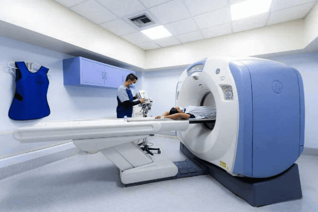
When you’re set for a skeletal scan, knowing what to expect can make things easier. At Liv Hospital, we focus on your comfort and safety during the scan.
Patient Preparation and Positioning
Getting ready for a skeletal scan is important. We ask patients to arrive early to fill out paperwork and ask questions. It’s key to remove metal objects, like jewelry or clothes with metal parts, as they can mess with the imaging. The type of scan will tell you what prep you need.
How you’re positioned during the scan matters too. You might lie on a table or stand against a special device. Our skilled techs will help you get comfy and in the right spot.
Duration and Comfort Considerations
The time it takes for a skeletal scan varies a lot. It can be just a few minutes for an X-ray or hours for a bone scan. The bone scan, for example, can take up to four hours, including time for the tracer to circulate and the scan itself. It’s important to stay very quiet during the scan to get clear images.
- For shorter scans, we make sure you’re comfortable with adjustable tables and pillows.
- For longer scans, we give regular breaks and make sure you’re positioned well to avoid discomfort.
Radiation Exposure and Safety Protocols
Concerns about radiation are normal. Our facilities use the latest tech to keep radiation low while getting great images. We follow strict safety rules to make sure the scan’s benefits are worth the risks.
As imaging tech gets better, we can diagnose bone health issues more accurately. We aim to give you top-notch care with advanced tech and caring service.
Bone Health Assessment Through Skeletal Imaging
Skeletal imaging is key to checking bone health. It gives a full view of how well our bones are doing. At Liv Hospital, we use top-notch imaging to check bone health.
Measuring Bone Mineral Density
Bone mineral density (BMD) is very important for bone health. DEXA scans help measure BMD. This gives doctors a clear picture of the risk of osteoporosis and fractures.
Doctors can spot trouble spots and plan the best treatment. This helps keep bones strong.
| Measurement | Description | Clinical Significance |
| Bone Mineral Density (BMD) | Quantitative measurement of bone density | Assesses risk of osteoporosis and fractures |
| T-score | Comparison to peak bone mass of a healthy young adult | Diagnoses osteoporosis and osteopenia |
| Z-score | Comparison to age-matched average bone density | Evaluates bone health in context of age |
Detecting Early Signs of Osteoporosis
Osteoporosis is a silent disease that can cause big problems if not caught early. Skeletal imaging helps doctors spot osteoporosis early. This means they can act fast to stop fractures and improve health.
Evaluating Fracture Risk
Knowing who might break a bone is important. Doctors look at BMD and other things to figure this out. This helps them make plans to keep bones safe.
Monitoring Treatment Effectiveness
Imaging also helps see if treatments are working. Doctors check BMD and other signs to see if treatments are helping. This way, they can change plans if needed to get the best results.
At Hospital, we focus on top-notch care that puts patients first. Our team works together to give patients the best care possible.
Detecting Bone Abnormalities and Diseases
Skeletal imaging is key in finding bone problems and diseases. It helps us understand our bone health better. Advanced imaging lets us spot many skeletal system issues.
Identifying Fractures and Microfractures
Fractures and microfractures are common bone issues. Early detection is key for good treatment and avoiding more problems. Skeletal scans can find even tiny fractures, helping doctors plan the best treatment.
- Fracture detection through skeletal imaging
- Microfracture identification
- Monitoring fracture healing progress
Diagnosing Bone Infections and Osteomyelitis
Bone infections and osteomyelitis need quick diagnosis and treatment. Skeletal imaging helps doctors see where the problem is. Accurate diagnosis is critical for the right treatment.
- Identifying signs of bone infection
- Diagnosing osteomyelitis through skeletal imaging
- Monitoring treatment response
Spotting Degenerative Joint Diseases
Degenerative joint diseases, like osteoarthritis, can be found and tracked with skeletal imaging. Doctors can see how much damage there is and plan treatment. Early detection helps a lot in improving patient results.
- Detecting early signs of degenerative joint diseases
- Monitoring disease progression
- Evaluating treatment effectiveness
Recognizing Metabolic Bone Disorders
Metabolic bone disorders, such as osteoporosis, can be spotted with skeletal imaging. Doctors can check bone density and structure to diagnose these conditions. Early recognition is essential for preventing serious issues and better patient care.
Through skeletal imaging, we get a full picture of bone health. It helps find many bone problems and diseases. Advanced imaging lets doctors give accurate diagnoses and effective treatments, improving patient care.
Cancer Detection and Monitoring Through Skeletal Scans
Skeletal scans have changed how we find and watch cancer. They help spot bone metastases and primary bone tumors. These scans show how far cancer has spread, helping us make treatment plans that fit each patient.
Identifying Bone Metastases
Bone metastases are common in cancers like breast, prostate, and lung. Skeletal scintigraphy is very good at finding these, often before X-rays can. It shows that up to 25% of patients with normal X-rays have metastases that scintigraphy can find.
Primary Bone Tumors Visualization
Primary bone tumors are less common but can be found with skeletal scans. CT and MRI give detailed pictures for diagnosing and planning treatment. Seeing these tumors clearly is key to surgery or other treatments.
Tracking Cancer Treatment Response
Skeletal scans are great for checking how well treatment is working. By comparing scans, doctors can see if bone metastases are getting smaller or bigger. This helps them know if the treatment is effective.
Statistical Significance: The 25% Detection Advantage
The role of skeletal scans in cancer detection is huge. Up to 25% of patients with normal X-rays have metastases on scintigraphy. This 25% advantage is vital for better patient care.
Skeletal Scans for Early Disease Detection
Skeletal scans have changed how we find diseases early. They help doctors catch problems early, leading to better care. These scans use advanced tech to look at bones closely, spotting issues before they get worse.
Detecting Abnormalities Before Standard X-rays
Skeletal scans can spot problems before X-rays can. This early catch is key for starting treatments that work well. It stops bone diseases from getting worse.
Benefits of Early Detection:
- Timely intervention
- Improved treatment outcomes
- Reduced risk of disease progression
Identifying Stress Fractures and Trauma
These scans are great for finding stress fractures and trauma that X-rays miss. This is vital for athletes or anyone who’s very active. Ignoring stress fractures can cause bigger injuries.
Early Signs of Arthritis and Joint Degeneration
Skeletal scans can also find early signs of arthritis and joint wear. Finding these problems early lets doctors start treatments that can slow them down. This helps patients live better lives.
| Condition | Early Detection Benefits | Typical Symptoms |
| Osteoarthritis | Early intervention, reduces pain | Joint pain, stiffness |
| Stress Fractures | Prevents further injury | Pain during activity |
| Arthritis | Slows disease progression | Joint inflammation, pain |
Forensic Applications of Skeletal Imaging
Skeletal imaging has greatly improved forensic science. It helps us understand human remains better. With new technology, we can make more accurate diagnoses, which helps in many investigations.
Age Determination Through Skeletal Analysis
One key use of skeletal imaging is figuring out how old someone was when they died. Experts look at bones to guess the age. They check things like bone growth and dental wear.
- Examining the degree of bone ossification
- Analyzing the fusion of epiphyseal plates
- Assessing dental development and wear
This helps forensic anthropologists guess the age range of the person. Knowing this is important for identifying them and understanding their background.
Sex and Ancestry Identification
Skeletal imaging also helps figure out if someone was male or female and their ancestry. Experts look at the pelvis, skull, and long bones. These clues tell us about the person’s sex and ancestry.
- The shape and size of the pelvis
- Cranial morphology
- Long bone dimensions
This information is key for understanding who the person was.
Trauma and Cause of Death Assessment
Modern imaging like CT and MRI scans let us see injuries in detail. Experts can spot fractures and understand the order of injuries. This helps piece together what happened to the person.
- Identify fractures and microfractures
- Assess the extent and nature of injuries
- Determine the sequence of traumatic events
This info is vital for figuring out how someone died.
Personal Identification Through Unique Skeletal Features
Skeletal imaging can also identify people by their unique bone features. Experts look for things like old injuries or special bone shapes. This helps match remains to a person’s identity.
- Previous fractures or surgical interventions
- Anatomical variations
- Other distinctive skeletal characteristics
By comparing these features, experts can confirm who someone is.
In summary, skeletal imaging is a critical tool in forensic science. It helps solve crimes and identify people. As technology gets better, we’ll be able to do this work even more accurately.
Technological Advancements Improving Diagnostic Accuracy
New technologies are changing how we look at bones, making diagnoses better. These changes are helping patients get better care.
Artificial Intelligence in Skeletal Image Analysis
Artificial intelligence (AI) is changing skeletal imaging. AI can spot small problems that humans might miss. This makes diagnoses more accurate.
Benefits of AI in Skeletal Imaging:
- Improved detection of early-stage diseases
- Enhanced precision in measuring bone density
- Reduced interpretation time for radiologists
3D Reconstruction and Visualization
New 3D technologies are helping us understand bones better. They give detailed views of bones and joints. This helps doctors make better plans for treatment.
3D visualization lets doctors see how bones fit together. This is very helpful in orthopedic and trauma cases.
Portable Scanning Technologies
Portable scanners are making it easier to get bone scans. They let doctors scan patients right where they are. This saves time and makes getting a diagnosis faster.
| Feature | Traditional Scanning | Portable Scanning |
| Location | Fixed Imaging Department | Point of Care |
| Accessibility | Limited by Location | Highly Accessible |
| Speed | Slower due to Transport | Faster, Immediate Results |
Future Directions in Skeletal Imaging
Technology will keep getting better, leading to even more accurate bone scans. We’ll see AI working with other tools, clearer images, and cheaper scanners.
These updates will make getting a bone scan easier and more accurate. This will help patients get better care and results.
Quality Standards in Skeletal Scanning Facilities
Skeletal scanning facilities must follow strict quality standards for the best patient care. At Liv Hospital, we focus on maintaining high standards in skeletal scanning. We aim for internationally competitive outcomes and keep our protocols up-to-date.
International Accreditation and Protocols
We see international accreditation as key to quality assurance in skeletal scanning. Our facilities hold accreditation from recognized international bodies. This ensures our protocols meet global standards.
Key aspects of our accreditation include:
- Regular audits to ensure compliance with international standards
- Continuous training for our staff to stay updated with the latest techniques
- Investment in state-of-the-art equipment to provide high-quality images
Multidisciplinary Approach to Skeletal Assessment
A multidisciplinary approach is vital for thorough skeletal assessment. Our team includes radiologists, orthopedic specialists, and other healthcare professionals. They work together to provide accurate diagnoses and effective treatment plans.
This collaborative approach enables us to:
- Share knowledge and expertise to improve patient outcomes
- Develop personalized treatment plans tailored to each patient’s needs
- Enhance patient care through coordinated multidisciplinary efforts
Patient-Centered Care in Diagnostic Imaging
We prioritize patient-centered care in diagnostic imaging. We know that skeletal scans can cause anxiety for many patients. We aim to make the process as comfortable and stress-free as possible.
Our patient-centered approach includes:
- Clear communication about the scanning process and what to expect
- Comfort measures to reduce anxiety and discomfort during the scan
- Prompt reporting and follow-up to address patient concerns
Case Study: Liv Hospital’s “5-Star Tourism Healthcare” Model
The “5-Star Tourism Healthcare” model offers an exceptional patient experience. It combines high-quality medical care with the hospitality of 5-star hotels. This model is great for international patients seeking advanced medical treatments.
Key features of our model include:
| Feature | Description | Benefit |
| Personalized Service | Dedicated patient coordinators | Enhanced patient satisfaction |
| Comfort and Amenities | State-of-the-art facilities and comfortable accommodations | Reduced patient anxiety |
| International Standards | Adherence to international accreditation standards | Assurance of high-quality care |
By combining these elements, we ensure our patients get top-notch skeletal scanning services. They also get a supportive and caring experience.
Conclusion: The Complete Picture of Skeletal Health and Identity
Getting a skeletal scan gives a full view of your bone health. It helps doctors spot many issues, like fractures and infections. We use top-notch imaging to understand your bones well.
This lets us make better treatment plans for you. It helps improve your health and well-being.
With skeletal scans, we check bone density and find early signs of osteoporosis. This helps us plan treatments that fit your needs. Our goal is to help you manage your bone health and feel good about yourself.
We aim to give our patients the best care possible. Our commitment to quality ensures you get the best results. As we keep improving in skeletal imaging, we’re here to support you in understanding your health and identity.
FAQ
What is a skeletal scan, and how does it work?
A skeletal scan is a test that shows the bones using different methods. It uses X-rays and special tracers to create detailed bone images. This helps doctors see the bones clearly.
What are the different types of skeletal scanning technologies available?
There are many skeletal scanning technologies. These include bone scans, DEXA scans, and X-rays. Each one is used for different bone-related issues.
How do I prepare for a skeletal scan?
Getting ready for a skeletal scan is important. You might need to remove metal items and wear a hospital gown. This helps get clear images.
What are the benefits of using skeletal imaging for bone health assessment?
Skeletal imaging is key for checking bone health. It helps doctors see bone density and find early signs of osteoporosis. This way, they can plan better treatments.
Can skeletal scans detect bone abnormalities and diseases?
Yes, skeletal scans can find many bone problems. They help doctors spot fractures, infections, and diseases early. This is important for treating these issues effectively.
How are skeletal scans used in cancer detection and monitoring?
Skeletal scans are vital for finding and tracking cancer. They help doctors see tumors and how treatments are working. This gives a clear picture of the cancer’s progress.
What are the forensic applications of skeletal imaging?
Skeletal imaging is used in forensic science, too. It helps identify age, sex, and ancestry of skeletal remains. It also aids in understanding trauma and identifying individuals.
How is artificial intelligence being used in skeletal image analysis?
Artificial intelligence is making skeletal imaging better. AI helps doctors spot problems more accurately. It analyzes data and finds patterns that humans might miss.
What are the quality standards for skeletal scanning facilities?
High-quality standards are essential for skeletal scanning. Facilities must follow international guidelines and work together with different teams. This ensures accurate diagnoses and care.
What is the significance of patient-centered care in diagnostic imaging?
Patient-centered care is very important in imaging. It means giving patients the best care possible. At Liv Hospital, we focus on making patients comfortable and supported during scans.
References:
- Kim, K., Ha, M., & Kim, S.-J. (2024). Comparative study of different imaging modalities for diagnosis of bone metastases of prostate cancer: A Bayesian network meta-analysis. Clinical Nuclear Medicine, 49(4), 312-318. https://pubmed.ncbi.nlm.nih.gov/38350066/


