Last Updated on December 1, 2025 by Bilal Hasdemir

Nearly 1.8 million new cancer cases are diagnosed every year in the United States. A big part of these are solid tumors. It’s very important to diagnose these tumors right for the best treatment. We’ll show you how to detect and diagnose solid tumors.
Figuring out if you have a solid tumor takes different tests and steps. This includes imaging and biopsies. We aim to make these methods easy to understand. We’ll explain the diagnostic process in simple terms solid tumor diagnosis.
Key points to remember include diagnostic tests for solid tumors, which encompass imaging studies and biopsies.
- Diagnostic tests for solid tumors include imaging studies and biopsies.
- Accurate diagnosis is key for good cancer treatment.
- Many procedures help find and diagnose tumors.
- A detailed approach leads to better results.
- Knowing how tumors are diagnosed can ease worries.
Understanding Solid Tumors
It’s important to know about solid tumors to improve cancer care. These are abnormal tissue masses that can appear anywhere in the body. They can be either benign or malignant, which affects how they are treated.
Definition and Classification of Solid Tumors
Solid tumors are made of abnormal cell growth. They are sorted by where they start and what the tumor cells are like. Knowing how to classify them helps us understand their behavior and how to treat them best.
There are different types of solid tumors, like carcinomas and sarcomas. Each type comes from different cells and behaves differently. This helps us decide the best treatment for each tumor.
| Tumor Type | Tissue of Origin | Characteristics |
| Carcinoma | Epithelial cells | Common in organs like breast, lung, and colon |
| Sarcoma | Connective tissue | Can occur in bone, fat, or muscle |
| Others | Varies | Includes tumors like gliomas and melanomas |
Common Locations and Organ Systems
Solid tumors can happen anywhere in the body, but some places are more common. Knowing where they often occur helps us catch them early.
The breast, lung, colon, prostate, and skin are common places for solid tumors. These tumors come from different cells in these organs, leading to many types.
Distinguishing Benign vs. Malignant Tumors
It’s key to tell the difference between benign and malignant tumors. This helps us know what to expect and how to treat them.
Benign tumors are not cancerous and don’t spread. Malignant tumors are cancerous and can spread. We look at how fast the tumor grows and its genetics to tell them apart.
We use imaging and biopsies to figure out if a tumor is benign or malignant. Getting it right is important for choosing the right treatment.
Warning Signs and Symptoms

Cancer symptoms can vary a lot. Knowing the common signs is important for early medical check-ups. We’ll talk about the usual symptoms of solid tumors and when to see a doctor.
General Cancer Warning Signs
There are some common warning signs for cancer. These include:
- Unexplained weight loss
- Persistent fatigue
- Changes in skin, such as new moles or changes in existing moles
- Persistent pain
These symptoms can also mean other health issues. But, if they last, see a doctor right away.
“The earlier cancer is detected, the better the chances of successful treatment. Being aware of the warning signs is the first step.”
Location-Specific Symptoms
Some symptoms are linked to where the tumor is. For example:
- Breast cancer may show as a lump or changes in the breast.
- Lung cancer can cause a long-lasting cough or trouble breathing.
- Colorectal cancer might lead to changes in bowel habits or blood in the stool.
When to Consult a Healthcare Provider

If you have any lasting or odd symptoms, see a doctor. Early detection is key for treating cancer well. Look for any of the signs we’ve talked about.
Your doctor will do a full check-up. This might include a physical exam and tests to find out what’s wrong.
The Solid Tumor Diagnosis Process
Diagnosing solid tumors involves a detailed process. It starts with an initial check-up and includes various tests. We’ll walk you through each step to help you understand the process.
Initial Consultation and Assessment
The first step is an initial consultation. A healthcare provider will check your overall health and talk about your symptoms. This meeting is key to planning further tests.
It’s important to share your medical history, symptoms, and any worries you have. This helps identify risk factors and choose the right tests.
Medical History and Risk Factor Evaluation
Looking at your medical history and risk factors is vital. This includes checking for environmental exposures, genetic risks, and lifestyle choices. Knowing these helps doctors understand test results and make good decisions.
Our team carefully reviews your medical history. We look for any risk factors that might affect your diagnosis and treatment.
Diagnostic Pathway Overview
The diagnostic pathway for solid tumors includes several tests. These tests help confirm the tumor’s presence, type, and stage. We’ll cover the common tests used and how they help in making an accurate diagnosis.
It’s important to understand the diagnostic pathway. This helps patients navigate their diagnosis journey. We’re here to provide clear guidance and support every step of the way.
Physical Examination Techniques
A thorough physical examination is key in finding solid tumors. It helps doctors spot issues that need more checking. We use these techniques to learn about tumors, helping make the next steps clear.
Inspection and Palpation Methods
Inspection and palpation are basic parts of a physical check-up. Inspection means looking at the patient for signs like swelling or color changes. Palpation is feeling with the hands for things like lumps or soft spots.
When feeling with their hands, doctors check the size, shape, and feel of any lumps. This helps figure out what the tumor might be.
Lymph Node Assessment
Checking lymph nodes is very important. Lymph nodes are checked for size, tenderness, and feel. If they’re big or hurt, it could mean a tumor or infection.
- Look at lymph nodes in the neck, armpits, and groin.
- Check for tenderness or pain in lymph nodes.
- See how big and firm the lymph nodes are.
Organ-Specific Examination Approaches
Each organ system needs its own way of checking. For example, checking the belly helps find issues in organs like the liver or spleen.
On the other hand, checking the brain or spine might involve looking at nerve function, muscle strength, and feeling. This helps spot tumors in these areas.
“A thorough physical examination is essential for detecting signs of solid tumors, facilitating early detection and treatment.” – Medical Expert
Laboratory Tests for Tumor Detection
Laboratory tests are key in finding and diagnosing solid tumors. They help doctors know if a tumor is there, what it is, and how big it is. This info helps decide the next steps in treatment. We use many tests to get all the details about the tumor.
Complete Blood Count and Chemistry Panels
A Complete Blood Count (CBC) and chemistry panels are basic tests. A CBC checks the blood for health issues like anemia or infection. Chemistry panels look at how organs work and can show if a tumor might be there.
Tumor Marker Blood Tests
Tumor marker blood tests find substances made by tumors or in response to them. These markers can spot certain cancers, see how well treatment is working, and find if cancer comes back. For example, Prostate-Specific Antigen (PSA) is a marker for prostate cancer.
Urinalysis and Other Bodily Fluid Tests
Urinalysis and other fluid tests can hint at a tumor’s presence. For example, urinalysis can find blood or odd cells in the urine. This might mean bladder or kidney cancer.
| Laboratory Test | Purpose |
| Complete Blood Count (CBC) | Assess overall health, detect conditions like anemia, infection, or leukemia |
| Chemistry Panels | Evaluate organ function, detect abnormalities suggesting tumor presence |
| Tumor Marker Blood Tests | Detect substances produced by tumors, monitor treatment response, detect recurrence |
| Urinalysis | Detect blood or abnormal cells in urine, indicating possible bladder or kidney cancer |
These tests help doctors get the info they need to diagnose and manage solid tumors well.
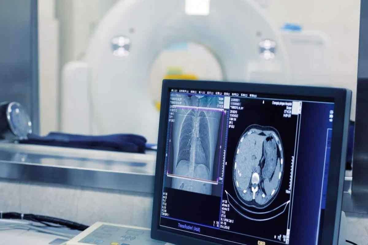
Basic Imaging in Solid Tumor Detection
Basic imaging is key for finding and diagnosing solid tumors. These methods give vital info on tumor size, location, and type. They help decide on further tests and treatments.
X-ray Examinations
X-rays are a basic tool for finding solid tumors. They work well for tumors in the lungs, bones, and other spots. Digital X-ray technology makes images better and cuts down on radiation.
Ultrasound Technology and Applications
Ultrasound uses sound waves to see inside the body. It’s great for finding tumors in organs like the liver, kidney, and thyroid. Ultrasound-guided biopsies help get tissue samples for more tests.
CT Scan Procedures and Protocols
CT scans are vital for finding solid tumors. They show detailed images of the body, pinpointing tumor locations and sizes. CT scan protocols change based on the body part and the needed diagnosis.
Advanced Imaging Techniques
We use advanced imaging to understand solid tumors better. This helps us create better treatment plans. These methods give us key info for diagnosing and staging tumors. This info helps doctors make smart choices.
MRI for Soft Tissue Tumor Visualization
Magnetic Resonance Imaging (MRI) is great for seeing soft tissue tumors. It shows details clearly and can tell different tissues apart. MRI helps see how far tumors spread into other areas. This is important for planning surgery.
Key benefits of MRI include:
- High-resolution imaging of soft tissues
- Ability to assess tumor extent and invasion
- Non-invasive and safe for patients
PET Scan for Metabolic Activity Assessment
Positron Emission Tomography (PET) scans are key for checking tumor activity. They show where tumors are using a lot of glucose. This helps tell if tumors are cancerous or not. It also shows how well treatments are working.
| Modality | Primary Use | Key Benefits |
| MRI | Soft tissue tumor visualization | High resolution, non-invasive |
| PET Scan | Metabolic activity assessment | Monitors tumor activity, guides treatment |
Nuclear Medicine Studies
Nuclear medicine studies, like scintigraphy, give more info on tumors. They help find out what kind of tumor it is. They also check if treatments are working.
By using these advanced imaging methods together, doctors get a full picture of solid tumors. This helps them make accurate diagnoses and plan treatments well.
Endoscopic Diagnostic Procedures
Endoscopy has changed how we fight cancer, making diagnosis and treatment more precise. It lets doctors see inside the body. This helps find problems and take tissue samples for tests.
Upper and Lower GI Endoscopy
Upper GI endoscopy looks at the esophagus, stomach, and duodenum. Lower GI endoscopy checks the colon and rectum. These are key for finding tumors in the gut. GI endoscopy is essential for spotting issues in the digestive system.
Bronchoscopy and Thoracoscopy
Bronchoscopy lets doctors see the airways and lungs. It helps find lung cancer and other lung problems. Thoracoscopy looks at the pleural space. It’s used to diagnose and treat lung and pleura issues.
Cystoscopy and Urological Procedures
Cystoscopy lets doctors see inside the bladder and urethra. It helps find bladder cancer and other urinary issues. Other tests might look at the upper urinary tract.
Endoscopic Ultrasound Applications
Endoscopic ultrasound (EUS) uses ultrasound with endoscopy. It gives detailed images of the digestive tract and nearby tissues. EUS is great for checking cancer stages and lymph nodes. The endoscopic ultrasound helps plan treatments.
Thanks to these endoscopic tests, doctors can better diagnose and treat solid tumors. This leads to better care for patients.
Biopsy Methods for Solid Tumor Diagnosis
Diagnosing solid tumors involves several biopsy methods. These methods help figure out what kind of tumor it is. A biopsy is key because it gives tissue samples for doctors to examine.
Fine Needle Aspiration (FNA)
Fine Needle Aspiration (FNA) is a small procedure to get cell samples. It uses a thin needle to take cells from areas that look suspicious. Then, these cells are checked cytologically.
Advantages of FNA:
- It’s a small procedure, which means less risk of problems
- It can be done with just local anesthesia or a little sedation
- Patients usually recover quickly
But, FNA might not always give clear results. This can happen if the sample is too small or if the area is too complex.
Core Needle Biopsy Techniques
Core Needle Biopsy (CNB) uses a bigger needle than FNA to get a piece of tissue. This method gives more tissue for doctors to study. This is important for diagnosing some tumors.
| Biopsy Method | Tissue Sample | Diagnostic Yield |
| FNA | Cellular aspirate | Cytological examination |
| CNB | Tissue core | Histological examination |
Excisional and Incisional Surgical Biopsies
Surgical biopsies are more involved. A surgeon takes part or all of a suspicious area. Excisional biopsy removes the whole area, while incisional biopsy takes just a piece.
“Surgical biopsies provide the most complete tissue samples. These are key for accurate diagnosis and treatment planning.”
Excisional biopsies are for smaller areas, while incisional biopsies are for bigger tumors. Both give valuable tissue for analysis. This helps doctors decide on the best treatment.
Pathological Analysis of Tumor Tissue
Getting a solid tumor diagnosed right is key. It helps doctors understand the tumor, decide on treatment, and predict how well a patient will do.
Histopathology Processing and Staining
Histopathology is all about looking at tissue under a microscope. It starts with taking a tumor sample, usually through a biopsy. The sample is then prepared, put in paraffin, and cut into thin slices.
These slices are stained with dyes like hematoxylin and eosin (H&E). This makes the cells stand out. A pathologist then looks at these slices to spot tumor features like cell shape and arrangement.
Histopathological examination is key for figuring out what kind of tumor it is and its grade. The grade shows how aggressive the tumor is. Knowing this helps doctors choose the right treatment.
Cytological Examination
Cytology looks at single cells or small groups of cells. It’s used for quick checks or when getting a biopsy is hard. Samples are taken through fine-needle aspiration (FNA) or other ways.
Cells are stained and checked for any odd features. Cytology is great for looking at tumors that are easy to reach. But, it might not give as much detail as histopathology.
Immunohistochemistry Markers
Immunohistochemistry (IHC) finds specific proteins in tumor cells. It uses antibodies that stick to these proteins, making them visible. IHC is super helpful for figuring out what kind of tumor it is and what it might do in the future.
Immunohistochemical markers can tell apart tumors that look similar under normal tests. For example, IHC can spot certain breast cancers by looking for estrogen or progesterone receptors. It can also find neuroendocrine tumors or lymphomas by their unique markers.
By using histopathology, cytology, and immunohistochemistry together, pathologists can give a full diagnosis. This helps doctors plan the best treatment for patients with solid tumors.
Molecular and Genetic Testing
Molecular and genetic testing are key in diagnosing and treating solid tumors. These tests give important details about tumor genetics and molecular traits. This info helps doctors create specific treatment plans.
Next-Generation Sequencing Technologies
Next-generation sequencing (NGS) is a cutting-edge tool. It quickly sequences large DNA or RNA amounts. This tech spots genetic changes in tumors, guiding treatment choices.
Benefits of NGS include:
- Comprehensive genetic analysis
- Identification of actionable mutations
- Personalized treatment planning
PCR and FISH Testing Methods
Polymerase Chain Reaction (PCR) and Fluorescence In Situ Hybridization (FISH) are methods for finding specific genetic changes in tumors.
| Technique | Description | Application |
| PCR | Amplifies specific DNA sequences | Detecting genetic mutations |
| FISH | Visualizes specific DNA sequences | Identifying chromosomal abnormalities |
Genomic Profiling for Precision Medicine
Genomic profiling looks at a tumor’s genetic makeup to find treatment targets. This method leads to precision medicine, where treatments match the patient’s genetic profile.
Genomic profiling can help:
- Identify genetic mutations associated with treatment response
- Develop personalized treatment plans
- Monitor treatment response and adjust as needed
By using molecular and genetic tests, doctors can give better, more targeted treatments for solid tumors.
Tumor Staging and Classification Systems
Tumor staging and classification systems are key in cancer diagnosis and treatment planning. They help us understand how big a tumor is and its impact on health. This information is vital for planning treatment and predicting outcomes.
TNM Staging Framework
The TNM staging system is a common way to classify tumors. It looks at the tumor’s size (T), nearby lymph nodes (N), and if it has spread (M). This system helps doctors describe how far cancer has spread.
TNM Staging Components:
- T (Tumor): Size and extent of the primary tumor.
- N (Node): Degree of lymph node involvement.
- M (Metastasis): Presence or absence of distant metastasis.
| TNM Stage | Description |
| T1, N0, M0 | Small tumor, no lymph node involvement, no metastasis. |
| T2, N1, M0 | Larger tumor, some lymph node involvement, no metastasis. |
| T3, N2, M1 | Large tumor, significant lymph node involvement, distant metastasis. |
Tumor Grading Criteria
Tumor grading is important in cancer classification. It looks at how aggressive a tumor is by its microscopic appearance. The grade is based on how much the tumor cells look like normal cells.
Tumor Grading Scale:
- Grade 1: Well-differentiated cells, less aggressive.
- Grade 2: Moderately differentiated cells.
- Grade 3: Poorly differentiated cells, more aggressive.
Organ-Specific Staging Systems
Some cancers have their own staging systems, aside from TNM. For example, brain tumors are staged differently because of their unique characteristics and locations.
Examples of Organ-Specific Staging:
- Brain Tumors: Staged based on tumor type, grade, and location.
- Lymphoma: Staged using the Ann Arbor Staging System.
Knowing these staging systems is key for effective treatment plans and predicting outcomes. Accurate staging and classification help us manage cancer better and improve patient care.
Specialized Approaches for Different Tumor Types
Diagnosing solid tumors needs a custom plan. Each tumor type, like those in the breast, lung, and more, needs its own way of being checked. This ensures the best care for each patient.
Breast Tumor Diagnostic Protocol
Checking for breast tumors uses imaging and biopsies. We start with mammography and ultrasound to see the tumors. Sometimes, MRI helps to see how big the tumor is.
For tissue samples, we use fine-needle aspiration and core needle biopsy. These help doctors look at the tissue under a microscope.
Lung Tumor Evaluation Methods
Lung tumor checks often use chest X-rays and CT scans. We also use PET scans to see how active the tumor is.
Bronchoscopy lets us see inside the airways and get tissue for biopsies. Sometimes, needle biopsy is used to reach lung tumors that are hard to get to.
Gastrointestinal Tumor Assessment
Diagnosing tumors in the GI tract uses endoscopy and imaging. We use upper GI endoscopy and colonoscopy to look inside and get samples.
CT scans and PET scans help us see how big the tumor is and if it has spread. Endoscopic ultrasound is used to check how deep the tumor is.
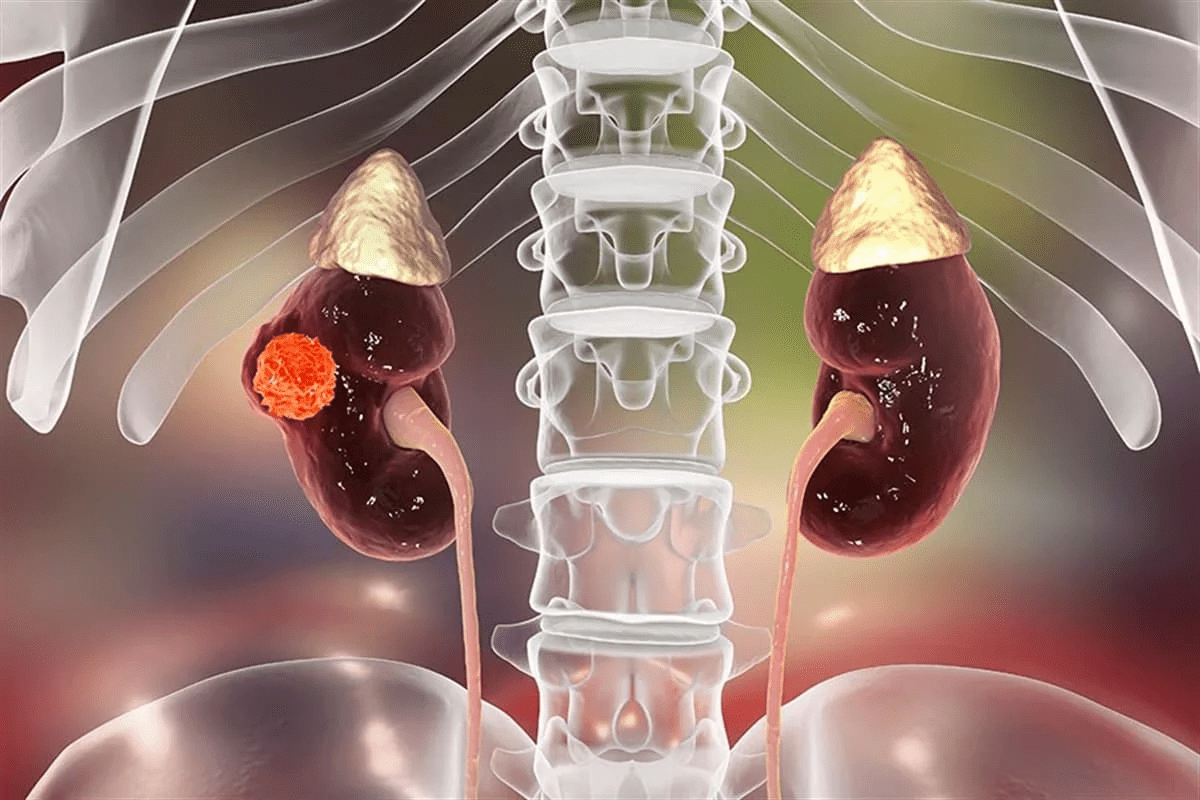
Genitourinary Tumor Diagnosis
Checking for tumors in the genitourinary system uses imaging and biopsies. We use ultrasound and CT scans to look at the kidneys, bladder, and prostate.
Cystoscopy lets us see inside the bladder and get samples. We also use PSA testing to check for prostate cancer.
Multidisciplinary Tumor Boards
Multidisciplinary tumor boards are groups of experts from different medical fields. They discuss and manage complex tumor cases together. This teamwork ensures patients get care that fits their needs perfectly.
Composition and Function
A tumor board usually has oncologists, surgeons, radiologists, and pathologists. The team can change based on the tumor type. For example, a breast cancer board might have breast surgeons and oncologists.
The main job of these boards is to review patient cases. They talk about diagnosis and treatment options. They decide on the best care plan together.
| Specialty | Role in Tumor Board | Contribution to Patient Care |
| Oncologist | Provides medical oncology expertise | Develops treatment plans, including chemotherapy and targeted therapy |
| Surgeon | Offers surgical expertise | Performs surgical resections, provides input on surgical options |
| Radiologist | Interprets imaging studies | Provides diagnostic information, assists in treatment planning |
Case Presentation and Review Process
At a tumor board meeting, cases are presented in detail. This includes medical history and current treatment plans. The team reviews and discusses each case, considering multiple perspectives and treatment options.
The team looks at diagnostic images and pathology results carefully. This helps them make a well-informed treatment plan.
Benefits of Collaborative Diagnosis
Multidisciplinary tumor boards have many benefits. They improve diagnosis, treatment planning, and patient outcomes. By working together, these boards ensure patients get the best care.
The benefits of collaborative diagnosis include:
- Improved diagnostic accuracy through the review of multiple experts
- Enhanced treatment planning, considering various perspectives and options
- Better patient outcomes, resulting from coordinated, compassionate care
Challenges and Advances in Tumor Diagnosis
Even with new diagnostic tools, finding solid tumors is hard. Tumors are complex, and they can look different in everyone. Current methods have their limits.
Addressing Inconclusive Results
When tests aren’t clear, it can cause worry and delay treatment. We’re looking into new ways to test and improve what we already have. This aims to make diagnoses more accurate.
One idea is to make imaging better. For example, using special agents in MRI or CT scans to see tumors more clearly.
Liquid Biopsy Technologies
Liquid biopsy is a big step forward. It lets us check for tumor DNA in blood without surgery. This gives us clues about tumors without the need for invasive tests.
This tech could help find tumors early, track how well treatments work, and spot when tumors become resistant. It could change how we fight cancer.
Artificial Intelligence in Diagnostic Imaging
AI is changing how we look at images in radiology. AI can scan lots of data fast and find things humans might miss.
AI helps doctors spot tumors sooner and get better at reading images. This means better care for patients.
Emerging Biomarkers and Detection Methods
New biomarkers and ways to find them are key to better tumor diagnosis. Technologies like nanotechnology and advanced proteomics are being looked at. They might help find new biomarkers and make tests more sensitive.
These new tools could help us find tumors sooner and more accurately. This could lead to better treatments.
Conclusion
Diagnosing solid tumors needs a mix of tests and procedures. We’ve looked at imaging, biopsies, and genetic tests. These show how teamwork is key in finding cancer.
Having a detailed plan for diagnosis is vital. It helps doctors find the best treatment. By using tests and scans, we can give better care and improve results.
Tests like blood tests and PET scans help doctors make accurate diagnoses. Working together, we ensure patients get the right treatment. As we keep improving, we’re dedicated to top-notch care for all patients.
FAQ
What is a solid tumor, and how is it diagnosed?
A solid tumor is an abnormal mass of tissue. It can be benign or malignant. To diagnose it, doctors use many tests and procedures. These include imaging studies, biopsies, and molecular testing.
What are the common symptoms of solid tumors?
Symptoms of solid tumors vary by location and type. Common signs include unexplained weight loss and pain. Changes in skin or bowel habits and unusual bleeding are also symptoms.
For example, esophageal tumors may cause difficulty swallowing. Bladder tumors might lead to hematuria.
What is the role of imaging tests in diagnosing solid tumors?
Imaging tests are key in finding and diagnosing solid tumors. Tests like x-rays, ultrasound, CT scans, MRI, and PET scans are used. They show the tumor’s size, location, and if it has spread.
What is a biopsy, and how is it used to diagnose solid tumors?
A biopsy removes tissue or cells from a tumor for examination. There are different biopsy methods, like fine needle aspiration and core needle biopsy. The sample is then analyzed to find cancer cells.
What is the TNM staging system, and how is it used?
The TNM staging system helps understand solid tumors. It looks at the tumor’s size and spread. It also checks nearby lymph nodes and distant metastasis.
What is the role of molecular and genetic testing in diagnosing solid tumors?
Molecular and genetic testing find specific genetic changes in tumors. Tests like next-generation sequencing and PCR are used. This info helps choose treatments and predict outcomes.
How are tumor markers used in diagnosing solid tumors?
Tumor markers are substances made by cancer cells. Blood tests can find these markers. They suggest certain cancers but are not definitive. They’re used with other tests for diagnosis.
What is the role of multidisciplinary tumor boards in diagnosing and treating solid tumors?
Multidisciplinary tumor boards include experts from various fields. They discuss and manage complex cases. This ensures patients get the best care.
What are some of the challenges in diagnosing solid tumors?
Diagnosing solid tumors can be tough. Factors like tumor location and size play a role. Inconclusive results and technology limits are also challenges. New technologies like liquid biopsy and artificial intelligence are being developed to help.
How is genetic testing used in the diagnosis and treatment of solid tumors?
Genetic testing finds specific genetic changes in tumors. This info helps choose treatments and predict outcomes. It also finds targets for therapy.
What is the significance of histopathology in diagnosing solid tumors?
Histopathology examines tissue structure and cell morphology. It helps diagnose and characterize solid tumors. This analysis informs treatment decisions and predicts outcomes.
References
- Huang, J., & Zhang, L. (2021). The role of surgery in the multidisciplinary management of solid tumors. Journal of Clinical Medicine, *10*(15), 3298. https://www.ncbi.nlm.nih.gov/pmc/articles/PMC8347462/


