Last Updated on December 1, 2025 by Bilal Hasdemir

Did you know nuclear medicine is key in finding and managing cancer? Many cancer diagnoses use SPECT tumor detection scans to spot and track the disease
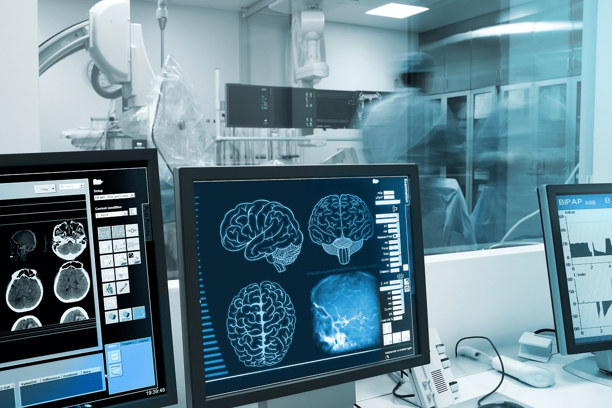
Single Photon Emission Computed Tomography (SPECT) is a top-notch imaging tool. It uses tiny amounts of radioactive tracers to see and check how tissues work. This is vital for cancer detection and planning treatments.
SPECT scans use special medicines that light up where tissues are not working right. This helps show where tumors are, how big they are, and how active they are.
Key Takeaways
- SPECT scans are used in nuclear medicine for cancer diagnosis.
- Radiopharmaceuticals are key in showing where tissues are not working right.
- The technology helps find where tumors are, how big they are, and how active they are.
- SPECT scans are important for planning and checking on cancer treatments.
- Nuclear medicine, including SPECT, is vital in managing cancer.
What is a SPECT Scan?

A SPECT scan is a way to see how tissues in the body work. It’s great for cancer diagnostic imaging. Doctors use it to see how tumors and other issues behave.
Definition and Basic Principles
SPECT, or Single Photon Emission Computed Tomography, uses a radiopharmaceutical to find active areas in the body. These areas might have tumors. The gamma camera picks up the radiation, making 3D images of these areas.
This method helps doctors understand how tumors work. It shows how active they are, which helps in planning treatment.
| Key Components | Function |
| Radiopharmaceutical | Accumulates in areas of high metabolic activity, emitting gamma rays |
| Gamma Camera | Detects gamma rays emitted by the radiopharmaceutical |
| Image Reconstruction Software | Constructs 3D images from the detected gamma rays |
How SPECT Differs from Other Nuclear Medicine Techniques

SPECT scans are different from PET scans. Both use radiopharmaceuticals to see how tissues work. But SPECT is better at showing specific areas, like tumors.
One big plus of SPECT scans is they can focus on nuclear medicine tumor imaging. They use special radiopharmaceuticals to target certain tumors. This makes them very useful for doctors.
The Science Behind SPECT Tumor Detection
SPECT tumor detection uses radiopharmaceuticals that tumors take up. This is seen with gamma camera technology. It helps doctors see and check tumor activity in the body.
Radiopharmaceuticals Used in Tumor Imaging
Many radiopharmaceuticals are used in SPECT tumor detection. Each one is chosen based on the tumor type and how it works. For example, Tc-99m sestamibi is often used for parathyroid adenomas and some cancers. I-131 is used for thyroid cancer.
| Radiopharmaceutical | Application |
| Tc-99m sestamibi | Parathyroid adenomas, certain cancers |
| I-131 | Thyroid cancer |
| Tc-99m MDP | Bone metastases, bone cancer |
Gamma Camera Technology and Image Reconstruction
The gamma camera is key in SPECT imaging. It finds the gamma radiation from the radiopharmaceuticals in tumors. Then, special algorithms make detailed 3D images of tumor activity.
The mix of radiopharmaceuticals and gamma camera technology makes SPECT great for cancer detection and tracking. Knowing how SPECT works helps doctors use it to help patients more.
How SPECT Scans Work to Identify Abnormal Tissue
SPECT scans are key in finding abnormal tissue by spotting changes in how cells work and blood flow. This is very important for finding and treating cancer.
Detecting Metabolic Activity in Tumors
Tumors have different metabolic activity than normal cells. SPECT scans use radiopharmaceuticals to find this difference. These substances go to cells that use a lot of glucose, which is common in cancer cells.
A tiny amount of radioactive material is given to the patient. It goes to the tumor. The SPECT scanner then shows detailed images of the tumor’s activity.
Visualizing Blood Flow Patterns in Cancerous Tissue
SPECT scans also show how blood flows in tumors. Cancerous tissues have different blood vessel and flow patterns. By using special radiopharmaceuticals, SPECT scans can show these patterns.
This info helps doctors understand the tumor, plan treatment, and check how well it’s working. For example, tumors with lots of blood flow might need stronger treatments.
| Characteristics | Normal Tissue | Cancerous Tissue |
| Metabolic Activity | Normal glucose metabolism | Altered, often increased glucose metabolism |
| Blood Flow | Normal blood vessel structure and flow | Abnormal blood vessel formation and flow patterns |
SPECT scans give a full picture of tumor biology by spotting metabolic and blood flow changes. This info is key for managing cancer, from finding it to treating it and checking on it later.
Types of Tumors Detectable with SPECT Imaging
SPECT imaging is key in finding cancers by using special medicines. It can spot tumors in many parts of the body.
Brain Tumors and Neurological Lesions
SPECT imaging is great for finding brain tumors and other brain issues. It uses medicines that can get past the blood-brain barrier. This gives info on how tumors work and how fast they grow.
Bone Tumors and Metastases
Bone tumor imaging with SPECT is very good at finding bone tumors and metastases. It uses medicines that stick to bone, like Technetium-99m methylene diphosphonate (Tc-99m MDP). SPECT shows where bone activity is high, which means tumor is present.
Other Cancer Types Detectable via SPECT
SPECT imaging can also find other cancer types, like some endocrine and neuroendocrine tumors. The right medicine is key, as it must match the tumor’s special traits. For example, SPECT can spot tumors based on receptors or metabolism.
In summary, SPECT imaging is a strong tool in fighting cancer. It can find many tumor types and gives important details. This makes it a key part of cancer care.
SPECT-CT: Enhanced Tumor Detection Capabilities
SPECT-CT imaging combines two powerful tools for better cancer diagnosis. It merges the strengths of SPECT and CT technologies. This gives a deeper understanding of tumor biology and anatomy.
Benefits of Combined Anatomical and Functional Imaging
The mix of SPECT and CT offers many advantages in finding and understanding tumors. Improved diagnostic accuracy is a key benefit. SPECT-CT gives detailed images of tumor metabolism and anatomy.
- Enhanced tumor localization
- Better differentiation between benign and malignant lesions
- More accurate staging of cancer
This technology helps reduce the need for extra tests. It makes the diagnostic process smoother. And it could lead to better patient outcomes.
Improved Localization of Tumors
SPECT-CT greatly improves tumor location accuracy. It combines SPECT’s functional data with CT’s anatomical detail. This lets healthcare providers precisely identify tumor locations and their relation to nearby structures.
- Accurate tumor localization aids in surgical planning
- Precise targeting for radiation therapy
- Better monitoring of treatment response
This precise localization is very useful in complex areas like the head and neck or pelvis. Accurate tumor definition is key for effective treatment planning.
Effectiveness of SPECT Tumor Detection in Clinical Practice
When we talk about SPECT’s role in finding tumors, we look at its sensitivity and specificity. These numbers tell us how well SPECT scans can spot tumors. This is key for making a diagnosis, planning treatment, and checking how well treatment is working.
Sensitivity and Specificity Rates
The success of SPECT in finding tumors depends on several things. This includes the type of tumor, the radiopharmaceutical used, and the imaging method. For some tumors, like brain tumors and certain bone metastases, SPECT can be very accurate. But, it might not always be right because of non-specific uptake of radiopharmaceuticals in other tissues.
A study in a Journal showed SPECT with Technetium-99m was 85% sensitive and 90% specific for some bone metastases. Another study with Thallium-201 for brain tumors reported 80% sensitivity and 85% specificity.
False Positives and False Negatives
SPECT, like any test, can make mistakes. False positives happen when a scan says there’s a tumor when there isn’t. This can cause worry, more tests, and wrong treatments.
False negatives are when a scan misses a tumor. This can mean a late start to treatment, which might hurt patient results. Small tumors, low activity, or SPECT system limits can cause these errors.
It’s important to know these possible mistakes when looking at SPECT scans. By using SPECT with other tests and medical knowledge, doctors can get better at finding and understanding tumors.
Comparing SPECT to Other Tumor Imaging Modalities
The world of cancer imaging is full of different tools like SPECT, PET, MRI, and CT. Each has its own strengths and weaknesses. Knowing these helps doctors pick the best tool for diagnosing cancer.
SPECT vs. PET for Cancer Detection
SPECT and PET are both used in cancer imaging. PET scans are great at showing how active cells are. But, SPECT is sometimes better because it’s more available and cheaper.
When deciding between SPECT and PET, doctors look at what they need to know. They consider the type of cancer and the specific questions they have.
SPECT vs. MRI for Tumor Visualization
MRI is amazing for showing soft tissues, like in the brain. SPECT adds functional info that MRI can’t. Together, they give a clearer picture of tumors.
SPECT vs. CT for Cancer Diagnosis
CT scans are great for seeing tumors in different parts of the body. SPECT-CT combines SPECT’s function with CT’s anatomy. This makes finding tumors more accurate.
CT is good for seeing structural changes. But, SPECT shows how active tumors are. This helps doctors understand tumors better.
In summary, each imaging tool has its role in fighting cancer. The choice between SPECT, PET, MRI, and CT depends on the situation. It also depends on the type of cancer and what doctors need to know.
The Patient Experience During a SPECT Tumor Detection Scan
Patients getting SPECT scans for tumors need to know what to expect. They must prepare and understand the safety steps. A good scan depends on the patient’s knowledge of the process.
Preparation and Procedure
Before a SPECT scan, patients get specific instructions. They might need to fast or avoid certain medicines. These steps help ensure the scan’s accuracy.
On scan day, patients bring needed papers and ID to the imaging center. They then get a radiopharmaceutical, a tiny bit of radioactive stuff. This helps doctors see the tumor area.
During the scan, patients lie on a table. The gamma camera moves around them to take images from different sides. It’s painless and can last from 30 minutes to hours, based on the scan’s complexity.
| Preparation Step | Description |
| Fasting | Patients may be required to fast for a certain period before the scan. |
| Medication Avoidance | Certain medications may need to be avoided before the scan to prevent interference. |
| Radiopharmaceutical Administration | A small amount of a radioactive substance is administered to visualize the area of interest. |
Radiation Exposure and Safety Considerations
One big worry for patients is radiation from SPECT scans. Though the dose is small, it’s key to know the risks and benefits.
The scan’s benefits usually outweigh the radiation risks. But, patients should talk to their doctor about any worries.
Safety measures are taken to lower radiation exposure. This includes using the least amount of radiopharmaceutical and checking the gamma camera’s work.
Knowing about SPECT scan preparation, procedure, and safety helps patients understand their diagnostic journey better.
Clinical Applications of SPECT in Oncology
SPECT imaging is key in oncology for managing cancer patients. It offers functional insights into tumors, essential at all stages of cancer care.
Initial Cancer Diagnosis
SPECT scans help spot tumors at the start. They use special drugs to show where cancer might be. This is because cancer cells act differently than normal cells.
It’s great for finding cancer in bones, for example. A bone SPECT scan can show where cancer might have spread.
Staging and Treatment Planning
Knowing how far cancer has spread is vital. SPECT helps by showing how active tumors are. This info helps doctors plan the best treatment.
By mixing SPECT data with CT or MRI images, doctors get a clearer picture. This helps in making more accurate treatment plans.
Monitoring Treatment Response
SPECT scans track how well treatments work. They watch for changes in tumor activity. This shows if the cancer is responding to treatment.
This info is key for tweaking treatment plans. If a tumor’s activity goes down, it might mean the treatment is working. This could lead to adjusting or continuing the current treatment.
| Application | Description | Benefits |
| Initial Cancer Diagnosis | Detection of tumors using specific radiopharmaceuticals | Early identification of cancer, guiding further diagnostic procedures |
| Staging and Treatment Planning | Assessment of tumor extent and metabolic activity | Accurate staging, informed treatment decisions |
| Monitoring Treatment Response | Evaluation of changes in tumor metabolic activity | Adjustment of treatment plans based on response |
Limitations of SPECT in Tumor Detection
SPECT scans are key in cancer diagnosis but have some limits. It’s important for doctors and patients to know these to make better choices about treatment.
Resolution Constraints
SPECT scans can’t see as much detail as MRI or CT scans. This makes it hard to spot small tumors or those that don’t show up much. It’s a big problem when tumors are in tricky spots or blend in with the background.
Impact of Resolution Constraints: The low detail can mess up how doctors plan treatments and check if they’re working.
Tumor Types Poorly Visualized by SPECT
Not every tumor shows up well on SPECT scans. Some tumors don’t take up the special dye used, or they blend in with the rest of the tissue. This makes it tough to find certain soft tissue tumors or those that don’t use much energy.
The table below shows which tumors SPECT can spot:
| Tumor Type | Detectability with SPECT |
| Brain Tumors | Moderate to High |
| Bone Metastases | High |
| Soft Tissue Sarcomas | Low to Moderate |
| Lymphoma | Moderate |
In summary, SPECT is a great tool in fighting cancer, but it has its downsides. We need better SPECT tech and new dyes to see tumors more clearly. This will help doctors diagnose better and treat more effectively.
Insurance Coverage and Costs for SPECT Tumor Scans in the US
The cost of SPECT scans, including insurance coverage, is a big deal for cancer patients. Knowing how coverage works can help patients and doctors make better choices about SPECT scans.
Insurance for SPECT scans changes a lot, with different plans and providers covering different things. In the US, Medicare and Medicaid are key in paying for SPECT scans for some reasons.
Medicare and Medicaid Coverage
Medicare usually pays for SPECT scans if they’re needed for diagnosing or managing some conditions, like cancer. Medicaid coverage can change by state, but it usually follows Medicare’s rules for SPECT scans.
| Insurance Provider | Coverage for SPECT Scans | Conditions Covered |
| Medicare | Generally covered when medically necessary | Cancer diagnosis and management |
| Medicaid | Varies by state; generally covers for certain conditions | Cancer and other specific conditions |
| Private Insurance | Varies by policy and provider | Depends on the specific policy |
Private Insurance Considerations
Private insurance for SPECT scans can change a lot, based on the policy and provider. Some plans might cover SPECT scans for certain reasons, while others might not. It’s key for patients to check their insurance and talk to their doctors to know what’s covered.
The cost of SPECT scans can be high, so knowing about insurance is important for managing costs. Patients should work with their doctors and insurance to figure out what’s covered and what they might have to pay for.
Advancements in SPECT Technology for Improved Cancer Imaging
Advances in SPECT technology are changing how we fight cancer. New research and development are key. They bring new radiopharmaceuticals and better hardware and software.
New Radiopharmaceuticals for Specific Tumor Types
New radiopharmaceuticals are a big deal for SPECT technology. They are made to find certain tumors better. This makes SPECT scans more accurate.
Targeted radiopharmaceuticals are a big step forward. They focus on specific cancer cells. This helps doctors tell different tumors apart and see how aggressive they are.
Hardware and Software Innovations
Improvements in SPECT hardware and software are also important. Modern gamma cameras are more sensitive and clear. This means better images and more accurate diagnoses.
Software updates, like new image algorithms, also help. They make SPECT images clearer and give more details about tumors.
Combining these advancements in hardware, software, and radiopharmaceuticals will greatly help SPECT in cancer care. As these technologies get better, they will help diagnose and treat cancer more effectively.
Conclusion: The Role of SPECT in Modern Cancer Care
SPECT is a key tool in cancer diagnosis and management. It gives insights into tumor biology and metabolism. As cancer care evolves, SPECT’s role will stay important, used with other imaging and tools.
Using SPECT for tumor imaging helps doctors understand tumor characteristics. This information aids in better treatment planning and monitoring. A SPECT scan can also spot cancer presence and extent, guiding treatment and improving patient results.
Healthcare providers can make the most of SPECT by knowing its strengths and weaknesses. This knowledge helps in diagnosing, planning, and monitoring cancer care. It improves the quality of care for cancer patients.
FAQ
What is a SPECT scan, and how does it detect tumors?
A SPECT scan is a way to see inside the body using tiny amounts of radioactive tracers. It helps find tumors by showing where the body’s activity is different. This is done with special medicines that light up tumors and are seen by a camera.
What types of tumors can be detected using SPECT imaging?
SPECT scans can find many kinds of tumors. This includes brain, bone, and some endocrine tumors. It depends on the medicine used and how the scan is done.
How does SPECT-CT enhance tumor detection capabilities?
Adding CT to SPECT makes it better for finding tumors. It combines the body’s activity seen by SPECT with detailed pictures from CT. This gives a clearer view of tumors.
What are the limitations of SPECT in tumor detection?
SPECT has some limits. It’s hard to see small tumors or those that don’t use much energy. Some tumors might not show up well because they don’t take up the medicine.
How does SPECT compare to other tumor imaging modalities like PET, MRI, and CT?
SPECT is one of many ways to look at tumors. PET shows how active tumors are, MRI gives soft tissue details, and CT shows anatomy. Each has its own strengths.
What is the patient experience like during a SPECT tumor detection scan?
Getting ready for a SPECT scan includes following certain rules. Then, patients lie down while the camera takes pictures from all sides. It’s safe and helps doctors a lot.
What are the clinical applications of SPECT in oncology?
SPECT is key in fighting cancer. It helps find cancer, see how far it has spread, plan treatments, and check if they’re working. It gives doctors important info.
Is SPECT tumor detection covered by insurance in the US?
In the US, Medicare and Medicaid pay for SPECT scans for some reasons. But private insurance varies. It’s important to know what’s covered to plan costs.
What advancements are being made in SPECT technology for improved cancer imaging?
SPECT is getting better thanks to new medicines and technology. Better cameras and software make pictures clearer. This helps doctors find and treat tumors better.
References
- Abikhzer, G., Keidar, Z., Frenkel, A., Israel, O., & Even-Sapir, E. (2014). SPECT/CT in tumor imaging: Technical aspects and clinical applications. European Journal of Nuclear Medicine and Molecular Imaging, 41(Suppl 1), S67“S80. https://pubmed.ncbi.nlm.nih.gov/23990144/


