
The human heart is a complex organ with four main chambers. These chambers work together to circulate blood throughout the body. It is located in the thoracic cavity, and its anatomy is key to understanding its function.
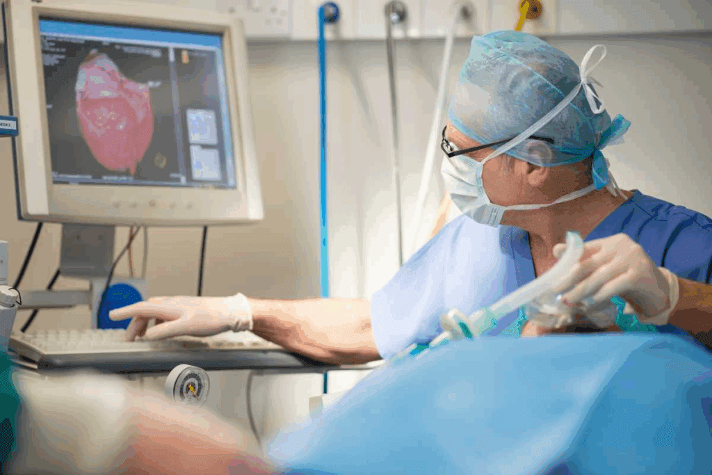
At Liv Hospital, we know how important it is to understand the heart’s internal view. Our patient-centered approach helps you learn about the heart structure in detail. We make sure everything is labeled clearly.
Knowing the structure of the heart is vital for accurate diagnosis and care. We will look at the 12 essential parts of the heart. This will give you a full understanding of its anatomy.
The human heart is a marvel of design. Its complex structure is key to our health. As we explore the heart’s anatomy, we see its vital role. Explore the structures of the heart with labeled internal diagrams and key functions.
The heart has four main chambers: two atria and two ventricles. It also has valves like the tricuspid, mitral, pulmonary, and aortic valves. These valves control blood flow. Knowing about these parts helps us understand how the heart works.
The heart sits in the thoracic cavity, a bit to the left in the chest. This spot is vital for its job. It’s linked to its role in the circulatory system.
Knowing the structure of the heart is key to understanding its function. It’s important for health. Knowing the heart’s anatomy helps in diagnosing and treating heart issues. It shows the heart’s internal structure is essential for its function.
The heart’s complex design is key to its function. Knowing its main parts helps us understand its role in the body. We’ll look at its four-chamber design, valves, heart layers, and blood vessel connections. These elements work together to keep blood flowing smoothly.
The heart has four chambers: the right atrium, left atrium, right ventricle, and left ventricle. These chambers separate blood based on its oxygen level. This ensures oxygen-rich blood reaches the body’s tissues and organs.
The right atrium gets deoxygenated blood from the body. The left atrium receives oxygen-rich blood from the lungs. The right ventricle sends blood to the lungs for oxygen. The left ventricle pumps oxygen-rich blood to the rest of the body.
The heart’s valves are vital for blood flow. There are four valves: the tricuspid, mitral, pulmonary, and aortic valves. These valves control blood flow between chambers and to the body, preventing backflow.
The tricuspid and mitral valves separate the right and left sides of the heart. The pulmonary valve controls blood flow to the lungs. The aortic valve regulates blood flow to the rest of the body.
The heart wall has three layers: the endocardium, myocardium, and epicardium. Each layer has a specific role in the heart’s function.
The endocardium lines the heart’s chambers and valves. The myocardium, made of cardiac muscle cells, pumps blood. The epicardium is the outer layer, protecting the heart.
The heart connects to major blood vessels. These vessels carry blood to and from the body. The pulmonary arteries and veins transport blood between the heart and lungs. The aorta and its branches distribute oxygen-rich blood to the body.
The superior and inferior vena cava bring deoxygenated blood to the heart. The pulmonary veins return oxygenated blood from the lungs. This completes the circulatory circuit.
The right atrium is the first chamber for deoxygenated blood. It gets blood from the body through the superior and inferior vena cava. Knowing how it works helps doctors diagnose and treat heart issues.
The right atrium has unique features and boundaries. It’s separated from the left atrium by the interatrial septum. Inside, you’ll find the crista terminalis and the fossa ovalis. The superior and inferior vena cava bring blood into it.
Inside the right atrium, there are important structures. The sinoatrial node is near the superior vena cava. It acts as the heart’s natural pacemaker. The right atrium also has the coronary sinus, which takes deoxygenated blood from the heart muscle.
The right atrium’s main job is to pump deoxygenated blood into the right ventricle. This is through the tricuspid valve. It’s key for blood to get oxygen in the lungs. The right atrium’s contraction helps blood flow well into the right ventricle, keeping circulation strong.
The left atrium is a key part of the heart. It gets oxygen-rich blood from the lungs. Knowing its role is important when we look at the heart’s structure.
The left atrium is made to hold oxygenated blood well. Its walls are thinner than the ventricles. This is because it doesn’t need to push blood as hard.
The heart has four chambers: the right atrium, right ventricle, left ventricle, and left atrium. This answers the question of how many cavities in the heart: four.
The left atrium connects to the lungs through the pulmonary veins. These veins bring oxygen-rich blood back to the heart. There are usually four pulmonary veins, two from each lung, that go into the left atrium.
This connection is key for the heart to circulate oxygenated blood around the body.
The left atrium is very important in the cardiac cycle. It pumps oxygenated blood into the left ventricle. The left ventricle then sends it to the body.
This process is vital for keeping the body’s oxygen levels up. The left atrium’s work is essential for good heart health.
Understanding the the structure of a heart and its parts, like the left atrium, is key. It helps us see how the heart and blood system work together.
The right ventricle is key to the heart’s function. It makes sure deoxygenated blood gets to the lungs for oxygen. Knowing how the right ventricle works is important for good care.
The right ventricle is different from other heart chambers. Its walls are thinner, fitting its role in pumping blood to the lungs. Its shape helps it pump blood well into the pulmonary trunk.
The right ventricle has two important parts: the moderator band and papillary muscles. The moderator band helps with electrical impulses. Papillary muscles keep the tricuspid valve closed during heart contraction.
The right ventricle’s main job is to pump blood into the pulmonary circuit. It pushes blood through the pulmonary valve into the pulmonary artery. This blood then goes to the lungs.
Understanding the heart’s structure, like the right ventricle, is key to top-notch healthcare. Knowing the heart’s details helps doctors make better diagnoses and treatments. This improves patient care.
The left ventricle is the heart’s main pumping chamber. It has thick muscular walls for high-pressure pumping. We will explore its structure and function in detail.
The left ventricle has the thickest walls of all the heart’s chambers. This is necessary for generating the high pressures needed to pump blood throughout the body. In contrast, the right ventricle pumps blood to the lungs and requires less pressure.
The muscular walls of the left ventricle are made of cardiac muscle cells. These cells are arranged in a specific pattern to maximize contractile force. This arrangement allows for efficient pumping of blood into the aorta, the largest artery in the body. The aorta then distributes oxygenated blood to various parts of the body.
Internally, the left ventricle contains several key structures that facilitate its function. The mitral valve, also known as the bicuspid valve, regulates blood flow from the left atrium into the left ventricle. The valve is supported by papillary muscles and chordae tendineae, which prevent the valve leaflets from being forced back into the atrium during ventricular contraction.
The left ventricle also features a smooth inner lining called the endocardium. This lining helps to reduce friction and prevent blood clotting within the chamber.
The primary role of the left ventricle is to pump oxygenated blood into the systemic circulation. It achieves this by contracting and generating high pressures. These pressures push blood through the aortic valve into the aorta.
The efficiency of the left ventricle in performing this function is critical. It ensures adequate blood supply to tissues and organs throughout the body. Any dysfunction in the left ventricle, such as heart failure or myocardial infarction, can have significant implications for overall health.
| Feature | Description | Importance |
| Thick Muscular Walls | Composed of cardiac muscle cells for high-pressure pumping | Essential for systemic circulation |
| Mitral Valve | Regulates blood flow from left atrium to left ventricle | Prevents backflow and ensures efficient pumping |
| Papillary Muscles and Chordae Tendineae | Supports the mitral valve during ventricular contraction | Prevents valve leaflets from being forced back into the atrium |
The heart valves are key to keeping blood flow in one direction. There are four valves in the heart. Each one has a special job to make sure blood moves well around the body.
The tricuspid valve is between the right atrium and ventricle. It lets blood move from the right atrium to the right ventricle but stops it from going back. The tricuspid valve has three leaflets that open and close with each heartbeat, making sure blood flows the right way.
The mitral valve, also known as the bicuspid valve, is between the left atrium and ventricle. It has two leaflets that control blood flow from the left atrium to the left ventricle. The mitral valve is key for the left side of the heart to work well.
The pulmonary valve is between the right ventricle and the pulmonary artery. It lets blood flow from the right ventricle to the pulmonary artery. The pulmonary valve prevents backflow into the right ventricle, ensuring that blood continues its journey to the lungs.
The aortic valve is between the left ventricle and the aorta. It controls the flow of oxygenated blood from the left ventricle into the aorta. The aortic valve is critical for maintaining the systemic circulation, making sure oxygenated blood reaches all parts of the body.
Knowing about the heart’s structures, including the valves, is important for diagnosing and treating heart issues. The detailed design of these valves makes sure blood flows in one direction. This keeps the circulatory system working efficiently.
| Valve | Location | Function |
| Tricuspid | Right atrium and ventricle | Regulates right-sided flow |
| Mitral | Left atrium and ventricle | Controls left atrial outflow |
| Pulmonary | Right ventricle and pulmonary artery | Gateway to the lungs |
| Aortic | Left ventricle and aorta | Entry to systemic circulation |
The heart wall has three key layers: the myocardium, endocardium, and epicardium. These layers work together to help the heart function. Knowing about these layers is key to understanding how the heart works and keeps us healthy.
The heart wall’s design is complex. It allows the heart to do its job well in the circulatory system.
The myocardium is the heart’s muscular middle layer. It’s responsible for pumping blood around the body. It’s the thickest layer and made of muscle cells that can contract strongly.
The myocardium’s structure is vital for pumping blood well. Its thickness changes in different heart chambers. The ventricles have thicker myocardium than the atria.
The endocardium is the heart’s innermost layer. It lines the chambers and valves. It’s a thin, smooth layer that reduces friction as blood moves through the heart.
The endocardium also helps with the heart’s electrical system. It’s connected to the blood vessel lining.
The epicardium is the heart’s outer layer, also known as the visceral pericardium. It’s a layer of connective tissue that covers the heart. It’s part of the pericardial sac around the heart.
The epicardium has blood vessels and nerves for the heart. It also makes pericardial fluid. This fluid reduces friction between the heart and other structures.
The heart’s three layers work together for proper function. A cardiologist notes, “The heart wall’s complex structure shows the body’s amazing ability to keep things balanced.” Each layer is essential for heart health and overall well-being.
Understanding the cardiac septa is key to knowing how the heart works. These walls divide the heart’s chambers, making sure blood flows right. This is important for the heart’s function.
The interatrial septum is a thin wall between the right and left atria. It’s vital for keeping oxygen-rich and oxygen-poor blood separate. A key feature is the fossa ovalis, a depression from fetal development.
The interventricular septum is thicker and separates the right and left ventricles. It’s a muscular wall that helps the heart pump blood. It works with the ventricular walls for efficient blood pumping.
Septal defects can cause serious health problems. Atrial septal defects (ASD) and ventricular septal defects (VSD) are common. These defects can lead to blood shunting, causing heart issues.
| Type of Defect | Description | Clinical Impact |
| Atrial Septal Defect (ASD) | A hole in the interatrial septum | May cause shunting of blood between atria, potentially leading to right heart overload |
| Ventricular Septal Defect (VSD) | A hole in the interventricular septum | Can cause shunting of blood between ventricles, potentially leading to left heart overload and pulmonary hypertension |
Knowing about septal defects is key for diagnosis and treatment. Accurate imaging is vital for finding these defects and planning treatment.
By understanding the heart’s structure, including the septa, doctors can give better care. This leads to more accurate diagnoses and effective treatments, improving patient outcomes.
The heart’s core is the cardiac conduction system. It sends out electrical impulses that make our heart beat in sync. This system is key for a normal heart rhythm, ensuring our heart pumps blood well.
The sinoatrial (SA) node is in the right atrium. It starts the heartbeat with electrical impulses. These impulses’ rate changes with factors like the nervous system and hormones.
Key Functions of the SA Node:
The SA node’s impulse goes to the atrioventricular (AV) node. It’s between the atria and ventricles. The AV node delays the impulse, letting the atria contract fully before the ventricles.
The impulse then goes down the Bundle of His. This is a group of fibers that sends the impulse to the ventricles. The Bundle of His splits into left and right branches, spreading the impulse to the ventricular muscle.
| Component | Function |
| SA Node | Initiates heartbeat |
| AV Node | Delays impulse for atrial contraction |
| Bundle of His | Conducts impulse to ventricles |
The Purkinje fibers are the last part of the system. They spread the electrical impulse through the ventricular muscle. This quick spread helps the ventricles contract together, pumping blood efficiently.
The SA node, AV node, Bundle of His, and Purkinje fibers work together. They make our heart pump blood well, meeting our body’s needs.
Seeing the heart’s parts through labeled diagrams is key for learning and practice. These diagrams and 3D models help us grasp the heart’s complex layout. They show us how different parts work together.
Looking at the heart’s inside and cross-sections gives us important details. These views help us see how the heart’s chambers, valves, and blood vessels connect. Labeled diagrams make it easier to understand the heart’s role.
Color-coding and labeling are important for clear heart diagrams. They help us tell different parts apart. For example, arteries are red, and veins are blue. This makes the heart’s anatomy easy to follow.
3D models and digital imaging have changed how we learn about the heart. They offer interactive and detailed views. We can see the heart from all sides, learning more about its parts. Digital imaging like MRI and CT scans give us detailed views of the heart’s inside.
Using both old and new methods, we get a full picture of the heart. This mix is vital for better learning and practice in medicine.
The human heart is a complex organ with four cavities. It has the right and left atria, and the right and left ventricles. These work together to circulate blood throughout the body.
Understanding the heart’s structure is key. It includes chambers, valves, and layers. This knowledge helps us see its importance in keeping us healthy.
The heart’s structure is shown in a detailed diagram. At Liv Hospital, we know how vital it is to understand the heart’s anatomy. This knowledge helps us provide better care.
The heart’s structures work together to pump blood efficiently. They supply oxygen and nutrients to our tissues and organs.
Healthcare professionals can better diagnose and treat heart conditions by understanding the heart’s parts. We are dedicated to delivering top-notch healthcare. We support international patients, ensuring everyone gets the care they need.
The heart has four chambers and a valvular system. It also has heart wall layers and blood vessel connections. The chambers are the right atrium, left atrium, right ventricle, and left ventricle.
Heart valves ensure blood flows only one way through the heart. Each valve has a special role to prevent backflow.
The heart wall has three layers: the myocardium, endocardium, and epicardium. Each layer plays a key role in the heart’s function.
The cardiac conduction system is the heart’s electrical system. It includes the sinoatrial node, atrioventricular node, Bundle of His, and Purkinje fibers.
Knowing the heart’s structure helps us understand its role in health. It’s also key for accurate diagnosis and care.
The heart has four chambers: the right atrium, left atrium, right ventricle, and left ventricle.
The right atrium receives deoxygenated blood from the body. It then pumps it into the right ventricle.
Cardiac septa are walls that divide the heart’s chambers. They are vital for understanding the heart’s anatomy and function.
Diagrams, internal views, and cross-sections clarify the heart’s structure. Tools like color-coding and 3D models aid in medical education and practice.
The left ventricle pumps oxygenated blood to the body. Its thick walls allow it to generate high pressures for circulation.