
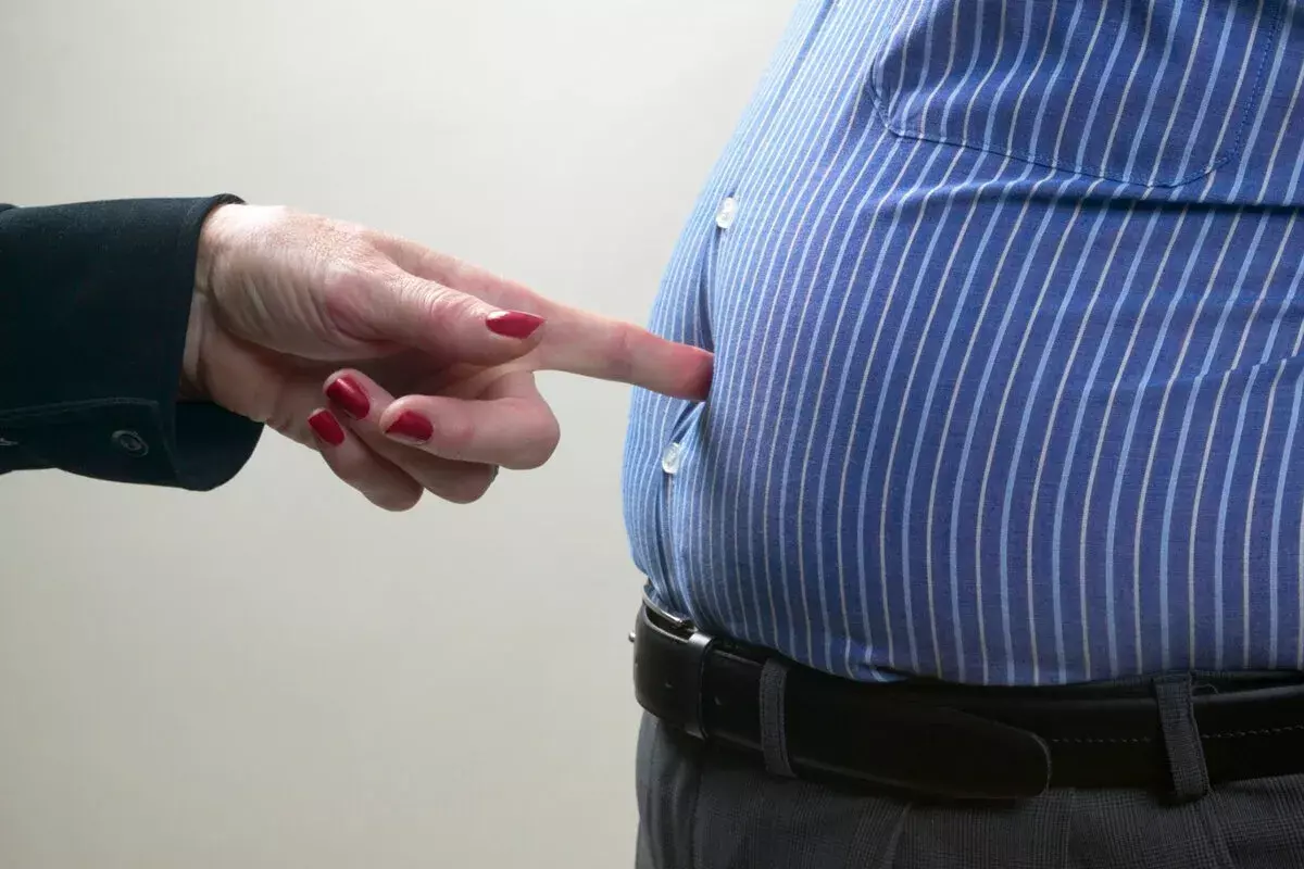
Medical imaging has changed how we find and treat health problems. At LivHospital, we are committed to providing world-class care with the newest radiology scans. We offer various types of body scans including X-rays, CT scans, MRI, PET scans, and ultrasounds, each suited for different diagnostic needs.
We understand the importance of accurate diagnosis. Our team uses many medical imaging methods. These include X-rays, CT scans, MRI, ultrasound, PET, and DEXA scans.
These scans help doctors see inside the body. They help find and manage many health issues. By using these advanced technologies, we can provide our patients with complete care and effective treatments.
Key Takeaways
- Medical imaging plays a key role in modern healthcare.
- Liv Hospital uses the latest in radiology scans.
- Many medical imaging techniques are used for diagnosis.
- Accurate diagnosis is key to good treatment.
- Advanced imaging technologies improve patient care.
The Critical Role of Medical Imaging in Modern Healthcare
Medical imaging is key in today’s healthcare, changing how we diagnose and treat. It lets doctors see inside the body, helping them find and fix health problems better.
Diagnostic imaging has transformed medicine by giving doctors a peek inside without surgery. A top medical journal says, “New imaging tech has made diagnoses more accurate and helped patients more.”
“Imaging technologies have become indispensable in modern medicine, giving insights we couldn’t imagine before.”
How Diagnostic Imaging Has Transformed Medicine
Diagnostic imaging has changed medicine a lot. It lets doctors:
- Find problems sooner and more right
- Watch how diseases grow and how treatments work
- Help with less invasive surgeries
These changes have made patients’ lives better. For example, CT scans and MRI are key in finding and treating serious issues like cancer and brain problems.
Overview of Imaging Technologies
Many imaging techs are used in healthcare today, each with its own uses and perks. Some top ones are:
- X-ray: good for bones and lungs
- CT scans: show detailed cross-sections
- MRI: great for soft tissues
- Ultrasound: safe and doesn’t use radiation
These techs have changed how we tackle health issues, giving us many choices for different needs.
X-Ray Scans: The Fundamental Imaging Technology
X-ray technology has changed how we see inside the body. For over a century, X-ray scans have been key in medical imaging. They give us important views of what’s inside us.
How X-Ray Technology Creates Images
X-ray technology uses X-rays to make images of the body’s inside. When X-rays hit the body, different parts absorb them differently. Dense things like bone absorb more, showing up white on the image. Softer parts show up in gray or black. This helps doctors see bones, organs, and more.
Common Applications in Bone and Chest Imaging
X-ray scans are often used for bone and chest imaging. They help quickly spot problems like fractures and lung issues. X-ray technology is also key in emergency rooms and regular check-ups.
Radiation Considerations and Safety Protocols
X-ray scans are useful but involve radiation. So, we follow strict safety rules to lower radiation exposure. We use lead aprons and adjust settings to use the least amount of radiation. We also think carefully about the risks, like for pregnant women and kids.
CT Scans: Advanced Cross-Sectional X-Ray Imaging
CT scans have changed how we diagnose and treat health issues. They use X-rays to make detailed images of the inside of the body. This gives us important information for diagnosis.
The Technology Behind Computed Tomography
CT scans mix X-rays and computer tech to show body parts inside. An X-ray machine moves around the body to take pictures from many angles. Then, a computer puts these images together into detailed cross-sections.
Key components of CT scan technology include:
- X-ray tube: Makes X-rays that go through the body.
- Detectors: Catch X-rays after they go through the body.
- Computer system: Makes images from the data it gets.
Key Diagnostic Applications
CT scans are used in many medical fields. They help diagnose:
| Condition | Diagnostic Use of CT Scans |
| Cancer | Staging and checking how treatment is working |
| Trauma | Finding internal injuries |
| Vascular diseases | Looking at blood vessel health |
The American College of Radiology says, “CT scans are great in emergencies when quick diagnosis is needed.”
“CT scans are key in today’s medicine, giving detailed insights for treatment plans.”
” Radiology Today
Balancing Benefits and Radiation Exposure
CT scans are very helpful, but also expose us to radiation. It’s important to weigh their benefits against the risks of radiation.
We follow the ALARA principle (As Low As Reasonably Achievable) to keep radiation doses low. This way, we get good images without too much radiation.
Ways to cut down radiation include:
- Using dose modulation techniques
- Employing iterative reconstruction algorithms
- Limiting scan ranges to the area of interest
MRI Scans: Detailed Soft Tissue Visualization
MRI scans are a big step forward in medical imaging. They show soft tissues in great detail. We use them to find and watch for many health problems. They make clear images without harmful radiation.
How Magnetic Fields Generate Detailed Images
MRI scans use strong magnetic fields and radio waves to show body parts inside. We tell patients they’ll be in a big magnetic field during an MRI scan. This field lines up hydrogen atoms in their body. Then, radio waves disturb these atoms, sending signals for detailed images.
Neurological, Musculoskeletal, and Organ Applications
MRI scans are great for soft tissues. They’re perfect for the brain, muscles, and organs. We use them to spot strokes, multiple sclerosis, and spinal injuries. They also check muscle injuries and organ function.
Contraindications and Safety Considerations
Even though MRI scans are helpful, there are things to watch out for. We tell patients with metal implants, like pacemakers, to tell us first. The magnetic field can mess with these devices. We also help those who are scared of tight spaces during the scan.
| Imaging Modality | Radiation Use | Soft Tissue Detail | Common Applications |
| MRI | No | High | Neurological, Musculoskeletal, Organ Imaging |
| CT Scan | Yes | Moderate | Trauma, Cancer, Internal Injuries |
| X-Ray | Yes | Low | Bone Fractures, Lung Conditions |
| Ultrasound | No | Moderate | Obstetrics, Gallbladder Disease, Vascular Conditions |
Ultrasound Imaging: Non-Radiation Real-Time Visualization
Ultrasound imaging uses high-frequency sound waves for safe and effective diagnosis. It lets us see inside the body in real-time, without using harmful radiation.
Sound Wave Technology Principles
Ultrasound imaging works by reflecting sound waves. When sound waves hit the body, they bounce back off internal structures. The ultrasound device catches these waves, creating detailed images.
The sound wave frequency can be changed for different depths and tissues. This makes ultrasound useful for many diagnostic needs.
Applications Beyond Obstetrics
Ultrasound is not just for pregnant women. It’s used to check on the health of abdominal organs and blood vessels. It also helps guide some medical procedures.
In sports medicine, ultrasound helps find tendon and ligament injuries. It’s also good for checking thyroid nodules and other surface structures.
Advantages of Radiation-Free Imaging
One big plus of ultrasound is that it doesn’t use harmful radiation. This is great for patients needing many scans or those who are more sensitive to radiation, like pregnant women.
Ultrasound’s real-time feature lets us see how things move and function. This is super helpful for guiding some medical procedures.
PET Scans: Metabolic and Functional Imaging
PET scans use radioactive tracers to show how the body works. They are key in modern medicine for diagnosing and tracking diseases.
Tracking Metabolic Activity with Radioactive Tracers
PET scans use a small amount of radioactive tracer. This tracer goes to areas where the body is most active. It then shows detailed images of these areas.
Key aspects of PET scan technology include:
- High sensitivity to metabolic changes
- Ability to detect diseases at an early stage
- Capability to monitor treatment response
Cancer Detection and Treatment Monitoring
PET scans are great for finding cancer and checking how treatments work. Cancer cells use more energy than normal cells. This makes them show up on PET scans.
| Application | Description |
| Cancer Detection | Identifying tumors based on increased metabolic activity |
| Treatment Monitoring | Assessing changes in tumor metabolism in response to therapy |
| Disease Staging | Determining the extent of cancer spread |
Neurological Applications in Alzheimer’s and Epilepsy
PET scans also help in neurology. They can spot brain activity linked to Alzheimer’s early on. For epilepsy, they help find where the brain is acting strangely, helping plan surgery.
“The use of PET scans in neurology has opened new avenues for understanding and managing complex brain disorders.”
As technology gets better, PET scans will play an even bigger role in treating cancer and brain diseases. They will help doctors find new ways to help patients.
DEXA Scans: Specialized Bone Density Assessment
Now, we can understand bone health better than ever before. This is thanks to DEXA scans that measure bone mineral density accurately. DEXA (Dual-Energy X-ray Absorptiometry) scans are key in diagnosing and managing bone-related conditions, like osteoporosis.
DEXA scans help us check bone health because they are non-invasive and very accurate. They measure bone mineral density (BMD). This is important for diagnosing osteoporosis, figuring out fracture risk, and seeing if treatments are working.
Measuring Bone Mineral Density
Bone mineral density shows how healthy our bones are. DEXA scans are the best way to measure BMD. They use two X-ray beams to find out the density of minerals like calcium in bones.
Key benefits of DEXA scans for BMD measurement include:
- High accuracy in detecting bone density
- Low radiation exposure
- Ability to monitor changes in bone density over time
| DEXA Scan Features | Benefits |
| Dual-Energy X-ray Technology | Accurate BMD measurement |
| Low Radiation Dose | Safe for repeated measurements |
| Quick Procedure | Minimally invasive, comfortable for patients |
Osteoporosis Screening and Fracture Risk Assessment
DEXA scans are vital for osteoporosis screening. They measure BMD in areas like the hip and spine. This helps doctors figure out the risk of fractures.
Figuring out fracture risk is complex. It involves BMD, age, and family history. DEXA scans give a key piece of this puzzle. They help target interventions to lower fracture risk.
Body Composition Analysis Applications
DEXA scans can also measure fat mass and lean mass. This is useful in clinical and research settings.
DEXA scans give a full view of bone health and body composition. They are a valuable tool for healthcare providers. They help manage conditions like osteoporosis and metabolic disorders.
Nuclear Medicine Scans: Functional Organ Imaging
Nuclear medicine scans give us a peek into the body’s inner workings. They use special substances to check how organs are doing. These scans are key to diagnosing many health issues.
How Radiopharmaceuticals Work in the Body
Radiopharmaceuticals are special compounds with tiny amounts of radioactive material. When they enter the body, they go to certain areas. As they break down, they send out gamma rays.
These rays are caught by cameras, making detailed images of the body’s inner workings. This lets doctors see how organs are working and if there are any problems.
Different radiopharmaceuticals target different parts of the body. For example, some go to the thyroid gland. Others check the heart or kidneys.
Thyroid, Heart, and Kidney Function Assessment
These scans are great for checking the thyroid, heart, and kidneys. They can spot issues like thyroid problems or heart damage. They also help find kidney problems.
“Nuclear medicine scans have changed how we diagnose and treat diseases,” making them very important in medicine.
Safety Protocol and Radiation Considerations
Nuclear medicine scans do involve some radiation, but they are often necessary. Strict safety rules are followed to keep exposure low. This includes using the least amount of radioactive material needed.
Patients are told how to reduce their radiation exposure after the scan. This includes drinking plenty of water and following their doctor’s advice. This way, doctors can get important information while keeping patients safe.
Comparing Different Types of Body Scans for Specific Conditions
There are many body scans, each good for different health issues. These include neurological disorders and cancer. The right scan depends on the condition, how detailed it needs to be, and the patient’s needs.
Choosing the Right Scan for Neurological Conditions
For brain problems like stroke or tumors, MRI scans are best. They show soft tissues clearly.
Optimal Imaging for Musculoskeletal Issues
For muscle, tendon, and ligament problems, ultrasound or MRI is used. Ultrasound is great for seeing how tendons move.
- Ultrasound for superficial structures and dynamic assessment
- MRI for detailed evaluation of soft tissues and complex injuries
- X-ray for bone fractures and degenerative changes
Cancer Detection and Staging: Which Scans Work Best
PET/CT scans are key to finding and checking cancer. They mix PET’s function info with CT’s body details.
| Condition | Preferred Scan | Reason |
| Neurological | MRI | Detailed soft tissue imaging |
| Musculoskeletal | Ultrasound or MRI | Dynamic assessment and soft tissue detail |
| Cancer | PET/CT | Combining functional and anatomical information |
Preparing for Your Medical Imaging Appointment
Getting ready for your medical imaging appointment is important. Knowing what to expect and how to prepare can greatly improve the quality of the images. It also makes your experience better.
General Preparation Guidelines
Follow these guidelines for a smooth appointment. Arrive 15 minutes early to fill out paperwork. Wear comfy clothes that are easy to take off if needed. Avoid jewelry or clothes with metal to prevent scan interference.
Tell your healthcare provider about any health issues, like claustrophobia or allergies to contrast materials. This helps them plan the best imaging for you and keeps you safe.
Scan-Specific Requirements
Each medical imaging scan has its own needs. For a PET scan, you might need to fast beforehand. For an MRI, remove all metal items, including jewelry and certain clothes.
| Scan Type | Preparation Requirement |
| CT Scan | May require contrast dye; inform your doctor about any allergies |
| MRI | Remove metal objects; may require contrast dye |
| PET Scan | Fast for a certain period; avoid strenuous exercise before the scan |
Insurance Coverage and Cost Considerations
Know your insurance and the costs of imaging to plan financially. Most plans cover necessary imaging, but check your coverage and any costs you might face.
Call your insurance before your appointment to confirm coverage and discuss costs. Our billing team can also help with costs and payment options.
Conclusion: The Future of Medical Imaging
Medical imaging is changing fast. New technologies and methods are making it better for diagnosing and treating diseases. This is a big step forward for healthcare.
Thanks to these advances, doctors can spot diseases sooner and more accurately. This leads to better health outcomes for patients. Artificial intelligence is also being used to improve how images are analyzed and diagnosed.
The future of medical imaging looks bright. We can expect even better image quality and less radiation. It will also be used more in personalized medicine, leading to more effective treatments.
FAQ
What are the different types of body scans used in medical imaging?
We use many types of body scans. These include X-ray, CT scans, MRI scans, ultrasound, PET scans, DEXA scans, and nuclear medicine scans. Each has its own use and benefits.
How do I prepare for a medical imaging appointment?
To get ready, remove jewelry and clothes that might get in the way. Also, follow specific instructions for your scan, like fasting or avoiding certain meds. It’s a good idea to check with your insurance about coverage and costs.
What is the difference between a CT scan and an MRI scan?
CT scans use X-rays to make detailed images. MRI scans use magnetic fields for soft tissue images. CT scans are good for emergencies and bones. MRI scans are better for soft tissues and the brain.
Are there any risks associated with medical imaging?
We take safety seriously and follow strict rules to reduce radiation. Some scans, like X-rays and CT scans, use radiation. Others, like MRI and ultrasound, don’t. We consider the benefits and risks for each patient and scan.
How do PET scans work in cancer detection and treatment monitoring?
PET scans use radioactive tracers to see metabolic activity. They help find cancer, check treatment progress, and track disease. They’re key in managing cancer and some neurological conditions.
What is a DEXA scan used for?
DEXA scans measure bone density. They help us check for osteoporosis risk, monitor bone health, and look at body composition. They’re important in preventive care and managing diseases.
Can I undergo a medical imaging scan if I have a medical implant or metal in my body?
Some scans, like MRI, might not be safe with implants or metal. We look at each case and suggest safe options.
How long does a typical medical imaging appointment take?
Time varies with the scan type and complexity. Allow time for prep, the scan, and any extra steps or talks.
Will I be able to see the results of my medical imaging scan immediately?
Sometimes, we can share results right after. But detailed analysis and diagnosis take longer. We’ll tell you when to expect the full results.
References
- Wiley Online Library. (2022). Modern diagnostic imaging technique applications and progress: Review. Journal of Healthcare Engineering, 2022, Article ID 5164970. https://onlinelibrary.wiley.com/doi/10.1155/2022/5164970


