The human body has many types of joints that let us move and do different things. Did you know we have over 360 joints in an adult body? These joints are key for our movement and health.
Knowing about the different joints helps us understand our body better. We’ll look at the five main types, where they are, and what they do. This will give us a deeper look into joint anatomy and why it matters.
Exploring musculoskeletal joint anatomy shows how our bodies are made for movement. This knowledge is important for doctors and anyone who wants to know more about their body.
Key Takeaways
- There are five main classifications of joints in the human body.
- Understanding joint anatomy is key for good musculoskeletal health.
- The average adult human body has over 360 joints.
- Joints help us move and stay flexible.
- Knowing about joint types is important for doctors and everyone else.
The Fundamental Role of Joints in Human Movement
Joints play a fundamental role in enabling movement and maintaining stability in the human body. They let us do everything from walking to playing music. This is because joints are complex structures that help us move in many ways.
How Joints Enable Body Mobility and Stability
Joints allow bones to move together, giving us flexibility and support. The range of motion in joints changes based on the joint type and where it is in the body. For example, the shoulder and hip can move a lot, while the elbow moves less.
Keeping joints stable is also vital. It makes sure bones stay in the right place when we move. Joint stability types depend on the bones’ shape, the presence of ligaments, and muscle tone.
Basic Components of a Joint System
A joint system has several important parts. These include bones, ligaments, tendons, synovial fluid, and articular cartilage. The synovial fluid function is key because it makes movement smooth by reducing friction.
Articular cartilage in joints helps absorb shock and spread out loads. Keeping these parts healthy is important for joint function and avoiding problems like osteoarthritis.
| Component | Function |
| Articulating Bones | Provide the structural framework for the joint |
| Ligaments | Connect bones to each other, providing stability |
| Synovial Fluid | Lubricates the joint, reducing friction |
| Articular Cartilage | Absorbs shock and distributes loads |
Joint Classification Systems in Anatomy
Anatomists use different ways to understand the many types of joints in our bodies. These systems help sort joints by their features. This makes it easier to see how they work and why they’re important in human anatomy.
Functional Classification Based on Movement
Joints are grouped by how much they can move. There are three main types: synarthroses (immovable), amphiarthroses (slightly movable), and diarthroses (freely movable). Synarthroses connect bones tightly, allowing little to no movement. Amphiarthroses let bones move a bit. Diarthroses allow for lots of movement and are found in our limbs.
Structural Classification Based on Tissue Type
Joints are also sorted by the tissue that holds bones together. There are three main types: fibrous, cartilaginous, and synovial joints. Fibrous joints are held together by dense tissue and include sutures, syndesmoses, and gomphoses. Cartilaginous joints have cartilage and include symphyses and synchondroses. Synovial joints have a synovial cavity and are the most mobile and complex.
The Relationship Between Structure and Function
The way a joint is built affects how it works. For example, joints with a fibrous structure are stable but not very mobile. On the other hand, joints with a synovial structure can move a lot. Knowing this helps us see how joints help us move and stay stable.
The 5 Types of Joints: Structure and Location
There are five main types of joints in the human body. They are categorized by their structure and function. Knowing about these types helps us understand how our bodies move and work.
Synovial Joints: The Most Mobile Joint Type
Synovial joints are the most common and most mobile in our bodies. They have a space between bones filled with synovial fluid. This fluid reduces friction, allowing for smooth movement. Examples include the knee, elbow, and shoulder joints.
These joints are divided into subtypes based on their structure and movement. This includes ball-and-socket, hinge, and pivot joints.
Fibrous Joints: Providing Stability and Protection
Fibrous joints, also known as synarthroses, are connected by dense tissue rich in collagen fibers. They offer strong connections between bones and have little to no movement. Examples include the skull’s sutures and the gomphoses that anchor teeth to the jawbone.
These joints are key in providing stability and protection to important structures.
Cartilaginous Joints: Offer Limited Movement
Cartilaginous joints are connected by cartilage and allow for limited movement. They are divided into synchondroses and symphyses based on the type of cartilage. Examples include the intervertebral discs and the pubic symphysis.
These joints balance stability and flexibility. They allow for slight movements while keeping strong connections.
Synarthroses: Immovable Joint Connections
Synarthroses are immovable or have very limited movement. They are divided into fibrous and cartilaginous types. These joints are vital for maintaining the integrity of certain body structures, like the skull.
Understanding synarthroses helps us appreciate the complex anatomy of the human body.
Synovial Joints: Anatomy and Physiological Function
Understanding synovial joints is key to grasping human movement and joint health. These joints, also known as diarthrodial joints, are the most common and movable in our bodies.
The Critical Role of Synovial Fluid
Synovial fluid is vital for synovial joints. It acts as a lubricant, reducing friction between cartilage and other tissues. This makes movement smooth. It also supplies oxygen and nutrients to cartilage, which has no blood supply.
Joint Capsule Structure and Protective Ligaments
The joint capsule is a fibrous sac that encloses the joint. It has two layers: an outer fibrous layer and an inner synovial membrane. The synovial membrane produces fluid, while the fibrous layer adds strength and stability. Ligaments, dense tissue bands, connect bones and support the joint.
Articular Cartilage and Its Role in Joint Health
Articular cartilage covers bone ends in synovial joints. It allows for smooth gliding and reduces friction. It’s made of chondrocytes in a matrix of collagen and proteoglycans. Keeping cartilage healthy is key to joint function and preventing osteoarthritis.
- Smooth Movement: Cartilage lets bones glide smoothly over each other.
- Shock Absorption: It absorbs shock and distributes loads evenly.
- Low Friction: Its smooth surface reduces friction, minimizing wear and tear.
Blood Supply and Innervation of Synovial Joints
Synovial joints get their blood supply from surrounding vessels. The synovial membrane is richly innervated with nerve fibers. This provides sensation, including pain. The blood and nerve supply are vital for joint function and response to injury or disease.
- The blood supply nourishes the joint capsule and synovial membrane.
- Nerve fibers provide pain sensation, which is critical for responding to injury.
- The vascular and neural supply supports the healing process in case of joint damage.
Ball and Socket Joints: Maximum Mobility in the Body
Ball and socket joints are special in the human body. They allow for movement in many directions. This is key for everyday actions and sports.
The Shoulder (Glenohumeral) Joint: Structure and Movement Range
The shoulder joint is a great example of a ball and socket joint. It connects the humerus head to the scapula’s glenoid cavity. This joint lets us move our arms in many ways.
It supports a wide range of motion. This includes moving our arms up, down, and around. The muscles and ligaments around the joint help keep it stable and flexible.
“The shoulder joint’s wide range of motion makes it one of the most versatile joints in the human body,” as noted by medical professionals. Yet, this flexibility also makes it more prone to injuries.
The Hip Joint: Balance Between Stability and Flexibility
The hip joint is another important ball and socket joint. It connects the femur to the pelvis’s acetabulum. It helps us walk, run, and jump by bearing our body’s weight.
The hip joint balances stability and flexibility. It’s supported by strong ligaments and muscles. This balance is key for moving and supporting our body.
Muscular Support Systems for Ball and Socket Joints
The muscles around ball and socket joints are complex. For the shoulder, the rotator cuff muscles are key. They help keep the joint stable. The hip joint has muscles like the gluteals and iliopsoas, which support it.
- The rotator cuff muscles stabilize the shoulder joint.
- The gluteals and iliopsoas support the hip joint.
Common Injuries and Conditions Affecting These Joints
Ball and socket joints can get injured or have conditions. The shoulder can get dislocated or have rotator cuff injuries. The hip can get osteoarthritis, fractures, or labral tears.
Prevention and treatment strategies include physical therapy, medicine, and sometimes surgery. A healthy lifestyle can help prevent some of these issues.
Hinge Joints: Controlled Unidirectional Movement
Hinge joints are key for controlled movements in our bodies. They allow movement in one direction, helping us do many things with precision. Hinge joints are great for bending and straightening, which we do every day.
The Elbow Joint: Anatomy and Functional Mechanics
The elbow hinge joint connects the humerus, radius, and ulna bones. It lets us bend and straighten our arms. This joint is stable thanks to ligaments and muscles, making sure our movements are controlled.
The Knee Joint: Structure of the Body’s Largest Hinge
The knee hinge joint is the biggest hinge joint in our bodies. It supports a lot of our weight and helps us walk and run. The knee is made of the femur and tibia bones, with the patella helping it work well. It has ligaments, menisci, and muscles that work together for smooth movement.
Ankle, Interphalangeal, and Temporomandibular Joints
Other hinge joints include the ankle hinge joint, interphalangeal joints in our fingers and toes, and the temporomandibular joint (TMJ). The ankle lets us move our feet up and down. Interphalangeal joints help us move our fingers and toes. The TMJ lets us open and close our mouths, important for eating and speaking.
Biomechanical Principles of Hinge Joint Movement
The biomechanics of hinge joint movement involve bones, ligaments, muscles, and tendons working together. This teamwork allows for controlled, one-way movement. Knowing these principles helps us understand and treat hinge joint problems, and helps in making good rehab plans.
Pivot Joints: Enabling Rotational Movement
Pivot joints are key for our ability to move in circles. They let us turn our heads, rotate our forearms, and more. We’ll look at how these joints work and why they’re important.
The Atlantoaxial Joint: Facilitating Neck Rotation
The atlantoaxial joint is a pivot joint between the first and second cervical vertebrae. It lets us turn our heads a lot. This joint is complex, with parts that help it move but also stay stable. This unique setup is vital for everyday life.
Proximal and Distal Radioulnar Joints: Forearm Rotation
In the forearm, the proximal and distal radioulnar joints help us rotate. The proximal one is near the elbow, and the distal is near the wrist. These joints help us do things like turn door handles. They work together for many daily tasks.
Biomechanics and Movement Limitations of Pivot Joints
Pivot joints let us move in circles around a single axis. But, they can only move so much because of ligaments and bones. For example, the atlantoaxial joint has strong ligaments to keep it from moving too much. Knowing how pivot joints work helps us treat injuries.
“The pivot joint is a type of synovial joint that allows for rotational movement around a single axis.” –
Orthopedic Anatomy
Clinical Significance and Common Disorders
Pivot joints can get hurt or have diseases like sprains, fractures, or arthritis. For instance, rheumatoid arthritis can harm the atlantoaxial joint. Quick diagnosis and treatment are key to keeping joints healthy.
We’ve seen how pivot joints are important for moving in circles. We’ve looked at their structure, how they work, and why they matter. Understanding these joints helps us appreciate human movement and the importance of taking care of them.
Saddle Joints: Complex Multi-axial Movement
Saddle joints are fascinating in the human body. They allow for complex movements. Their unique shape helps in a wide range of motion while keeping things stable.
The Carpometacarpal Joint of the Thumb: Structure and Function
The carpometacarpal joint of the thumb is a key example. It’s between the trapezium bone and the first metacarpal bone. This joint lets the thumb move in many ways, like grasping objects.
This joint’s design gives both flexibility and strength. It’s vital for daily tasks like writing and gripping.
The Sternoclavicular Joint: Connecting Arm to Axial Skeleton
The sternoclavicular joint connects the collarbone to the breastbone. It’s important for moving the arm and shoulder. This joint helps in attaching the upper limb to the body’s main skeleton.
It allows the clavicle to move in several ways. This movement helps in the overall mobility of the shoulder.
Range of Motion and Movement Capabilities
Saddle joints are great at multi-axial movements. The thumb’s joint, for example, enables circular movements. This is key for thumb opposability. The sternoclavicular joint also enhances upper limb movement.
Their unique shape is what makes these movements possible. The articular surfaces fit together perfectly.
Evolutionary Significance of Saddle Joints
Saddle joints have been important in human evolution. They’ve helped in thumb opposability and shoulder mobility. These abilities have aided in tool use and motor development.
The sternoclavicular joint has also played a big role. It allows for various upper limb movements. This is helpful for activities like throwing and climbing.
Plane Joints: Facilitating Gliding Motion
Plane joints, also known as gliding joints, are key for smooth movement between flat bones. They have flat or slightly curved surfaces. This allows for limited movement in different directions.
Intercarpal and Intertarsal Joints: Location and Function
Intercarpal joints are in the wrist, between carpal bones. Intertarsal joints are in the foot, between tarsal bones. Both types of joints help with gliding movements.
Intercarpal joints help with wrist adjustments, improving grip and dexterity. Intertarsal joints help the foot adapt to surfaces, improving balance and movement.
Vertebral Facet Joints: Enabling Spinal Movement
Vertebral facet joints are between vertebrae’s articular processes. They help with spinal movement like flexion, extension, and rotation.
These joints allow for gliding and rotational movements. This helps keep the spine flexible and stable. It’s important for posture and movement.
Acromioclavicular and Other Plane Joints
The acromioclavicular joint is between the scapula’s acromion and the clavicle. It allows for small movements important for shoulder actions.
Other plane joints include those between vertebrae’s articular processes and some intermetatarsal joints. Each joint helps with the body’s mobility and function.
Functional Importance in Daily Activities
Plane joints are vital for daily tasks, from gripping objects to walking or running. They help with smooth transitions between positions.
For example, during walking, intertarsal joints adjust the foot. Vertebral facet joints help the spine move smoothly. Intercarpal joints fine-tune hand movements, important for precision tasks.
Fibrous Joints: Locations and Structural Importance
Fibrous joints, also known as synarthroses, connect bones with dense tissue rich in collagen fibers. Knowing about fibrous joints helps us understand the body’s structure.
These joints are usually immovable or have very little movement. They provide stability and support to the skeletal system. We will look at the different types of fibrous joints and their role in human anatomy.
Cranial Sutures: Connecting Skull Bones
Cranial sutures are a key example of fibrous joints. They connect the skull bones. These sutures are vital during birth, allowing the skull to move and change shape.
Syndesmoses: The Tibiofibular and Radioulnar Examples
Syndesmoses are fibrous joints held by ligaments or interosseous membranes. The ankle’s tibiofibular and the forearm’s radioulnar syndesmoses are examples. They allow some movement, giving flexibility while keeping things stable.
Gomphoses: Specialized Joints Securing Teeth
A gomphosis is a special fibrous joint that holds a tooth in its socket. The periodontal ligament, a fibrous tissue, keeps the tooth in place. It allows for slight tooth movement during chewing.
Developmental Changes in Fibrous Joints
Fibrous joints change throughout life. In infancy and childhood, they are more flexible for growth. As we age, some, like cranial sutures, may become more rigid or fuse.
Understanding these changes is key for diagnosing and treating fibrous joints issues. Knowing their role and how they change helps healthcare professionals manage related conditions.
Cartilaginous Joints: Strategic Locations in the Body
Cartilaginous joints are key in our skeletal system. They offer support and flexibility. These joints are made of cartilage and are vital for growth, development, and movement.
Symphyses: The Pubic Symphysis and Intervertebral Discs
Symphyses are cartilaginous joints with hyaline cartilage covering the bones. They are connected by a fibrocartilaginous disc. The pubic symphysis and intervertebral discs are two main examples.
The pubic symphysis is between the two pubic bones. It’s important for the pelvic structure. It allows slight movements, helping with childbirth in females.
Intervertebral discs are between vertebrae in the spine. They act as shock absorbers. They help distribute the load and make the spine flexible.
Synchondroses: Growth Plates and Costal Cartilages
Synchondroses are cartilaginous joints with hyaline cartilage forming growth plates or connecting bones. Growth plates are key for the growth of long bones. They let bones lengthen as we grow.
Costal cartilages link the ribs to the sternum. They make the rib cage flexible. This helps with breathing movements.
The Role of Cartilaginous Joints in Growth and Development
Cartilaginous joints are vital for our body’s growth and development. They help bones develop and mature properly. This ensures our skeletal system grows right.
- Enable bones to grow longer through growth plates.
- Give flexibility and support to body structures.
- Help the spinal column and other bones develop.
Age-Related Changes in Cartilaginous Structures
As we age, cartilaginous joints change a lot. For example, intervertebral discs can degenerate. This can cause degenerative disc disease.
Also, costal cartilages can calcify with age. This might affect breathing.
“The degeneration of cartilaginous joints with age can lead to various health issues, stressing the need for a healthy lifestyle to support joint health.”
It’s important to understand these changes. This helps manage age-related conditions and keeps our skeletal system healthy.
Joint Health and Common Pathological Conditions
Keeping our joints healthy is key for moving well and feeling good. As we get older or face other challenges, our joints can get sick. This makes it hard to move freely.
Many things can affect our joint health. Genetics, the environment, and our lifestyle play big roles. Diseases like osteoarthritis and rheumatoid arthritis are common and affect many people.
Osteoarthritis and Rheumatoid Arthritis: Impact on Different Joint Types
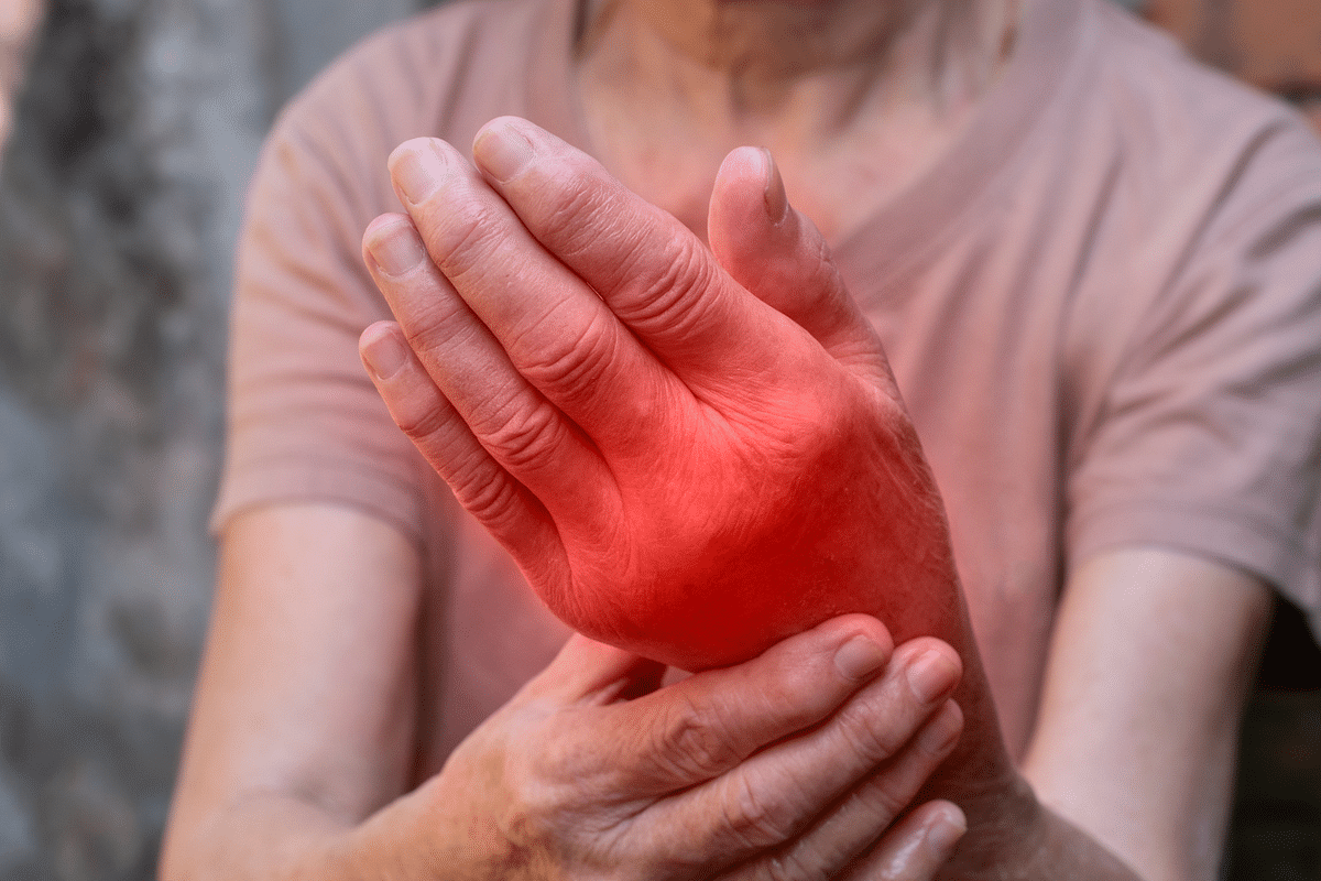
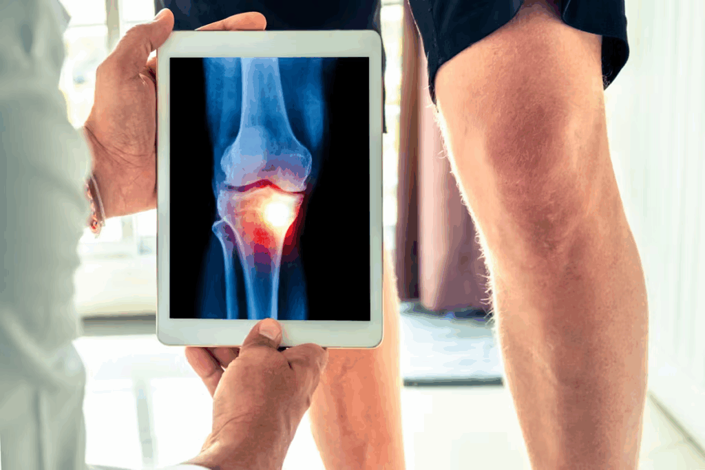
Osteoarthritis (OA) is a disease that wears down cartilage. This causes bones to rub together, leading to pain. It often hits the hips, knees, and hands.
Rheumatoid arthritis (RA) is an autoimmune disease. It causes inflammation and pain in the joints. RA usually affects the hands, feet, wrists, and knees.
| Condition | Primary Joints Affected | Key Characteristics |
| Osteoarthritis | Hips, Knees, Hands | Degenerative, Cartilage Breakdown |
| Rheumatoid Arthritis | Hands, Feet, Wrists, Knees | Autoimmune, Inflammatory |
Traumatic Injuries Specific to Each Joint Classification
Traumatic injuries can happen in any joint. They often come from accidents, sports injuries, or sudden moves. For example, the knee and shoulder are more likely to get sprains or tears.
Fibrous joints, like the skull sutures, are usually stable. But, they can get hurt from severe head trauma.
“The integrity of our joints is key for keeping us mobile and pain-free. Knowing the risks and taking steps to prevent injuries can really help.” – An Orthopedic Specialist
Inflammatory and Degenerative Joint Diseases
Inflammatory diseases, like RA, make the immune system attack the joints. This leads to inflammation and damage. Degenerative diseases, like OA, come from wear and tear. They cause cartilage loss and pain.
Preventative Measures for Maintaining Joint Health
Keeping our joints healthy requires lifestyle changes and proactive steps. Regular exercise, a healthy weight, and avoiding strain can help prevent joint problems.
- Do regular, low-impact exercises to strengthen joint muscles.
- Keep a healthy weight to ease pressure on joints.
- Avoid repetitive strain and take breaks if your job is repetitive.
- Eat a balanced diet with omega-3s and antioxidants to fight inflammation.
By knowing about joint diseases and taking steps to prevent them, we can improve our joint health. This makes our lives better overall.
Modern Clinical Approaches to Joint Disorders
Today, we have new ways to treat joint problems. Medical tech has improved, and we know more about keeping joints healthy. This means we can help patients with joint issues better than before.
Advances in Joint Replacement Technologies
Joint replacement surgeries are now common and work well. Minimally invasive techniques and advanced prosthetics have made things better for patients. For example, new materials like ceramic and metal alloys make prosthetics last longer and move better.
| Material | Durability | Complications Rate |
| Metal Alloys | High | Low |
| Ceramic | Very High | Very Low |
| Polyethylene | Moderate | Moderate |
Regenerative Medicine and Tissue Engineering
Regenerative medicine is changing how we treat joint problems. Stem cell therapy and platelet-rich plasma (PRP) therapy help fix or grow back damaged tissues. This might mean fewer surgeries for some patients.
Rehabilitation Protocols for Different Joint Types
Good rehab is key to getting better after injuries or surgeries. Personalized rehab plans are made for each patient. They consider the joint type, injury, and overall health.
- Ball and socket joints like the shoulder and hip need rehab to get strong and move well.
- Hinge joints, like the elbow and knee, focus on stability and controlled movement in rehab.
Future Directions in Joint Treatment and Research
The future of treating joint disorders looks bright. Research on biologics and gene therapy is promising. These could lead to better, more targeted treatments for joint problems.
As we learn more and improve treatments, our goal is to help patients more. We aim to make their lives better and their joints healthier. With new tech and care, we’re moving towards a future where joint issues are managed well, and patients can live better lives.
Conclusion
The human body has many types of joints, each with its own structure and function. These joints help us move and stay stable. Knowing about synovial, fibrous, and cartilaginous joints is key to understanding how they work.
Keeping our joints healthy is vital for our overall well-being. By learning about joint anatomy and taking care of our joints, we can avoid many problems. This knowledge helps us understand how conditions like osteoarthritis affect our joints.
As we learn more about joints, we can find better ways to treat and prevent joint issues. This will help people with joint problems live better lives. It’s all about improving our health and well-being.
FAQ
What are the main types of joints in the human body?
The human body has three main types of joints. These are synovial, cartilaginous, and fibrous. They are classified based on their structure and function.
What is the function of synovial fluid in joints?
Synovial fluid reduces friction in joints. It helps the articular cartilage move smoothly. This reduces wear and tear.
What are ball and socket joints, and where are they located?
Ball and socket joints are found in the shoulder and hip. They allow for movement in many directions. This gives maximum mobility.
How do hinge joints differ from other types of joints?
Hinge joints, like the elbow and knee, move in only one plane. They provide controlled movement in one direction.
What is the role of articular cartilage in joint health?
Articular cartilage covers the ends of bones in synovial joints. It reduces friction and absorbs shock. This keeps joints healthy and moving smoothly.
What are the common pathological conditions affecting joints?
Common joint problems include osteoarthritis, rheumatoid arthritis, and injuries. These can affect different joints and need different treatments.
How are joint disorders treated using modern clinical approaches?
Modern treatments for joint disorders include new joint replacement technologies. They also include regenerative medicine and tissue engineering. Rehabilitation plans are tailored for each joint type.
What are the preventative measures for maintaining joint health?
To keep joints healthy, maintain a healthy weight and exercise regularly. Avoid repetitive strain injuries. Manage conditions like arthritis.
What is the significance of understanding joint anatomy and function?
Knowing about joint anatomy and function is key. It helps prevent injuries and develop effective treatments for joint disorders.
How do different joint types contribute to overall mobility and stability?
Different joints, like synovial, cartilaginous, and fibrous, work together. They provide a range of movements. This keeps the body mobile and stable.
References
- National Institute of Arthritis and Musculoskeletal and Skin Diseases. (2020). Osteoarthritis. National Institutes of Health. https://www.niams.nih.gov/health-topics/osteoarthritis


