
Endometriosis affects more than 5.5 million American women. It causes big physical, emotional, and reproductive problems. This condition is hard to diagnose because it shows up in many ways.what does endometriosis look likeWhat Is Excision Surgery for Endometriosis and How Does It Work?
Endometriosis is when tissue like the uterine lining grows outside the uterus. This leads to many symptoms. At Liv Ho, we know how important it is to see what endometriosis looks like. This helps find it early and plan treatment.
We want to help by showing what the different stages of endometriosis look like. Knowing about endometriosis helps doctors make better treatment plans for each person.
Key Takeaways
- Endometriosis affects over 5.5 million American women.
- The condition is characterized by tissue growing outside the uterus.
- Understanding the visual appearance of endometriosis is key for diagnosis.
- A visual guide helps doctors make good treatment plans.
- Finding endometriosis early is important for managing symptoms.
Understanding Endometriosis: The Basics

To understand endometriosis, we first need to know what it is and how it shows up. It’s a chronic condition where tissue like the uterus lining grows outside the uterus. This causes pain and other issues.
Definition and Prevalence in the United States
Endometriosis affects about 1 in 10 women of childbearing age in the U.S. That’s around 6.5 million women. It’s marked by tissue outside the uterus, leading to inflammation and scarring.
The exact cause of endometriosis is not known. But hormones, genetics, and environment play a role in its development.
How Endometriosis Develops Outside the Uterus
Endometriosis happens when tissue like the uterus lining grows outside the uterus. It often appears on ovaries, fallopian tubes, and pelvic tissue. This tissue thickens and bleeds with the menstrual cycle but can’t leave the body, causing pain and inflammation.
Several factors contribute to endometriosis outside the uterus. These include:
- Coelomic metaplasia: Cells lining the pelvic cavity turn into endometrial-like tissue.
- Retrograde menstruation: Menstrual blood flows back through the fallopian tubes, carrying endometrial cells into the pelvic cavity.
- Hormonal influences: Estrogen promotes the growth of endometrial tissue.
Common Symptoms and Their Correlation to Visual Findings
Symptoms of endometriosis vary but often include pelvic pain, heavy or irregular bleeding, and infertility. The appearance of endometriosis can range from small, clear vesicles to large, dark “powder-burn” lesions.
Symptom | Visual Finding |
Pelvic pain | Inflammation and adhesions |
Heavy menstrual bleeding | Large endometrial implants |
Infertility | Adhesions and scarring on reproductive organs |
Knowing these connections is key to diagnosing and treating endometriosis well.
The Importance of Visual Diagnosis

Visual diagnosis is key in treating endometriosis. Seeing endometrial lesions and adhesions helps doctors understand the disease’s spread. This knowledge guides treatment choices.
Why Visual Identification Matters for Treatment
Seeing endometriosis through laparoscopy lets doctors directly check the pelvic organs. They can spot endometrial implants, adhesions, and cysts. This info is essential for deciding on treatment, whether surgery or medicine.
Endometriosis looks different in each person, with red, black, and white lesions. Laparoscopic pictures of endometriosis show how widespread the disease is. They help doctors tailor treatments to each patient’s needs.
Diagnostic Challenges and Delayed Diagnosis
Diagnosing endometriosis is often hard because its symptoms are not clear. Many patients see several doctors and have many tests before getting a correct diagnosis.
Diagnostic Method | Advantages | Limitations |
Laparoscopy | Direct visualization, high accuracy | Invasive, requires surgical expertise |
Ultrasound | Non-invasive, widely available | Limited sensitivity for certain lesions |
MRI | High sensitivity, detailed imaging | Expensive, not always available |
The Role of Laparoscopy as the Gold Standard
Laparoscopy is the best way to diagnose endometriosis. It lets doctors see the pelvic cavity directly. Endometriosis laparoscopy pictures are very helpful for figuring out how much disease is present and for planning surgery.
The pictures from laparoscopy help doctors not just diagnose but also plan surgery. They can see adhesions, endometriomas, and other lesions that might need surgery.
What Does Endometriosis Look Like? Visual Characteristics
Endometriosis shows up in many ways, making it hard to diagnose. The different looks of endometrial lesions depend on their appearance and the disease’s stage.
Red Lesions: Early Active Implants (1-3mm)
Red lesions are signs of early active implants, usually 1-3mm big. They look like small, red or pink spots on pelvic organs. These spots are active because they have a lot of blood, showing they’re working hard.
Black “Powder-Burn” Nodules and Plaques
Black “powder-burn” nodules and plaques are bigger, older lesions. They look black or dark brown because of hemosiderin, a byproduct of hemoglobin. They show up in areas where endometriosis has been around for a while.
White and Yellow Non-Pigmented Lesions
White and yellow non-pigmented lesions are another type of endometrial implant. They look like white or yellow patches and are linked to scarring and fibrosis. They’re harder to spot because they don’t have the usual color of endometriosis.
Blue and Brown Lesions in Advanced Cases
In advanced endometriosis, lesions can be blue or brown. This is because of blood and its breakdown products. These bigger, more complex lesions can go deeper and cause adhesions or cysts.
Knowing how to spot these visual signs is key for diagnosing and staging endometriosis. The look of lesions can tell us a lot about the disease’s spread and activity. This helps doctors make better treatment plans and can lead to better results for patients.
The Endometriosis Staging System Explained
The endometriosis staging system is a key tool for doctors to see how far the disease has spread. It helps them understand how severe it is. This is important for choosing the right treatment and care plan.
The ASRM Classification System
The American Society for Reproductive Medicine (ASRM) has a system for staging endometriosis. It breaks the disease into four stages. These stages are based on where and how deep the lesions are, and how bad the adhesions are.
Point System and Scoring Methodology
The ASRM system uses points to measure how bad endometriosis is. Points are given for the size and location of lesions, and the severity of adhesions. The total points tell the stage of endometriosis, from Stage 1 (least severe) to Stage 4 (most severe).
Stage | Description | Score Range |
Stage 1 | Minimal Disease | 1-5 points |
Stage 2 | Mild Disease | 6-15 points |
Stage 3 | Moderate Disease | 16-40 points |
Stage 4 | Severe Disease | >40 points |
Limitations of the Current Staging System
The ASRM system is helpful but has its downsides. It doesn’t always match up with how bad symptoms are or how well treatments work. It mainly looks at what doctors see during a laparoscopy, which might miss some parts of the disease, like deep infiltrating endometriosis.
We know that knowing the stage of endometriosis is key for diagnosis and treatment. The ASRM system is useful, but we need to think about its limits to give the best care.
Stage 1 Endometriosis: Visual Guide
Stage 1 endometriosis is the first stage and has minimal disease. It has small, superficial lesions with little to no scar tissue. Knowing what Stage 1 endometriosis looks like is key for early diagnosis and treatment.
Minimal Disease Characteristics
Stage 1 endometriosis has small implants, usually less than 1-3 mm, and little adhesions. These early lesions are hard to spot because they are small and not very noticeable. The American Society for Reproductive Medicine (ASRM) rates Stage 1 as 1-5 points, showing it’s a mild case.
Typical Locations and Subtle Appearances
Early endometriotic lesions can be found in the ovaries, uterosacral ligaments, and peritoneal surfaces. They might look red, clear, or white and are tricky to see. We often find them during a laparoscopy because they can be missed easily.
Visual Examples and Clinical Significance
Seeing examples of Stage 1 endometriosis helps doctors and patients understand it better. For example, a laparoscopy might show small, red spots on the ovaries or scattered in the pelvic area. Finding these early is important for treating the disease before it gets worse.
Characteristics | Description | Clinical Significance |
Lesion Size | Typically 1-3 mm | Difficult to detect, requires thorough examination |
Lesion Appearance | Red, clear, or white implants | Subtle appearances can be easily overlooked |
Common Locations | Ovaries, uterosacral ligaments, peritoneal surfaces | Requires laparoscopy for accurate diagnosis |
ASRM Score | 1-5 points | Reflects minimal disease extent |
Understanding Stage 1 endometriosis helps doctors diagnose and treat it early. This can lead to better outcomes for patients. Pictures and visual guides are important for teaching both doctors and patients about the early signs of endometriosis.
Stage 2 Endometriosis: Visual Guide
Understanding Stage 2 endometriosis means looking closely at its visual signs. At this stage, the disease has grown beyond the small lesions of Stage 1. It shows more noticeable and complex features.
Mild Disease Characteristics
Stage 2 endometriosis has larger or deeper lesions than Stage 1. It also has minimal scar tissue. The lesions can be red, black, or white on the pelvic surfaces. The American Society for Reproductive Medicine (ASRM) says Stage 2 scores between 6-15 points.
Key Features:
- Lesions are more numerous and slightly larger than in Stage 1.
- Minimal adhesions may start to form.
- Ovarian endometriomas may begin to develop.
Common Visual Presentations and Patterns
In Stage 2 endometriosis, visual signs can differ a lot among patients. Common patterns include:
Visual Feature | Description |
Red Lesions | Active implants that are typically small and flat. |
Black “Powder-Burn” Nodules | Indicative of older, more established lesions. |
White Lesions | Represent areas of scarring and healing. |
These signs can be seen through laparoscopy, the best way to diagnose endometriosis.
“The visual diagnosis of endometriosis requires a thorough understanding of its various appearances and the ability to identify subtle lesions.” – Expert in Gynecologic Surgery
Distinguishing Features from Stage 1
The main difference between Stage 1 and Stage 2 endometriosis is the size and number of lesions. Stage 2 has more and deeper lesions, and more adhesions. Stage 1 has only small, surface-level lesions.
Knowing these visual signs helps doctors diagnose and treat Stage 2 endometriosis better. This improves patient care.
Stage 3 Endometriosis: Visual Guide
Exploring Stage 3 endometriosis, we see a mix of deep lesions, adhesions, and ovarian endometriomas. This stage has a moderate disease severity. It includes many deep lesions and can affect the ovaries.
Moderate Disease Characteristics
Stage 3 endometriosis has many deep lesions and small cysts on the ovaries. It also has thin scar tissue bands called adhesions. These adhesions and ovarian cysts set Stage 3 apart from earlier stages.
Key Features:
- Multiple deep lesions
- Possible small ovarian cysts
- Thin bands of scar tissue (adhesions)
Adhesions and Their Characteristic Appearance
Adhesions in Stage 3 endometriosis are thin and filmy. They look like transparent or translucent bands. These bands can link different pelvic structures, like the ovaries to the pelvic sidewall or the uterus to the bowel.
These adhesions cause a lot of pain and discomfort. They are a key sign of moderate endometriosis.
Ovarian Endometriomas and Their Visual Identification
Ovarian endometriomas, or “chocolate cysts,” are common in Stage 3 endometriosis. They look like cysts filled with thick, dark fluid, similar to chocolate.
Visual Identification:
- Cysts are typically 3-5 cm in size
- Filled with thick, dark fluid
- Can be present on one or both ovaries
Ovarian endometriomas are a big sign of Stage 3 endometriosis. Seeing them during laparoscopy is key for a correct diagnosis.
Stage 4 Endometriosis: Visual Guide
Stage 4 endometriosis is very severe. It has many deep implants and lots of adhesions in the pelvic area. This stage is the most advanced and causes a lot of pain and discomfort.
Severe Disease Characteristics
Stage 4 endometriosis scores over 40 points under the ASRM system. It has:
- Many deep endometrial implants
- Big ovarian endometriomas (>5cm)
- Extensive adhesions in the bowel, bladder, and reproductive organs
These signs show a complex and severe disease. It needs a detailed treatment plan.
Deep Infiltrating Endometriosis Appearances
Deep infiltrating endometriosis (DIE) is a key feature of Stage 4. It grows deep into the pelvic structures. During laparoscopy, you can see:
- Nodular lesions on the uterosacral ligaments
- Bowel and bladder wall involvement
- Significant scarring and adhesions
Large Ovarian Cysts and Their Presentation
Stage 4 endometriosis often has big ovarian endometriomas, or “chocolate cysts.” They are filled with old blood, making them dark. These cysts can be:
- Over 5cm in size
- Attached to nearby structures like the bowel or pelvic sidewall
- A major source of pain and discomfort
Extensive Adhesions Involving Bowel, Bladder, and Reproductive Organs
Stage 4 endometriosis is marked by extensive adhesions. These adhesions can:
- Change the shape of the pelvic area
- Lead to a lot of pain and discomfort
- Make surgery more complicated
Knowing these visual signs is key to diagnosing and treating Stage 4 endometriosis well.
Imaging Techniques for Visualizing Endometriosis
Imaging techniques are key in diagnosing and managing endometriosis. They help doctors see how far the disease has spread. This information guides treatment and improves patient care.
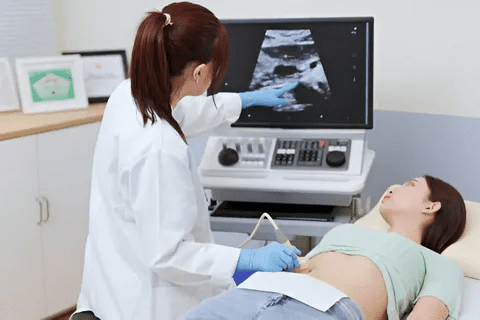
Transvaginal Ultrasound Findings and Limitations
Transvaginal ultrasound is a common tool for checking endometriosis. It’s good for spotting endometriomas and deep infiltrating endometriosis. It’s non-invasive, affordable, and shows images in real-time. But, it depends on the skill of the person doing the scan. It might miss small or surface lesions.
Key findings on transvaginal ultrasound include:
- Endometriomas, which look like ground-glass echogenicity cysts
- Deep infiltrating endometriosis, seen as hypoechoic lesions with irregular edges
- Adhesions, hinted at by fixed organs or “kissing ovaries”
MRI Characteristics of Different Endometriosis Lesions
Magnetic Resonance Imaging (MRI) gives detailed views of endometriosis. It’s great for complex cases and planning surgery.
Characteristic MRI findings include:
- Endometriomas, hyperintense on T1-weighted images and hypointense on T2-weighted images
- Deep infiltrating endometriosis, low-signal intensity on T2-weighted images
- Adhesions, seen as low signal intensity or anatomy distortions
Emerging Technologies in Non-Invasive Visual Diagnosis
New technologies are changing how we diagnose endometriosis. Contrast-enhanced ultrasound and advanced MRI sequences are being tested. They might help us see endometriosis better without invasive methods.
Future directions include:
- Improved ultrasound techniques, like contrast-enhanced ultrasound
- Advanced MRI sequences, including diffusion-weighted imaging
- Artificial intelligence to improve image analysis and accuracy
Atypical and Rare Visual Presentations
Endometriosis can show up in many ways, making it hard to diagnose and treat. It includes atypical and rare visual signs that doctors need to know about. This knowledge helps in giving better care to patients.
Clear Vesicular Lesions
Clear vesicular lesions are an unusual sign of endometriosis. These are clear or translucent and hard to spot during exams. Clear vesicular lesions are important because they might show early signs of endometriosis that could be missed.
Research shows these lesions can be active and cause symptoms. Accurate identification of these lesions is key for managing and treating the condition effectively.
Polypoid and Nodular Formations
Endometriosis can also show up as polypoid or nodular formations. These are different from the usual forms. They can appear in the pelvic area and cause serious symptoms.
- Polypoid lesions can look like other conditions, making it important to make the right diagnosis.
- Nodular formations are often linked with deep infiltrating endometriosis.
Looking at the body and using tools like ultrasound or MRI are key in spotting these unusual signs. Knowing how to identify polypoid and nodular formations is vital for correct diagnosis.
Extrapelvic Endometriosis Appearances
Extrapelvic endometriosis means endometrial tissue is found outside the pelvic area. This rare type can be in places like the abdomen, thorax, or even the brain. The look of extrapelvic endometriosis can change a lot based on where and how deep the lesions are.
For example, skin endometriosis might look like skin lesions or nodules. Intestinal endometriosis might cause symptoms related to the bowel. Knowing the different ways extrapelvic endometriosis can show up is important for quick and effective treatment.
Doctors need to think about endometriosis when they see unusual or unexplained symptoms, even outside the usual areas. Knowing about these unusual signs can help improve patient care.
Conclusion
We’ve looked into the different stages and what endometriosis looks like. This condition affects millions globally. Knowing how to spot it is key for getting the right treatment.
Images of endometriosis show a variety of lesions and implants. These can range from red and black to white and yellow. Spotting these differences is vital for doctors to treat it well.
Looking at endometriosis images helps us see how complex it is. This knowledge lets doctors create treatment plans that really work for each patient.
As we learn more about endometriosis, seeing it is more important than ever. We need to teach doctors and those with the condition about spotting endometriosis images. This will help improve care for everyone.
FAQ
What are the common visual characteristics of endometriosis lesions?
Endometriosis lesions can look like red, black, white, yellow, blue, or brown growths. They vary in size and shape. You can find them on the ovaries, fallopian tubes, and other pelvic areas.
What do red lesions in endometriosis look like?
Red lesions are small, usually 1-3mm. They are early signs of active implants. They look like tiny, red or pink spots on pelvic organs.
How do black “powder-burn” nodules and plaques appear in endometriosis?
Black “powder-burn” nodules and plaques show up as dark, burnt-looking areas. They have a “powder-burn” look on pelvic surfaces. This is a sign of more established endometriosis.
What is the significance of ovarian endometriomas in endometriosis?
Ovarian endometriomas, or “chocolate cysts,” are common in endometriosis. They look like dark cysts filled with old blood. This is seen on imaging studies.
How is endometriosis staged, and what does it mean?
Endometriosis is staged using the ASRM system. It has four stages based on disease extent. The system looks at lesion size, location, and depth. It also considers adhesions and ovarian endometriomas.
What are the characteristics of Stage 4 endometriosis?
Stage 4 endometriosis has severe disease. It includes extensive adhesions, large ovarian endometriomas, and deep infiltrating endometriosis. It’s the most advanced stage.
Can endometriosis be visualized using imaging techniques?
Yes, imaging like transvaginal ultrasound and MRI can show endometriosis. They help spot ovarian endometriomas, adhesions, and deep infiltrating endometriosis.
What are atypical and rare visual presentations of endometriosis?
Atypical and rare presentations include clear vesicular lesions, polypoid and nodular formations, and extrapelvic endometriosis. These are hard to diagnose and need a keen eye.
What do laparoscopic pictures of endometriosis show?
Laparoscopic pictures show lesions, adhesions, and ovarian endometriomas. Laparoscopy is the best way to diagnose endometriosis.
Can stage 4 endometriosis photos help in understanding the condition?
Yes, stage 4 endometriosis photos help understand the condition’s severity. They provide valuable visual information for diagnosis and treatment planning.
How do images of endometriosis on ultrasound help in diagnosis?
Ultrasound images can spot ovarian endometriomas, adhesions, and other signs of endometriosis. While it has limits, ultrasound is useful in diagnosing endometriosis.
References
National Center for Biotechnology Information. Endometriosis: Visual Guide to Stages, Symptoms, and Diagnosis. Retrieved from
https://www.ncbi.nlm.nih.gov/pmc/articles/PMC1924845/

