At Liv Hospital, we know how tricky arteriovenous malformations (AVMs) can be. These are unusual links between arteries and veins. They can pop up in different parts of the body, like the skin and legs. This can cause a bunch of symptoms and problems.
Handling AVMs right means starting with a good diagnosis. We use top-notch tools to figure out how big the problem is. This lets us create a treatment plan that fits each patient perfectly.
Our team is known worldwide for caring for patients with complex vascular issues. We use the newest AVM therapy to help our patients live better lives.
Key Takeaways
- Arteriovenous malformations are abnormal connections between arteries and veins.
- AVMs can affect different body parts, including the skin and legs.
- Proper diagnosis is key for managing AVMs well.
- Advanced diagnostic tools help us make treatment plans that work.
- Liv Hospital offers patient-focused care for tough vascular problems.
Understanding Arteriovenous Malformations (AVMs)
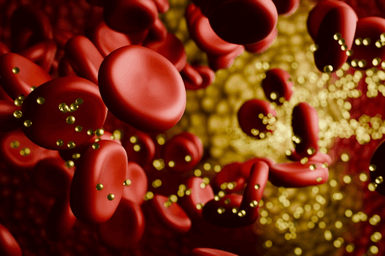
Arteriovenous malformations, or AVMs, are a mix of blood vessels. They connect arteries directly to veins, skipping capillaries. This can cause health problems, based on where and how big the AVM is.
Definition and Basic Anatomy
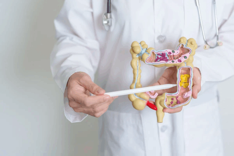
AVMs are a mix of blood vessels that don’t form properly. They connect arteries to veins, leading to blood flow issues. The anatomy of an AVM includes a nidus, feeding arteries, and draining veins.
Knowing how AVMs are structured is key to treating them. The nidus is the core, where arteries and veins connect abnormally. Arteries bring blood to the nidus, and veins take it away.
What Does AVM Stand for in Medical Terms
AVM stands for Arteriovenous Malformation in medical terms. It’s a connection between arteries and veins that shouldn’t be there.
“AVMs can occur anywhere in the body, but they are most commonly found in the brain, spine, and neck. They can also occur in other areas, such as the leg or skin.” – Medical Expert
| Key Components of AVMs | Description |
|---|---|
| Nidus | The central part of the AVM where abnormal connections occur |
| Feeding Arteries | Arteries that supply blood to the nidus |
| Draining Veins | Veins that carry blood away from the nidus |
Causes of Arteriovenous Malformation
It’s important to know what causes arteriovenous malformations (AVMs) to manage and treat them well. AVMs are complex blood vessel problems that can greatly affect a person’s life.
Congenital Factors
Most AVMs are congenital, meaning they are present at birth. Research shows that being born with AVMs is common. It’s thought that something goes wrong with blood vessel formation in the womb, causing an AVM.
Genetics also play a part in AVMs. Some genetic syndromes can make getting an AVM more likely. We’ll look into the genetic side of AVMs more.
Acquired Causes
But, not all AVMs are born with. Some can be acquired due to trauma, infection, or other blood vessel issues.
Studies hint that the environment might also play a role in AVMs. Yet, more research is needed to understand this fully.
Knowing the causes of AVMs is key to effective treatment. Doctors can tailor treatments based on whether an AVM is congenital or acquired.
Are AVMs Genetic? Hereditary Links Explained
Many people wonder if arteriovenous malformations (AVMs) are caused by genes. We look into the genetic side of AVMs and their possible family ties. This helps us grasp these complex blood vessel issues better.
Genetic Predisposition
Some studies link AVMs to genetic conditions. For example, certain genetic disorders might make someone more likely to get an AVM. We dive into how genetics play a part in AVMs, showing their role in their creation.
Research has found certain genetic mutations linked to AVMs. This means genetic tests could help find people at higher risk of AVMs.
Familial Patterns
AVMs also show up in families, suggesting a genetic link. When many family members have AVMs, it points to a possible inherited factor.
We look at data on AVMs in families to see how genetics play a role. Knowing this helps figure out the risk for families with AVM history.
The table below highlights key points about AVMs and their genetic and family ties:
| Genetic Factor | Description | Impact on AVM Development |
|---|---|---|
| Genetic Mutations | Specific mutations associated with AVMs | Increased risk of AVM formation |
| Familial History | Presence of AVMs in family members | Potential hereditary link |
| Genetic Screening | Identification of genetic risk factors | Early detection and prevention |
Grasping the genetic and family ties of AVMs is key to better treatment plans. We keep studying these areas to help patients more.
Common Locations of AVMs in the Body
Arteriovenous malformations (AVMs) can be found in many parts of the body. These abnormal connections between arteries and veins pose different challenges in each location.
AVM Leg: Characteristics and Concerns
AVMs in the leg can cause a lot of problems. They can lead to pain, swelling, and make it hard to move the limb. In severe cases, they can cause varicose veins, skin discoloration, and ulcers.
A study in the Journal of Vascular Surgery found that leg AVMs often cause pain (71%), swelling (64%), and limb length discrepancy (21%) in patients.
“The management of AVMs in the leg requires a multidisciplinary approach, including vascular specialists, orthopedic surgeons, and sometimes dermatologists for skin-related issues.”
| Symptoms | Frequency |
|---|---|
| Pain | 71% |
| Swelling | 64% |
| Limb Length Discrepancy | 21% |
AVM Skin: Visual Identification
AVMs on the skin look different. They often appear as red or purple lesions due to the abnormal blood vessels. The size of the avm skin can vary greatly.
Being able to spot skin AVMs early is key. It helps in getting treatment before problems like bleeding or cosmetic issues arise.
Other Locations: Brain, AVM Heart, and Arm AVM
AVMs can also appear in other important areas. AVM heart or cardiac AVMs are rare but serious. Brain AVMs can cause neurological symptoms and are considered high-risk.
Arm AVM can also happen, causing pain, swelling, and limited mobility. Treating AVMs in these areas needs the expertise of skilled healthcare professionals.
In summary, AVMs can be found in many parts of the body, like the leg, skin, brain, heart, and arm. Knowing the specific issues with each location is important for proper care and management.
Symptoms and Signs of Arteriovenous Malformations
It’s important to know the symptoms of arteriovenous malformations (AVMs) early. AVMs can show different symptoms based on their size, location, and how severe they are.
General Symptoms
AVMs can cause pain, swelling, and changes in skin color or temperature. Some people might feel a lump or a full feeling in the affected area. In serious cases, AVMs can cause bleeding or ulcers.
The symptoms of AVMs can be hard to pinpoint. But knowing them can lead to further checks.
| Symptom | Description |
|---|---|
| Pain | Aching or sharp pain in the affected area |
| Swelling | Visible swelling due to the malformation |
| Skin Changes | Changes in skin color or temperature |
| Palpable Mass | A noticeable lump or mass |
AV Malformation in Leg Symptoms
AVMs in the leg can cause pain, swelling, and varicose veins. They might also lead to skin ulcers or changes in skin color. People might feel their leg is heavy or tired.
Arteriovenous Malformation Skin Manifestations
Skin AVMs can show up as lesions, port-wine stains, or skin discolorations. They can cause bleeding, ulcers, or pain. Skin AVMs might also lead to discomfort.
Spotting skin AVMs early is important for managing them well and avoiding problems.
We know dealing with AVM symptoms can be tough. Our team is here to offer full care and support to those with arteriovenous malformations.
Diagnosing Arteriovenous Malformations
Getting an accurate diagnosis for AVMs is key to planning treatment. This is done through a mix of physical checks and advanced imaging.
Physical Examination
The first step in diagnosing AVMs is a detailed physical check. We look for signs like swelling, warmth, or a lump in the affected area. Visible lesions or varicose veins in the leg or skin are also clues.
Understanding a patient’s medical history is also vital. It helps spot any family patterns or past symptoms that might point to an AVM.
MRI AVM: The Gold Standard for Diagnosis
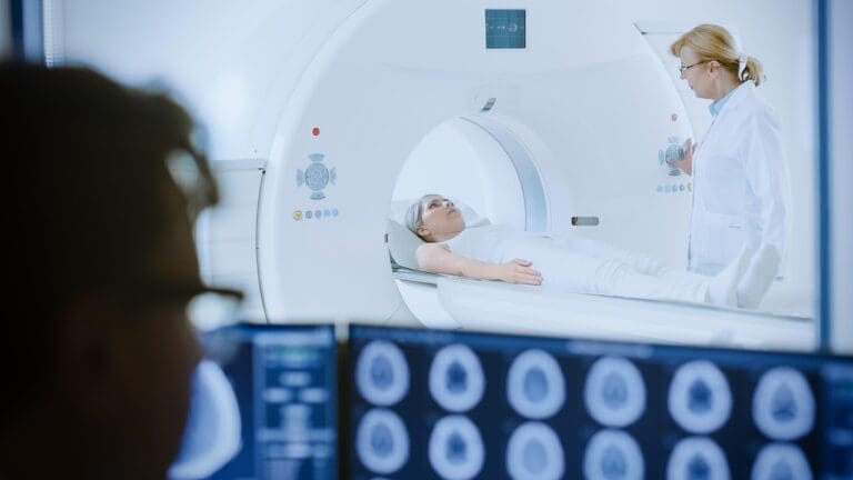
Magnetic Resonance Imaging (MRI) is the top choice for diagnosing AVMs. It gives us detailed info on the malformation’s size, location, and type. This is essential for planning treatment.
With MRI, we can see the AVM’s vascular structure. This includes the arteries that feed it and the veins that drain it. Knowing this helps us choose the best treatment.
Other Imaging Techniques
While MRI is the top choice, other methods like ultrasound, CT angiography, and conventional angiography are also used. They help in diagnosing AVMs.
| Imaging Technique | Advantages | Limitations |
|---|---|---|
| Ultrasound | Non-invasive, quick, and cost-effective | Limited depth penetration, operator-dependent |
| CT Angiography | High-resolution images, fast acquisition | Involves radiation, contrast required |
| Conventional Angiography | Detailed vascular anatomy, therapeutic options | Invasive, requires contrast and radiation |
Each imaging method has its own strengths and weaknesses. The choice depends on the AVM’s specifics and the patient’s situation.
Complications of Untreated AVMs
Arteriovenous Malformations (AVMs) can be serious if not treated. They can rupture and cause chronic vascular problems. These issues can greatly affect your life and even be life-threatening.
Rupture AVM: Risks and Consequences
AVM rupture is a major concern. It can cause severe bleeding. This can damage tissues and organs nearby.
“The rupture of an AVM can be a catastrophic event, leading to significant morbidity and mortality,” say vascular specialists. The damage depends on where and how big the malformation is.
Bleeding AVM: Management and Prevention
It’s important to manage and prevent bleeding in AVMs. Close monitoring and regular check-ups are key. Treatments like embolization or surgery can help prevent bleeding.
For AVMs in critical areas like the brain or spinal cord, the risk is higher. In these cases, early treatment is often advised to lower the risk of bleeding.
Long-term Vascular Damage
AVMs can also cause long-term vascular damage. The abnormal blood flow can lead to vascular remodeling. This can result in conditions like venous hypertension or arterial steal syndrome.
Over time, the blood vessels may become dilated or tortuous. This can lead to more problems. Treating AVMs early can prevent these changes and reduce complications.
AVM Therapy: Complete Treatment Plans
AVM therapy needs a mix of treatments, based on each patient’s needs. Arteriovenous malformations are complex, so treatments vary. They range from simple monitoring to more serious interventions.
Conservative Management
For some, watching and waiting is the best first step. This means regular check-ups to see how the AVM is doing. It’s good for AVMs that don’t cause problems or symptoms.
Here are some parts of conservative management:
- Regular visits to a doctor
- Imaging tests to check the AVM
- Medicine to handle symptoms
- Changes in lifestyle to lower risks
Medical Treatments
When watching and waiting isn’t enough, medical treatments are needed. These can be medicines or more serious procedures. The goal is to make the AVM smaller or get rid of it.
| Treatment Option | Description | Indications |
|---|---|---|
| Embolization | A procedure to stop blood flow to the AVM | Large AVMs, AVMs causing big symptoms |
| Sclerotherapy | Injecting a substance to close the AVM | AVMs with a big venous part |
| Surgical Resection | Removing the AVM surgically | AVMs that are easy to reach, causing severe symptoms or at risk of rupture |
When to Seek Treatment
Knowing when to get medical help is key. Pain, swelling, or skin color changes around the AVM mean it’s time to see a doctor. Also, if the AVM gets bigger or causes problems, treatment is needed.
We suggest patients talk to their healthcare team about their treatment options. Understanding the different treatments helps patients make better choices for their care.
Embolization of AVM: A Minimally Invasive Solution
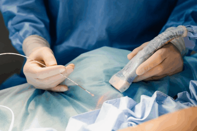
The embolization procedure is a top choice for treating AVMs. It’s less invasive than surgery. This method blocks the bad blood vessels with materials like coils or glue. This helps avoid rupture and other AVM problems.
The Embolization Process
An interventional radiologist does the embolization. They use imaging to guide a catheter to the AVM. Then, they use embolic materials to stop the bad blood flow.
This method can be used alone or with other treatments. It aims to block the AVM or make it smaller for easier treatment.
Candidates for Embolization
Not every AVM patient is right for embolization. The choice depends on the AVM’s size, location, and health of the patient. We look at each case carefully to decide if it’s the best option.
Embolization is often chosen for its less invasive nature. It can lower the risk of complications. Mayo Clinic says it’s effective for some AVMs, often with other treatments.
Recovery and Outcomes
Recovery from embolization varies by person and procedure. It’s usually quicker than surgery. We watch patients closely after to manage side effects and check if the treatment worked.
Many patients see big improvements after embolization. But, success depends on the AVM and the patient’s health.
Surgical Options for Arteriovenous Malformations
The way we treat AVMs has changed a lot. Now, we use new techniques to make treatments better and safer. Surgery is chosen when other treatments don’t work or can’t be used.
Traditional Surgical Approaches
Older surgery methods for AVMs mean removing the malformation directly. Microsurgical techniques have made these surgeries more precise and safe. Doctors use special tools and images to find and remove the AVM without harming nearby tissue.
Choosing traditional surgery depends on the AVM’s size, location, and how complex it is. Sometimes, doctors might suggest a mix of embolization and surgery.
| Factors Influencing Surgical Approach | Description |
|---|---|
| AVM Size and Location | Larger AVMs or those in critical areas may require more complex surgical planning. |
| AVM Complexity | The presence of multiple feeding vessels or draining veins can complicate surgery. |
| Patient Health | Underlying health conditions can impact the choice of surgical approach. |
Microsurgery Techniques
Microsurgery has changed how we treat AVMs. It lets doctors remove the malformation more precisely. Advanced microsurgical tools help them work more accurately, lowering the chance of problems.
Microsurgery is great for AVMs in hard-to-reach or delicate spots. High-resolution images and monitoring during surgery make these procedures safer and more effective.
Post-Surgical Care
After surgery, it’s important to watch patients closely for any issues. Managing pain is a big part of care. Doctors also check on the healing process and remove any stitches or staples.
Some patients might need to go through rehab, depending on where the AVM was or if there were any problems during surgery. A team of doctors works together to help the patient recover and meet any ongoing needs.
Specialized Treatments for AV Malformation Skin and Leg
Treating arteriovenous malformations (AVMs) in the leg and skin needs special care. These conditions have unique challenges. So, treatments must be tailored to manage symptoms and prevent problems.
Tailored Approaches for Leg AVMs
Leg AVMs need treatments that think about the blood flow and how they affect movement. Endovascular embolization is a common method. It involves injecting materials to block blood flow, helping to reduce symptoms.
Surgical excision is another option. It means surgically removing the AVM. This is often used for AVMs that are closer to the surface and have clear boundaries.
Dermatological Interventions for Skin AVMs
Skin AVMs are tricky because they can be seen and might bleed. Laser therapy is a treatment that can make skin AVMs less visible and manage symptoms.
Sclerotherapy is also an option. It involves injecting a solution to close off the abnormal blood vessels.
Combined Treatment Strategies
For complex AVMs, mixing treatments can be the best approach. For example, embolization followed by surgical excision can be used.
A team of experts is key in finding the right mix of treatments for each patient.
| Treatment Option | Description | Applicability |
|---|---|---|
| Endovascular Embolization | Blocking blood flow to the AVM | Leg AVMs, Complex AVMs |
| Surgical Excision | Surgical removal of the AVM | Superficial AVMs, Leg AVMs |
| Laser Therapy | Reducing appearance and symptoms | Skin AVMs |
| Sclerotherapy | Closing off abnormal blood vessels | Skin AVMs, Small AVMs |
“The key to successful AVM treatment lies in a tailored approach, considering the unique characteristics of each malformation and the patient’s overall health.” – AVM Specialist
Living with and Managing Arteriovenous Malformations
Managing arteriovenous malformations (AVMs) often needs a long-term plan. This includes regular check-ups and possibly many treatments over time. We know how important it is to manage AVMs well.
Regular monitoring and check-ups with doctors are key. This helps catch problems early and adjust treatment plans as needed. Taking care of AVMs long-term means working together with many medical experts.
People with AVMs can create a personal care plan with their doctors. This plan should cover managing symptoms and avoiding complications. Our aim is to support those living with AVMs, helping them manage their condition well.
With the right care, people with AVMs can live full and active lives. We’re dedicated to top-notch healthcare for international patients. We want to make sure they get the best care for their AVMs.
What does AVM stand for in medical terms?
AVM stands for Arteriovenous Malformation. It’s a birth defect where arteries and veins connect abnormally.
Are AVMs genetic?
AVMs might have a genetic link. Some are linked to genetic syndromes or run in families.
What are the symptoms of an AVM in the leg?
Symptoms include pain, swelling, and visible varicose veins. Skin discoloration or ulcers can also occur.
How is an AVM diagnosed?
Diagnosis starts with a physical exam. Then, imaging tests like MRI or angiography are used. MRI is often the best choice.
What are the risks of leaving an AVM untreated?
Untreated AVMs can rupture, bleed, or cause long-term damage. They may also cause pain or discomfort.
What is embolization of AVM, and how does it work?
Embolization is a treatment that blocks blood flow to the AVM. It uses materials to stop blood flow. This can reduce symptoms and prevent complications.
When is surgical treatment necessary for an AVM?
Surgery is needed for AVMs causing symptoms or at risk of rupture. The decision depends on the AVM’s location and size.
Can AVMs be treated with conservative management?
Yes, some AVMs can be managed without surgery. This is for those not causing symptoms or at low risk of complications. Regular monitoring is part of this approach.
What is the recovery process like after AVM embolization?
Recovery varies based on the AVM’s location. Patients may feel pain or discomfort, but this can be managed with medication.
How can I manage my AVM over time?
Managing an AVM involves regular check-ups and tests. This includes imaging and physical exams to watch for changes or complications.
References
- Gilbert, P. (2017). New treatment approaches to arteriovenous malformations. Frontiers in Neurology, (article) PMC5615391. Retrieved from https://pmc.ncbi.nlm.nih.gov/articles/PMC5615391/


