Last Updated on November 27, 2025 by Bilal Hasdemir

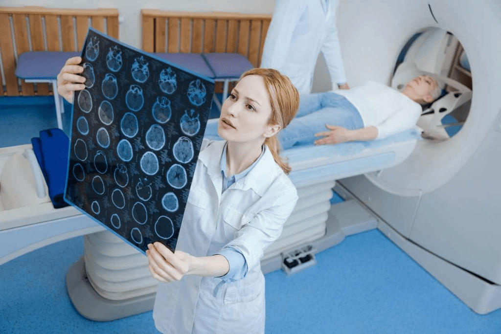
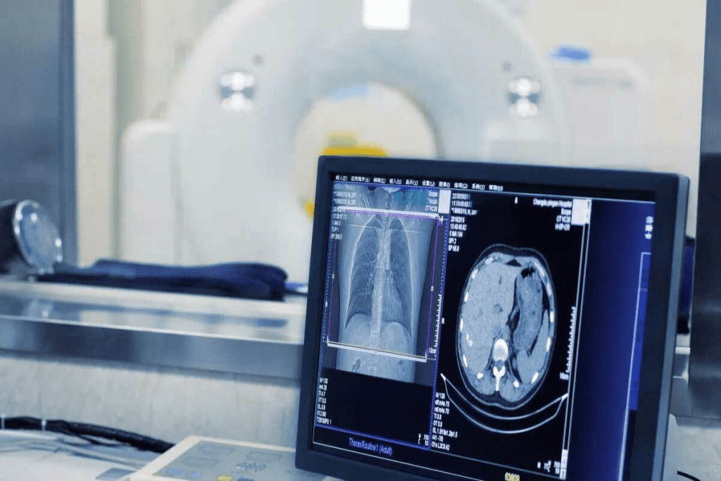
Understanding the differences between MRI and CT scans is key for diagnosis and care. Magnetic Resonance Imaging (MRI) and Computed Tomography (CT) scans are two unique ways to see inside the body. Many patients ask what is difference between mri scan and ct scan, and the main distinctions are that MRI uses magnetic fields and radio waves without radiation, while CT uses X-rays and is faster, making each better suited for different medical situations.
MRI scans use strong magnetic fields and radio waves to show soft tissues in detail. On the other hand, CT scans use X-rays to take pictures of the body’s inside from different sides. Experts say the right choice depends on the health issue.
MRI scans are great for soft tissues. But, CT scans are better in emergencies because they’re faster and show bones and injuries well.
Key Takeaways
- MRI scans use magnetic fields and radio waves to image soft tissues.
- CT scans use X-rays to capture images from multiple angles.
- The choice between MRI and CT scans depends on the medical condition.
- MRI scans are ideal for soft tissue imaging.
- CT scans are preferred in emergency situations.
The Fundamentals of Medical Imaging

Medical imaging is key in today’s medicine. It helps doctors see inside the body without surgery. This is thanks to tools like MRI and CT scans.
Both MRI and CT scans give detailed images. These images help doctors make better choices.
For more on MRI and CT scans, check out this article.
The Evolution of Diagnostic Imaging
Diagnostic imaging has changed a lot over time. From X-rays to MRI and CT scans, technology has improved a lot. This has made it easier to see inside the body and treat more conditions.
MRI scans use magnetic fields and radio waves to show soft tissues. CT scans use X-rays to show internal structures. Both are safe, noninvasive, and generally well-tolerated. They are very important in today’s medicine.
The Role of Imaging in Modern Medicine
In today’s medicine, imaging is very important. It helps find diseases early, guides treatment, and checks if treatments work. MRI and CT scans are great for getting detailed, high-resolution images. These images are key for diagnosing many conditions.
Choosing between MRI and CT scans depends on the condition, the patient’s health, and what the doctor needs. Knowing the difference between CT scan and MRI helps both doctors and patients make good choices.
Medical imaging will keep getting better. This will help healthcare even more. MRI and CT technologies will keep improving, leading to better care for patients.
How MRI Scans Work

MRI scans use advanced technology and science to create detailed images of the body. They are a key tool in diagnosing many health issues.
The Science Behind Magnetic Resonance Imaging
MRI scans use nuclear magnetic resonance to create images. Hydrogen atoms in the body, found in water and fat, are aligned by a strong magnetic field. Radio waves then disturb these atoms, causing them to send signals as they realign. These signals help the MRI machine make detailed images.
The science behind MRI involves the magnetic field, radio waves, and hydrogen atoms. This interaction helps create images that doctors use to diagnose many conditions.
MRI Equipment and Technology
MRI scans use a large cylindrical magnet, a radiofrequency coil, and a computer. The strength of the magnetic field is key, with most machines having a field strength of 1.5T or 3T. This provides high-quality images.
Over time, MRI technology has improved. It now offers faster scans and clearer images. Advances in coil technology and pulse sequences have also helped MRI’s diagnostic abilities.
The MRI Scanning Process
During an MRI scan, the patient lies on a table that slides into the machine. The scanning time varies based on the type of scan and the body part being imaged. It’s important for patients to stay very quiet and not move.
The MRI scanning process includes several steps. First, there’s preparation, which might involve removing metal objects or using a contrast agent. Then, the actual scanning happens. The patient is positioned in the machine, where the magnetic field and radio waves capture the necessary images.
How CT Scans Work
CT scans use X-rays and computers to show the body’s inside. This method is key in medical diagnosis. It helps doctors see what’s going on inside us.
The Science Behind Computed Tomography
CT scans use X-rays to take pictures from many angles. A device spins around the body, gathering data. This data is then turned into detailed images by computers.
CT scans work because different tissues absorb X-rays differently. This lets them show bones, muscles, and fat clearly. It helps doctors understand the body’s inner workings.
CT Equipment and Technology
Today’s CT scanners are very advanced. They have a big, doughnut-shaped part for X-rays and detectors. There’s also a table for the patient and a computer for the images. These scanners are faster, show more detail, and use less radiation.
New tech in CT scanners includes spiral and multi-detector scanners. Spiral scanners scan continuously, and multi-detector scanners take many images at once. This makes scans quicker and more detailed.
The CT Scanning Process
Getting a CT scan is easy. The patient lies on a table that slides into the scanner. The scanner starts taking pictures as it moves around the body.
Patients must stay very quiet and not move during the scan. Sometimes, a special dye is used to make certain areas clearer. After the scan, doctors look at the images to find and diagnose health issues.
What Is the Difference Between an MRI Scan and a CT Scan?
MRI and CT scans are both used to help doctors diagnose problems. But they work in different ways. MRI scans use strong magnetic fields and radio waves to see soft tissues inside the body. CT scans, on the other hand, use X-rays to show bones and internal bleeding.
Fundamental Technology Differences
The main difference is in how they work. MRI scans use nuclear magnetic resonance to create images. CT scans combine X-rays from different angles to make images.
Key Technological Differences:
- MRI: Uses magnetic fields and radio waves; ideal for soft tissue imaging.
- CT Scan: Employs X-rays; better for bone and internal injury imaging.
Image Quality and Detail Comparison
Both MRI and CT scans have their own strengths. MRI scans are great for soft tissues like organs and tendons. They show detailed information about these tissues. CT scans, on the other hand, are better for bones and detecting injuries.
Image Quality Comparison:
| Imaging Modality | Soft Tissue Imaging | Bone Imaging |
| MRI | Excellent | Good |
| CT Scan | Fair | Excellent |
Time and Cost Considerations
Another big difference is the time and cost. MRI scans take longer, from 15 to 90 minutes. CT scans are much quicker, taking just a few minutes. MRI scans are also more expensive because of the advanced technology.
Comparison of Time and Cost:
| Factor | MRI | CT Scan |
| Procedure Time | 15-90 minutes | A few minutes |
| Cost | Generally more expensive | Generally less expensive |
Clinical Applications of MRI Scans
MRI scans have many uses in medicine, helping us diagnose and treat diseases. They are safer than CT scans because they don’t use harmful radiation. MRI machines use a strong magnetic field and radio waves to create images.
Neurological Disorders and Brain Imaging
MRI scans are great for looking at the brain and spinal cord. They help doctors find problems like:
- Stroke
- Multiple sclerosis
- Brain tumors
- Spinal cord injuries
These scans give doctors clear pictures. They help figure out how bad the damage is and what treatment to use.
Musculoskeletal Conditions and Soft Tissue Injuries
MRI scans are also good for checking muscles and soft tissues. They help doctors see:
- Torn ligaments and tendons
- Herniated discs
- Soft tissue tumors
These scans show soft tissues clearly. This helps doctors plan treatments without needing surgery.
Cancer Detection and Monitoring
MRI scans are key in finding and tracking cancer. They help see tumors and how far cancer has spread. MRI is great for soft tissue cancers like:
- Breast cancer
- Prostate cancer
- Liver cancer
MRI scans are safe and detailed. They are vital for cancer patients who need many scans.
Clinical Applications of CT Scans
CT scans are key in medical imaging for their quick and accurate diagnoses. They are very useful in many clinical settings, where time is very important.
Trauma and Emergency Situations
CT scans are often the first choice for trauma and emergency situations. They are fast and can show complex injuries well. This helps doctors quickly see how bad the injuries are and start treatment right away.
In emergencies like head trauma, CT scans can spot hemorrhages or fractures fast. This allows for quick action to save lives. They also help diagnose acute conditions like appendicitis or internal bleeding.
Bone Fractures and Structural Abnormalities
CT scans are great at showing bones and structural abnormalities. They are better than X-rays for complex fractures or bone diseases. This is because they show more detail, which is important for the spine, pelvis, or complex joints.
They also help find bone degeneration or cancer in bones. Their clear images help doctors plan surgeries or track bone conditions.
Lung and Chest Imaging
CT scans are very important for lung and chest imaging. They give detailed views of the lungs, airways, and more. They help diagnose many conditions, like pneumonia, pulmonary embolism, or lung cancer.
CT scans can spot small changes in lung tissue, which is key for early treatment. They also help guide biopsies and other procedures in the chest. This makes them very useful for both diagnosis and treatment.
When comparing CT scans to MRI, each has its own strengths. MRI might be better for soft tissue, but CT scans are better for bones and quick emergency checks. Knowing what can an MRI show that a CT scan cannot and vice versa helps choose the right imaging for each case.
Safety Considerations and Risks
Both MRI and CT scans are important for diagnosing health issues. But, they also have safety concerns and risks. It’s key to understand these risks to ensure patient safety and get the best results from scans.
Radiation Exposure in CT Scans
CT scans use a small amount of ionizing radiation. The risk from one scan is usually low. But, getting many scans over time might raise the risk of cancer. Doctors must carefully consider the benefits and risks, mainly for young patients or those needing many scans.
Radiation Dose Management is a big deal in CT scans. New CT scanners aim to use less radiation while keeping image quality high. Tools like automatic exposure control and iterative reconstruction help achieve this goal.
MRI Safety Concerns and Contraindications
MRI scans don’t use ionizing radiation but have their own safety issues. The strong magnetic field can harm certain metal implants, like pacemakers. People with metal implants or foreign bodies might not be able to have an MRI.
Patient Screening is critical before an MRI. A detailed questionnaire and screening help find out if someone can safely have an MRI. This includes checking for metal implants, claustrophobia, or other risks.
Patient Screening Protocols
Good patient screening is key for both MRI and CT scans. For MRI, it’s about asking about metal implants, surgeries, and claustrophobia. For CT scans, it’s about deciding if contrast agents are needed and checking kidney function to avoid kidney problems.
| Safety Aspect | MRI Scan | CT Scan |
| Radiation Exposure | No | Yes |
| Magnetic Field Risks | Yes | No |
| Contrast Agent Risks | Rarely | Yes |
| Claustrophobia Concerns | Yes | Less Common |
Preparing for Your Imaging Procedure
Getting ready for an MRI or CT scan involves several steps. These steps help make the imaging process smooth and successful. Always follow the specific instructions given by your healthcare provider or the imaging facility.
Before Your MRI Scan
Before your MRI, tell your technician about any metal implants or devices. Also, remove any metal objects like jewelry or glasses before the scan.
Key steps to prepare for an MRI scan include:
- Informing your technician about any claustrophobia or anxiety
- Removing any metal objects or devices
- Following any specific dietary instructions
Before Your CT Scan
For a CT scan, you might need to wear a hospital gown. Remove any jewelry or objects that could interfere with the scan. Also, tell your technician about any allergies or sensitivities, if contrast dye is used.
Preparation steps for a CT scan may include:
- Informing your technician about any allergies or sensitivities
- Following dietary instructions, such as fasting
- Removing any jewelry or other objects
Addressing Claustrophobia and Anxiety
If you have claustrophobia or anxiety, there are ways to help. Open MRI machines or sedation might be suggested for severe cases.
Talk to your healthcare provider or the imaging facility about your concerns. They can help find the best solution for you.
Technological Advances and Future Developments
The world of medical imaging is on the verge of a big change. This is thanks to new tech in MRI and CT scans. These updates are making images clearer and making patients more comfortable and safe.
Innovations in MRI Technology
Recently, MRI tech has made big strides. Now, we have high-field MRI systems that give us sharper images. We also use functional MRI (fMRI) to see how the brain works.
- Advances in coil technology for better image quality
- Development of open MRI machines to reduce claustrophobia
- Improved software for faster image processing and analysis
Advances in CT Technology
CT tech has also improved a lot. Now, we have multi-detector CT scanners that scan faster and give clearer images. Plus, iterative reconstruction techniques help cut down on radiation while keeping image quality high.
- Development of dual-energy CT for better tissue characterization
- Advances in CT angiography for vascular imaging
- Improved portable CT scanners for emergency and critical care
The Role of Artificial Intelligence in Image Interpretation
Artificial intelligence (AI) is playing a big role in improving how we read MRI and CT scans. AI can spot problems, track disease, and make diagnoses more accurate.
- AI-assisted detection of lesions and abnormalities
- Quantification of disease severity and progression
- Integration of AI with imaging modalities for real-time analysis
As these techs keep getting better, we’ll see even better results in diagnosing and treating patients. The mix of AI with MRI and CT scans is very exciting. It could lead to more precise and tailored medicine.
Conclusion
Knowing the difference between MRI and CT scans is key for both patients and doctors. These scans are vital in today’s medical world. They offer unique benefits based on what the doctor needs.
MRI scans use magnetic fields and radio waves to show detailed images. They’re great for soft tissue, spinal issues, and inflammation. CT scans, on the other hand, use x-rays for quick images. They’re best for emergencies, finding bone fractures, and spotting internal bleeding.
In short, knowing the MRI and CT scan differences helps in making better diagnostic choices. By understanding their strengths and weaknesses, patients can make informed decisions about their care.
FAQ
What is the main difference between an MRI and a CT scan?
What does an MRI show that a CT scan doesn’t?
Are MRI and CT scans used for the same purposes?
Is a CT scan or an MRI more expensive?
Do CT scans use magnets?
Can an MRI detect something a CT scan can’t?
Are there any risks associated with MRI and CT scans?
How can I prepare for an MRI or CT scan?
Can artificial intelligence improve the interpretation of MRI and CT scans?
What are the latest advancements in MRI and CT technology?
References
- Functional Imaging: CT and MRI. (n.d.). https://pmc.ncbi.nlm.nih.gov/articles/PMC2435287/
- Comparison of computed tomography and magnetic resonance imaging in evaluation of skull lesions. (2022). Egyptian Journal of Radiology and Nuclear Medicine. https://ejrnm.springeropen.com/articles/10.1186/s43055-022-00745-9


