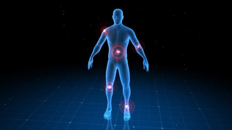
FDG PET scans represent a revolutionary advancement in nuclear medicine imaging, significantly transforming the landscape of oncology. These scans are key for finding and planning treatment for cancer. FDG, or fluorodeoxyglucose, is a sugar molecule that cancer cells gobble up more than normal cells do.
The organ most affected by FDG is important. It shows the good and bad sides of PET scans. Knowing this is vital for and patients.
Key Takeaways
- FDG PET scans are essential for finding and treating cancer.
- The organ most affected by FDG is key to understanding PET scan risks and benefits.
- Nuclear medicine imaging has greatly improved oncology.
- FDG is taken up by cells, with cancer cells using it more.
- It’s important for and patients to understand the role of FDG.
FDG PET scans represent a revolutionary advancement in nuclear medicine imaging, significantly transforming the landscape of oncology.
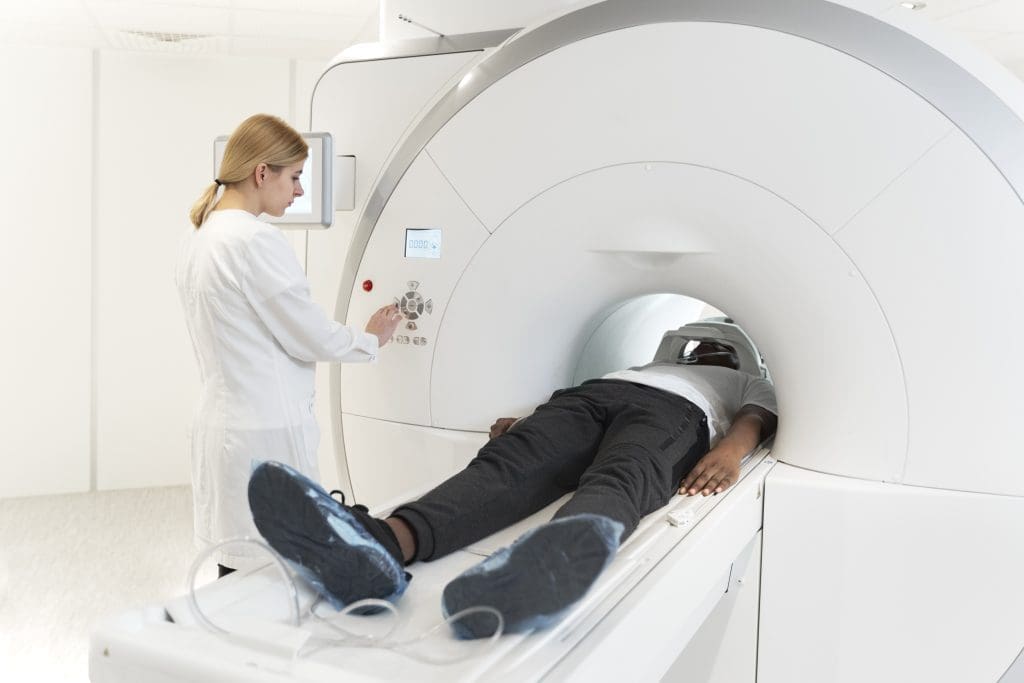
FDG PET scans are key in medical diagnostics. They use Fluorodeoxyglucose, a special compound. This compound is vital for finding and managing cancers.
FDG works like glucose, which cells use for energy. This makes it great for checking how active cells are. It’s perfect for looking at tissues that use a lot of glucose, like some tumors.
What is FDG (Fluorodeoxyglucose)?
Fluorodeoxyglucose (FDG) is a glucose-like substance used in PET scans. It has a fluorine-18 (F-18) atom instead of a hydroxyl group. This lets it enter cells like glucose but can’t be broken down further.
How FDG Functions as a Radiotracer
As a radiotracer, FDG emits positrons. These positrons meet electrons and create gamma photons. The PET scanner picks up these photons. The strength of the signal shows how active the tissue is.
| Characteristics | Description |
| Chemical Structure | Glucose analog with F-18 substitution |
| Cellular Uptake | Mimics glucose uptake, trapped after phosphorylation |
| Detection Mechanism | Positron emission and gamma photon detection |
FDG’s special features make it very useful in nuclear medicine. It helps see how tissues work and find diseases.
The Basics of FDG PET Scan Technology
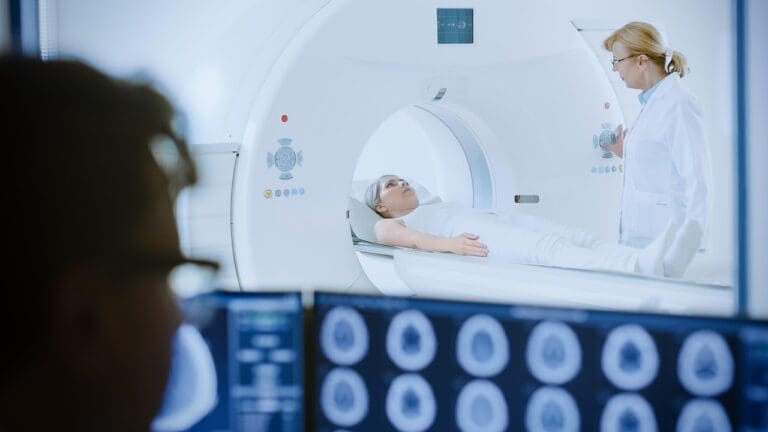
FDG PET scan technology is key in today’s medicine. It helps diagnose and manage diseases, like cancer. It combines PET’s metabolic info with CT’s body details for a full view.
Principles of Positron Emission Tomography
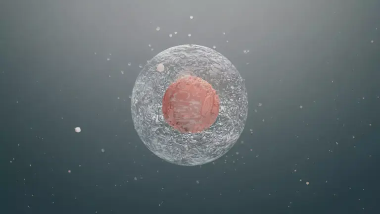
PET scans show how active cells are in the body. They use a special molecule, Fluorodeoxyglucose (FDG), to do this. FDG is a sugar molecule with a radioactive tag.
The steps are:
- FDG is injected into the patient’s blood.
- Cells, like cancer ones, take up the FDG.
- FDG’s positrons meet electrons, making gamma rays.
- The PET scanner catches these rays, showing where cells are active.
Integration with CT Scanning
Combining PET with CT is a big step forward. It’s called hybrid imaging. This mix gives both metabolic and body structure info at once.
The perks are:
- It’s easier to find where FDG is taken up.
- Diseases, like cancer, are spotted early.
- It’s clearer to tell if something is cancer or not.
| Technology | Primary Use | Benefits |
| PET | Metabolic activity imaging | Early disease detection, monitoring treatment response |
| CT | Anatomical imaging | Detailed structural information, useful for staging and planning treatment |
| PET/CT | Combined metabolic and anatomical imaging | Enhanced diagnostic accuracy, improved patient outcomes |
Critical Organs in FDG PET Imaging
When we talk about FDG PET imaging, knowing about critical organs is key. These organs are most at risk from radiation because of their function or sensitivity.
Definition of “Critical Organ” in Nuclear Medicine
In nuclear medicine, a critical organ is one that’s very sensitive to radiation or is very important for our body’s function. Knowing which organs are critical helps us understand the risks of radiation during tests like FDG PET scans.
The Bladder as a Primary Critical Organ
The bladder is a main critical organ in FDG PET imaging. It holds urine with the radioactive tracer FDG, so it gets a lot of radiation. The bladder’s job in holding and getting rid of the tracer makes it key when we talk about radiation risks. To lower radiation to the bladder, we use hydration and voiding protocols.
Other Organs of Concern
Other organs also get a lot of radiation during FDG PET imaging. These include the brain, heart, and parts of the gut, depending on where the FDG goes. Knowing where FDG goes in the body is important for reading PET scan results and understanding radiation exposure.
Each organ reacts differently to radiation. How much radiation they get can change based on the FDG dose and the patient’s health.
Radiation Dose Distribution in FDG PET Scans
Radiation dose in FDG PET scans is key for patient safety and good images. How FDG (Fluorodeoxyglucose) spreads in the body after it’s given affects the radiation to different organs.
How FDG Distributes Throughout the Body
FDG goes to cells in the body, mainly those that use a lot of energy, like cancer cells. The spread of FDG is not even; it builds up in some organs and tissues. This changes the radiation dose they get.
The radiation dose distribution depends on several things. These include the FDG dose given, how long it takes to scan after injection, and the patient’s health and body type.
Organs Receiving the Highest Radiation Exposure
Some organs get more radiation because of how they take up FDG or how they get rid of it. The brain, heart, liver, and bladder are among the organs that get a lot of radiation.
| Organ | Average Radiation Dose (mSv/MBq) |
| Brain | 0.017 |
| Heart | 0.019 |
| Liver | 0.021 |
| Bladder | 0.033 |
The bladder gets one of the highest doses because it excretes FDG through urine. Knowing how FDG PET scan works helps make it safer and reduce radiation to sensitive areas.
The Half-Life of FDG and Its Implications
Knowing the half-life of FDG is key for better PET scans and less radiation risk. The half-life of a radiotracer is how long it takes for its radioactivity to cut in half. This is very important for medical imaging.
FDG, or Fluorodeoxyglucose, has a half-life of 110 minutes. This short half-life means its radioactivity drops fast. This is important for both safety and when to do PET scans.
Understanding FDG’s 110-Minute Half-Life
FDG’s 110-minute half-life is just right. It lets us do PET scans quickly and safely. This half-life helps us see how the body works without too much radiation.
- The short half-life means we have to plan PET scans carefully for the best images.
- It lets us give enough FDG for great images without too long exposure.
Impact on Radiation Exposure and Imaging Timing
The half-life of FDG affects how much radiation we get and when to do PET scans. Radiation safety protocols help keep everyone safe by considering FDG’s decay rate.
When to do scans is very important. We wait a bit after giving FDG to let it spread in the body. The uptake period is key for good PET scan images.
- Optimizing the uptake period for the best FDG distribution in tissues.
- Planning scans based on FDG’s half-life for quality images and safety.
By understanding FDG’s half-life, healthcare can make PET scans safer and more effective. This improves both safety and results of diagnostic imaging.
Complete Guide to FDG PET Scan Procedures
The success of an FDG PET scan depends on several factors. These include patient preparation and the scanning protocol used. A well-conducted FDG PET scan is key for accurate diagnostic results.
Patient Preparation Guidelines
Proper patient preparation is essential for a successful FDG PET scan. This includes:
- Fasting for a specified period, usually 4-6 hours, to minimize glucose uptake in the body
- Avoiding strenuous exercise for a certain period before the scan
- Staying hydrated by drinking plenty of water
- Informing the healthcare provider about any medications, allergies, or medical conditions
Key Considerations: Patients with diabetes need special consideration. Their glucose levels can affect the FDG uptake. The healthcare provider will provide specific instructions on managing diabetes before the scan.
FDG Administration Process
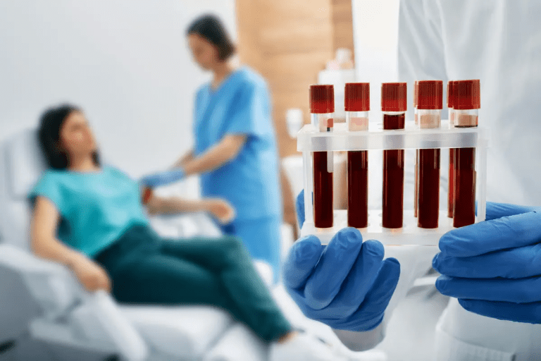
The administration of FDG is a critical step in the PET scan procedure. FDG is injected into a vein, usually in the arm. The dose is carefully calculated based on the patient’s weight and the specific requirements of the scan.
Important: The injection should be done in a quiet, comfortable environment. This is to minimize stress and anxiety, which can affect the distribution of FDG.
The Scanning Protocol
The scanning protocol involves several steps:
- The patient is positioned on the scanning table, usually in a supine position.
- The PET scanner detects the gamma rays emitted by the FDG, creating detailed images of the body’s metabolic activity.
- The scan typically takes around 30 minutes to an hour, depending on the area being scanned and the specific protocol.
Optimizing the Scan: The scanning protocol can be adjusted based on the patient’s condition and the specific diagnostic requirements. This may include adjusting the scan time or using additional imaging techniques.
Comparing Radiation Exposure in Medical Imaging
It’s important to know about radiation exposure when looking at different medical imaging methods. These methods help diagnose and treat many health issues. But, we must think about the radiation each one uses.
PET Scan vs. Other Imaging Modalities
PET scans are great for finding tumors, but they do use radiation. When we compare PET scans to CT scans, X-rays, and MRI, we learn about their safety levels.
| Imaging Modality | Typical Effective Dose (mSv) |
| PET Scan | 7-10 |
| CT Scan (Abdomen and Pelvis) | 10-20 |
| X-ray (Chest) | 0.1 |
| MRI | 0 |
The table shows the typical doses of radiation for each imaging method. PET and CT scans have higher doses than X-rays. MRI doesn’t use any ionizing radiation.
Cumulative Radiation from Combined PET/CT Procedures
Many PET scans are done with CT scans together. This gives both the function and structure of the body. But, it means more radiation for the patient.
Cumulative Radiation Exposure: The total radiation from a PET/CT scan is the sum of each scan’s dose. For example, a PET/CT scan might have a total dose of 15-25 mSv.
Implications for Patient Care: High doses of radiation are a big worry, mainly for patients who have many scans. It’s important to use the least amount of radiation needed for good results.
Radiation Dose Management in FDG PET Imaging
Managing radiation dose is key in FDG PET imaging to keep patients safe. As these scans are used more, making sure they are safe is very important.
Strategies to Reduce Radiation Exposure
To cut down on radiation in FDG PET scans, several steps can be taken. These include:
- Adjusting the amount of FDG given based on the patient’s weight and scan details.
- Using new algorithms to make images better at lower doses.
- Using the latest PET scanners that are more sensitive and clear.
Optimizing scan protocols is also vital. This means picking the right scan range, time, and other settings. It helps get good images while keeping doses low.
Optimizing Scan Parameters
Getting the right balance between image quality and radiation is key. Important factors include:
| Parameter | Optimization Strategy | Impact on Radiation Exposure |
| Scan Duration | Adjusting scan time based on patient condition and scanner capabilities. | Shorter scans mean less radiation. |
| FDG Dose | Adjusting the FDG dose based on patient weight and needs. | Lower doses mean less radiation. |
| Scan Range | Limiting the scan to only the needed area. | This reduces radiation to other parts. |
Technological advancements in PET scanners and image algorithms help too. New scanners and algorithms let us get great images at lower doses.
By using these strategies and keeping up with new tech, healthcare can manage radiation doses better. This makes patients safer and helps get better results.
Applications of FDG PET Scans
FDG PET scans are used in many medical fields like oncology, neurology, and cardiology. This makes them a key tool in today’s medicine.
Oncological Applications
In oncology, FDG PET scans find tumors, check how far they’ve spread, and see if treatments are working. They’re great at spotting tumors because they show where cells are most active.
Tumor Detection and Staging: These scans help find the main tumor and any spread, helping plan treatment.
Monitoring Treatment Response: They track how tumors change, showing if treatments are effective.
Neurological Applications
FDG PET scans also check brain activity and function in neurology. They’re key in diagnosing and treating brain diseases.
- Diagnosing neurodegenerative diseases such as Alzheimer’s
- Evaluating seizure disorders
- Assessing brain function after injury or infection
Cardiac Applications
In cardiology, FDG PET scans look at heart health and function. This info is vital for treating heart disease.
Myocardial Viability Assessment: They spot healthy heart areas, helping decide on treatments.
FDG PET scans have many uses in medicine. They give important metabolic info, improving care in many areas.
Interpreting FDG PET Scan Results
Understanding FDG PET scans is complex. It involves knowing about SUV and FDG uptake patterns. These scans need a detailed look at the patient’s history and the scan’s specifics.
Understanding SUV (Standardized Uptake Value)
The SUV is key in FDG PET imaging. It shows how much FDG is taken up by tissues. It’s found by measuring activity in a certain area and comparing it to the dose and body weight.
SUV Calculation: The SUV formula is: SUV = (Activity in ROI per unit mass) / (Injected dose / Patient body weight). This gives a semi-quantitative look at FDG uptake. It helps see how active tissues are metabolically.
| SUV Value | Interpretation |
| Low SUV (<2.5) | Typically indicates benign or low metabolic activity |
| Moderate SUV (2.5-5) | Suggests potentially malignant or inflammatory processes |
| High SUV (>5) | Often associated with highly metabolic or malignant tissues |
Common Patterns of FDG Uptake
FDG uptake patterns offer insights into lesions or tissues. There are three main types: focal, diffuse, and symmetric uptake.
Focal Uptake: Focal areas of high FDG uptake usually mean active lesions, like tumors. The intensity and details of focal uptake help tell if something is benign or malignant.
Diffuse Uptake: Diffuse FDG uptake is seen in inflammation or some cancers. The spread and intensity of this uptake are key for correct interpretation.
Knowing these patterns and SUV values is vital for understanding FDG PET scan results. By using this knowledge, can make better decisions about diagnosis and treatment.
Potential Risks and Side Effects of FDG PET Scans
It’s important for patients and to know about the risks of FDG PET scans. These scans are useful for diagnosis but involve radiation. This can affect health.
Short-term Effects of Radiation Exposure
Short-term effects of radiation from FDG PET scans are usually mild. They might include feeling tired, having headaches, or feeling sick. These symptoms are short-lived and go away by themselves.
Radiation Exposure Comparison
| Imaging Modality | Typical Effective Dose (mSv) |
| FDG PET Scan | 7-10 |
| CT Scan (Abdomen/Pelvis) | 10-20 |
| Chest X-ray | 0.1 |
Long-term Considerations
Long-term risks of FDG PET scans include the chance of radiation causing cancer. But, the risk is low. The scan’s benefits often outweigh the risks.
need to think carefully about ordering FDG PET scans. This is true for younger patients or those needing many scans.
Patients should talk to their about their risks and worries. This helps make informed choices about their health care.
Special Considerations for Different Patient Populations
Different patient groups need special care when they get FDG PET scans. This ensures the scan is safe and works well for them.
Pediatric Patients and FDG PET Scans
Kids are more sensitive to radiation and its long-term effects. Adjusting the FDG dose based on the child’s weight and using pediatric-specific protocols helps reduce radiation.
FDG PET scans are used in kids only when the benefits are clear. This is for things like cancer staging or disease extent.
Pregnant and Breastfeeding Women
Pregnant women face risks from FDG PET scans. The scan’s benefits must be weighed against the risks. Other tests should be tried first.
There’s worry about radioactive material in breast milk for breastfeeding women. Guidelines suggest stopping breastfeeding for a while after FDG to protect the baby.
Healthcare providers must talk to pregnant and breastfeeding women. This helps them make the best choices for their care.
Skull Base to Mid-Thigh vs. Whole Body PET Scanning
PET scans can cover different areas, from just the skull base to mid-thigh to the whole body. Choosing the right scan depends on what need to see.
Differences in Scan Coverage
Skull base to mid-thigh scans are often used for cancer checks. They focus on the torso, where many cancers are found. But, they might miss other areas.
Whole-body scans, on the other hand, look at everything from head to toes. They give a full view, which is key for some diagnoses or checking cancer spread.
Key differences in scan coverage include:
- The extent of body area covered
- The ability to detect abnormalities outside the primary scan region
- The need for extra scans for areas not in the first scan
Impact on Radiation Exposure
Choosing between these scans also affects how much radiation you get. Whole-body scans give more radiation because they scan more area. A study found that whole-body scans give a higher dose than scans of just the skull base to mid-thigh.
“The radiation exposure from PET scans is a critical consideration, especialy for patients who need many scans or are getting a full check-up.”
FDG PET scans represent a revolutionary advancement in nuclear medicine imaging, significantly transforming the landscape of oncology.
It’s important to pick the right scan to get the most info while keeping radiation low. must think about the patient’s health, what they need to see, and the risks of more radiation.
Advancements in FDG PET Scan Technology
FDG PET scan technology has seen big changes. These changes aim to improve image quality, lower radiation doses, and boost diagnostic accuracy.
Improved Detectors and Resolution
New advancements in FDG PET scans have led to better detector technology. Modern scanners have detectors that are more sensitive and precise. This means can spot and understand lesions more clearly, leading to better diagnoses.
Key Features of Advanced Detectors:
- Higher sensitivity to detect smaller lesions
- Better spatial resolution for more accurate localization
- Improved image quality for enhanced diagnostic confidence
Dose Reduction Technologies
There’s also a big push for reducing radiation doses in FDG PET scans. New technologies aim to cut down radiation without losing image quality. These include better detectors, advanced algorithms, and smarter scanning methods.
| Technology | Description | Impact on Dose Reduction |
| Advanced Detectors | High sensitivity detectors that capture more PET events | Significant reduction in required FDG dose |
| Reconstruction Algorithms | Improved algorithms that enhance image quality | Allows for lower dose scans while maintaining image quality |
| Optimized Scanning Protocols | Protocols tailored to minimize dose while maximizing image quality | Reduces overall radiation exposure to patients |
These updates in FDG PET scan tech are key to better patient care. They help make more accurate diagnoses and reduce radiation exposure. As tech keeps getting better, we can look forward to even more improvements in medical imaging.
Regulatory Guidelines for FDG PET Scan Radiation
Strict rules control the use of FDG PET scans to keep everyone safe. These rules are key to making sure FDG PET scans are used safely. They help lower the risk of radiation for both patients and medical staff.
FDA and International Radiation Safety Standards
The FDA and other global groups have set tough rules for FDG PET scans. These rules cover how to handle the radioactive materials, prepare patients, and use the PET scan machines.
Key parts of these rules are:
- Safe ways to handle and give FDG
- Steps to cut down radiation for patients and workers
- Keeping PET scan machines in good shape and checking their quality
Monitoring Cumulative Radiation Exposure
It’s important to watch how much radiation patients get from FDG PET scans. This means tracking the doses they receive and changing scan settings if needed to lower exposure.
Good ways to keep track include:
- Keeping detailed records of patient radiation doses
- Using new tech and methods to reduce doses
- Telling patients about radiation safety and risks
Following these rules and keeping an eye on radiation helps use FDG PET scans safely and well.
Conclusion
Knowing which organ is key for FDG PET scans is vital for safety and better imaging. The bladder is a main organ because it handles FDG excretion.
Managing radiation doses is key to protect patients and staff. By adjusting scan settings and using new FDG PET tech, can get better images with less radiation.
FDG PET scans are important for many health issues. Following rules and safety standards is critical. This ensures scans are used safely and effectively, helping patients get better care.
Radiation safety in FDG PET scans is very important. By focusing on this, can provide top-notch imaging while keeping risks low.
FAQ
What regulatory guidelines govern FDG PET scan radiation?
Rules from the FDA and global safety standards guide FDG PET scans. They help ensure scans are safe, including keeping track of how much radiation patients get.
What advancements have been made in FDG PET scan technology to improve image quality and reduce radiation dose?
New tech includes better detectors and ways to reduce the dose. These improvements make images clearer while using less radiation.
How does skull base to mid-thigh PET scanning compare to whole-body PET scanning in terms of radiation exposure?
PET scans from the skull to the mid-thigh use less radiation. This is because they cover a smaller area of the body.
What are the possible risks and side effects of FDG PET scans?
Risks include feeling tired from radiation and a small chance of getting cancer later. But these are rare.
How do PET scans compare to other imaging modalities in terms of radiation exposure?
PET scans do involve radiation, but the dose can be similar to or even less than other scans like CT scans. It depends on the specific scan used.
How can radiation exposure be managed and reduced in FDG PET imaging?
To lower radiation, scan settings can be optimized. New technologies can also help. The dose of FDG can be adjusted based on the patient’s needs.
What is the half-life of FDG, and how does it impact radiation safety?
FDG’s half-life is about 110 minutes. This means its radiation dose halves every 110 minutes. It affects when PET scans are done and how safe they are.
How does FDG distribute throughout the body after administration?
After it’s given, FDG spreads all over the body. It builds up in areas that use a lot of glucose. The bladder, brain, and heart get the most radiation.
What is the critical organ for FDG PET scans?
The bladder is the main organ of concern for FDG PET scans. It holds and gets rid of the tracer, leading to a lot of radiation exposure.
What is FDG and how is it used in PET scans?
FDG, or Fluorodeoxyglucose, is a special tracer used in PET scans. It helps show how active different parts of the body are. This is very helpful in finding cancer because cancer cells use more glucose.


