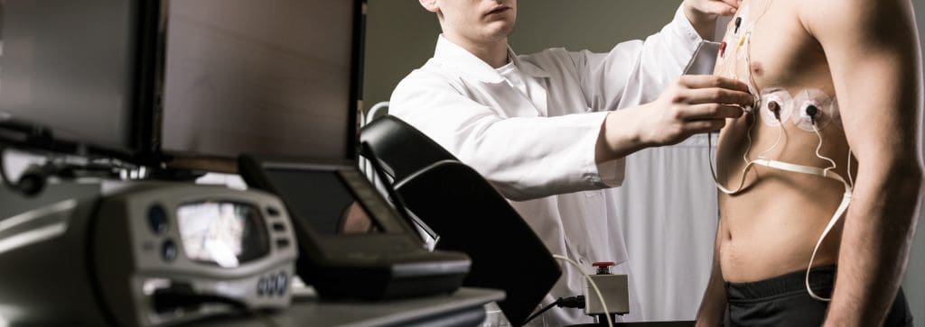
A nuclear stress test is a key tool for checking how well blood flows to the heart. It measures blood flow to the heart during rest and physical activity. This test helps doctors spot coronary artery disease, where the heart’s arteries get narrowed or blocked.
This test uses a tiny bit of radioactive material to make pictures of the heart. Doctors can see where blood flow is limited. Many patients then ask, “What is the next test after a nuclear stress test? since understanding the results often determines what follow-up steps are needed. Knowing the results of this test is important for figuring out what to do next for the patient.

A nuclear stress test is used to check for coronary artery disease. It shows how well the heart works when stressed, either through exercise or medicine.
Nuclear stress tests use tiny amounts of radioactive tracers like thallium or Cardiolite. These tracers help see the heart’s blood flow. Images are taken at rest and after stress to compare blood flow to the heart muscle.
There are two main types of nuclear stress tests. Exercise stress tests use physical activity, like walking on a treadmill. Pharmacological stress tests use medicines like Lexiscan for those who can’t exercise.
Nuclear stress tests can spot coronary artery disease and its severity. They help figure out the best treatment. But, they might not catch all heart issues or give full details on the heart’s structure. More tests might be needed for a full heart health picture.
Knowing what nuclear stress tests do helps patients prepare and understand their results better.
Understanding nuclear stress test results is key to knowing how well your heart works. These tests show if your heart gets enough blood flow when you’re active and when you’re not.
If your test shows a normal result, it means your heart muscle gets enough blood flow. But, abnormal results might point to heart disease or other heart issues. These issues can be either reversible or irreversible.
| Result Type | Description | Implication |
| Normal | Adequate blood flow at rest and during stress | No significant coronary artery disease |
| Reversible Defect | Blood flow is inadequate during stress but normal at rest | Possible coronary artery disease; ischemia |
| Irreversible Defect | Blood flow is inadequate both at rest and during stress | Possible scar tissue from previous heart attack |
Sometimes, test results are unclear. This can happen if the stress level isn’t high enough, if there are technical problems, or if the patient has obesity.
Doctors look at many things to decide if more tests are needed. They consider your medical history, symptoms, and the test results. Additional testing helps them understand your heart better and figure out the best treatment.
Coronary angiography is the top choice for finding coronary artery disease. It’s often used after a nuclear stress test to check findings. This test uses a special dye and X-ray imaging to see the coronary arteries.
A nuclear stress test might show coronary artery disease. But it doesn’t show the coronary arteries clearly. Coronary angiography is suggested when the test results are unclear or show big blockages. It helps doctors see how bad the blockages are and where they are.
In cardiac catheterization, a thin tube is put into an artery in the leg or arm. It’s then guided to the coronary arteries. A contrast dye is injected, and X-ray images are taken. The test is done under local anesthesia, and patients are awake but relaxed.
Coronary angiography is mostly safe, but there are risks like bleeding and allergic reactions. The big plus is it gives a clear diagnosis of coronary artery disease. This lets doctors treat it right. Recovery is quick, with most people back to normal in a day.
For those with worrying nuclear stress test results, cardiac catheterization offers clear insights and treatment options. This method involves putting a catheter into the heart. It lets doctors see the coronary arteries up close and check their health.
Nuclear stress tests can show if there’s a problem with the heart’s blood flow. If the test shows something off, doctors might suggest a cardiac catheterization. This helps confirm the issue and decide on the best treatment.
A catheter is put into an artery in the leg or arm and guided to the heart during cardiac catheterization. The procedure is done under local anesthesia, with some sedation. Patients might feel a bit of discomfort when the catheter is inserted, but most find it tolerable.
Cardiac catheterization also allows for immediate interventions. If a blockage is found, doctors can do an angioplasty to open the artery. They might also place a stent to keep it open. This can greatly improve blood flow to the heart, easing symptoms and possibly preventing a heart attack.
| Procedure | Description | Benefits |
| Cardiac Catheterization | Insertion of a catheter into the heart to visualize coronary arteries | Diagnostic clarity, immediate intervention possible |
| Angioplasty | Opening blocked arteries using a balloon | Improves blood flow, reduces symptoms |
| Stent Placement | Placing a stent to keep the artery open | Keeps the artery open, prevents narrowing |
Cardiac catheterization is a key tool for diagnosing and treating heart issues. It offers a detailed look at the heart and allows for treatments during the same visit. Knowing what to expect can help patients prepare for this important part of their heart care.
Cardiac CT Angiography is a less invasive way to see the coronary arteries compared to traditional angiography. It’s great for patients who need more checks after a nuclear stress test.
Traditional angiography uses a catheter to see blockages in the arteries. On the other hand, Cardiac CT Angiography uses X-rays and dye to show the heart and blood vessels. This method is safer because it doesn’t need a catheter.
CCTA is good for those at low risk of heart disease or who can’t have invasive tests. It’s a fast and easy way to check the heart’s arteries.
If a nuclear stress test shows odd blood flow, CCTA is often chosen. It gives clear images of the arteries without needing a catheter.
Doctors might pick CCTA for unclear test results or to see the arteries better.
Before a CCTA, patients can’t eat or drink for hours and might skip some meds. During the test, dye is injected, and the patient must stay very quiet while the CT scanner takes pictures.
The test itself is short, lasting 10-15 minutes. But getting ready and recovering can take longer.
Unlike nuclear stress tests, Cardiac MRI gives a detailed look at heart tissue. It helps understand heart health better.
Cardiac MRI, or heart MRI, shows the heart’s structure and function in detail. It spots scar tissue, inflammation, and fibrosis. These issues might not show up on nuclear stress tests.
MRI cardiology is key for cardiologists. It offers insights for better treatment and heart condition management.
A Cardiac MRI is suggested after a nuclear stress test for more detailed heart tissue info. It’s used to check cardiac damage after a heart attack, evaluate cardiomyopathy, and diagnose myocarditis.
| Condition | How Cardiac MRI Helps |
| Cardiac Damage Post-Heart Attack | Assesses the extent of scarring and damage |
| Cardiomyopathy | Evaluates the heart muscle’s structure and function |
| Myocarditis | Diagnoses inflammation of the heart muscle |
The cardiac imaging process in a Cardiac MRI is non-invasive. It takes about 30 to 60 minutes inside an MRI machine. It’s safe for most, but some metal implants or pacemakers might be a problem.
Some might feel claustrophobic and need sedation. Tell your doctor about any kidney issues. Some MRI contrast agents might not be good for you.
Echocardiograms give us a close look at how well the heart works and its structure. They are non-invasive tests that use sound waves to create heart images. This helps doctors see if the heart is healthy.
After a nuclear stress test, doctors might use different echocardiograms. Transthoracic echocardiography (TTE) is the most common. It uses an ultrasound probe on the chest. Transesophageal echocardiography (TEE) involves a probe in the esophagus for clearer images.
Stress echocardiography combines ultrasound with stress testing to check heart function. It’s different from nuclear stress testing, which looks at blood flow to the heart. Stress echocardiography focuses on heart function and structure.
Echocardiograms offer more insights into heart health. They show how well heart valves work and if there are any heart structure issues. They also check the heart’s pumping efficiency. This info is key for deciding what to do next for the patient.
Rhythm monitoring is key in diagnosing and managing heart rhythm disorders after an abnormal nuclear stress test. It helps find arrhythmias that might not show up in a standard test.
Abnormal nuclear stress test results can point to heart conditions like coronary artery disease or cardiomyopathy. Rhythm monitoring helps link arrhythmias with heart defects. This gives a clearer picture of the heart’s health.
Holter monitors are small, portable devices that record the heart’s rhythm for 24 to 48 hours. They’re great for catching arrhythmias that happen often. Event recorders are used for up to 30 days and are turned on by the patient when symptoms appear.
Both devices offer important insights into the heart’s electrical activity. This helps doctors diagnose and manage arrhythmias well.
| Device | Duration | Activation | Usefulness |
| Holter Monitor | 24-48 hours | Continuous | Frequent arrhythmias |
| Event Recorder | Up to 30 days | Patient-activated | Intermittent symptoms |
Implantable loop recorders (ILRs) are small devices implanted under the skin, usually near the collarbone. They can monitor the heart’s rhythm for years, giving long-term data on arrhythmias.
“ILRs have revolutionized the diagnosis of unexplained syncope and palpitations by providing prolonged monitoring without the need for external devices.”
It’s important to link arrhythmias with heart defects found in nuclear stress tests. Rhythm monitoring devices help doctors see if arrhythmias are caused by reduced blood flow in the heart.
This detailed approach helps healthcare providers create better treatment plans. It improves patient outcomes.
The nuclear stress test procedure has several steps, from getting ready to the actual test. Knowing these steps can help reduce anxiety. This test is key for checking how well the heart works, mainly for those with heart disease.
Before the test, you might be told not to eat certain foods or take some medicines. This is to make sure the test results are accurate. During the test, a special dye is put into your blood. A camera then takes pictures of your heart when it’s stressed and when it’s not.
Getting ready is important for good test results. You might be asked to wear loose clothes and not eat a big meal before. During the test, you’ll lie on a table while the camera takes pictures of your heart.
| Test Stage | Procedure | What to Expect |
| Before | Avoid certain medications and foods | Instructions from healthcare provider |
| During | Injection of radioactive tracer | Images taken by a special camera |
| After | Rest and hydration | Minimal side effects, results analyzed |
For those who can’t do physical stress, Lexiscan is used. It makes blood flow to the heart like exercise does. This lets doctors get clear images of the heart.
One worry about nuclear stress tests is radiation. But the dose is usually safe. After the test, drinking lots of water and avoiding close contact with others for a bit is advised. This helps keep others safe from radiation.
Knowing about the nuclear stress test and its safety can make you feel more at ease. It’s a vital tool for checking heart health.
Choosing the next test after a nuclear stress test is a detailed process. Doctors look at the test results and the patient’s health. This way, they make sure the next test fits the patient’s needs perfectly.
Doctors study the nuclear stress test results to find out about heart disease. Abnormal test results might mean they need to do more tests. For example, if the test shows a lot of damage, they might want to do a more detailed check.
The kind of test needed next depends on what the nuclear stress test found. If there’s a big blockage, coronary angiography might be suggested. It gives a clearer view of the heart’s arteries.
Each patient’s health history and current symptoms are important. These factors help decide the best test. For example, patients with kidney disease might need special tests to avoid problems.
Sometimes, one test isn’t enough. This is true if the first test doesn’t give clear answers or if the patient’s health is complex.
Doctors consider both the test results and the patient’s health to plan the best course of action. This ensures they get the right diagnosis and treatment.
A nuclear stress test is key for checking heart health and finding heart disease issues. The test’s results help decide on more tests and treatments.
After a nuclear stress test, doctors might suggest tests like coronary angiography or cardiac MRI. These help doctors get a clear picture of the heart and plan the best treatment.
Knowing how important nuclear stress tests and follow-up tests are helps keep the heart healthy. These tools help doctors find problems early. This can prevent serious issues and improve health outcomes.
In short, a nuclear stress test is vital for diagnosing and managing heart disease. It lets doctors give care that fits each patient’s needs.
A nuclear stress test is a test that uses a small amount of radioactive material. It checks how well the heart works and blood flows to the heart muscle.
It usually takes 3-4 hours to finish a nuclear stress test. But the actual test time is about 30-60 minutes.
A nuclear stress test uses a radioactive tracer to see the heart. A chemical stress test uses medicine to stress the heart without radioactive material.
It shows if parts of the heart don’t get enough blood. This can mean coronary artery disease or other heart issues.
The radioactive material leaves your body in a few hours to days. This depends on the tracer used.
Nuclear stress tests are mostly safe. But, there are risks like radiation exposure, allergic reactions, and complications from the test.
The next test depends on the results. It might be coronary angiography, cardiac catheterization, or cardiac CT angiography.
Doctors pick the next test based on the nuclear stress test results and patient factors.
A nuclear stress test checks blood flow to the heart muscle. An echocardiogram looks at the heart’s structure and function.
Sometimes, a nuclear stress test can find arrhythmias. But, it’s not mainly for this. Tests like Holter monitors or event recorders are used instead.
Lexiscan is a medicine used in nuclear stress tests. It’s for patients who can’t exercise.
Subscribe to our e-newsletter to stay informed about the latest innovations in the world of health and exclusive offers!