Last Updated on November 27, 2025 by Bilal Hasdemir

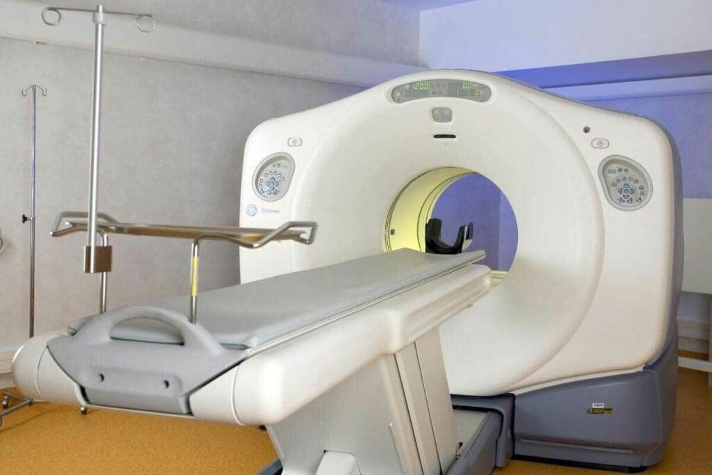
Positron Emission Tomography (PET) has changed medical imaging a lot. This technology lets doctors see how the body works inside. It’s key to finding and treating serious diseases. If you’re wondering who invented the PET scanner, it was created in 1973 by Edward Hoffman, Michael M. Ter-Pogossian, and Michael E. Phelps at Washington University.
There have been big changes in PET technology over the years. It’s now a vital tool in medicine. Thanks to PET scanning, doctors can make better diagnoses and treatments. At Liv Hospital, we use this tech to help our patients. We focus on the patient and follow international standards.
Key Takeaways
- The first PET camera for human studies was developed in 1973.
- Edward Hoffman, Michael M. Ter-Pogossian, and Michael E. Phelps were pioneers in PET technology.
- PET scanning has revolutionized medical imaging and disease diagnosis.
- Liv Hospital integrates PET scanning with a patient-centered approach.
- Advancements in PET technology have improved diagnostic accuracy and treatment outcomes.
The Origins of Medical Imaging Technology

Wilhelm Conrad Roentgen discovered X-rays in 1895, starting a new era in medical imaging. This led to the creation of Positron Emission Tomography (PET) technology. This breakthrough changed how doctors diagnose diseases and opened doors for more nuclear medicine innovations.
Precursors to Nuclear Medicine Imaging
In the early 20th century, nuclear medicine imaging started with radioactive isotopes for medical use. George de Hevesy, a Hungarian radiochemist, introduced the idea of using tracers in the 1920s. This idea was the start of PET imaging.
As nuclear medicine grew, so did the technology to find and image radioactive tracers. The gamma camera by Hal Anger in the 1950s was a big step. It lets people see where radioactive materials are in the body.
Early Experiments with Positron Detection
Positron detection, key to PET imaging, began in the mid-20th century. Robert Hofstadter and Stanford’s team worked on it in the 1960s. They figured out how positrons and electrons create gamma rays, a key part of PET tech.
“The discovery of positron emission and the subsequent development of PET scanning represent a significant advancement in our ability to diagnose and treat diseases at the molecular level.”
These early studies and tech improvements led to PET scanning. Today, it’s a key part of nuclear medicine.
Who Invented the PET Scanner: Hoffman and Phelps’ Breakthrough

Professor Edward J. Hoffman and an expert teamed up at Washington University in the 1970s. They created the PET scanner, a key medical technology. Their work started the era of modern medical imaging.
Professor Edward J. Hoffman’s Scientific Background
Professor Edward J. Hoffman was a top physicist in the field. He worked on the PET scanner, using his knowledge of nuclear physics. His focus was on detecting gamma rays and creating imaging methods.
Hoffman’s skills were key in solving the technical hurdles of the PET scanner. His work in nuclear medicine has greatly helped us see inside the body.
A medical expert’s Research Journey
A medical expert was a leading biophysicist in creating the PET scanner. He started by studying the body’s biochemical processes. His goal was to develop imaging that could show these processes.
Phelps’s creativity and commitment to medical imaging were vital. His work has changed how we diagnose and treat diseases, improving patient care.
The Historic Collaboration at Washington University
The partnership between Hoffman and Phelps at Washington University was groundbreaking. Their knowledge in physics and biophysics led to the first practical PET scanner in 1974. This was a major step forward in nuclear medicine.
Their work showed the power of PET scanning in giving doctors important information. It has helped us understand many diseases better. Hoffman and Phelps’ work continues to shape medical research and care today.
The First Practical PET Scanner in 1974
In 1974, the first practical PET scanner was developed. This was a big step forward in medical imaging. Professor Edward J. Hoffman and a medical expert at Washington University, worked together to make it happen. Their work improved medical technology and helped doctors diagnose better.
Technical Design and Innovation
The first PET scanner was a big technical achievement. Hoffman and Phelps created it to spot positron emissions from special substances. These substances help doctors see how the body works.
The design was complex, needing advanced engineering. A study on the National Center for Biotechnology Information (NCBI) website says PET technology was a big leap in nuclear medicine.
Key innovations included using coincidence detection to spot gamma rays at the same time. This is key for PET imaging. It showed how different parts of the body work, a big step up from old imaging methods.
“The introduction of PET scanning revolutionized the field of nuclear medicine by providing a powerful tool for diagnosing and managing diseases.”
First Clinical Tests and Results
The first tests of the PET scanner were exciting. The results were good, showing it could tell different tissues apart. This was the start of PET scanning being used in hospitals, mainly for cancer, heart, and brain diseases.
These early tests showed PET scanning could help patients by giving doctors better information. As the medical expert and his team kept improving it, PET scanning became a key tool in medicine.
Understanding the Science of Positron Emission Tomography
PET scanning uses the physics of positron emission and detection. It helps doctors see how the body works by using a special substance. This substance, called a radiotracer, emits positrons that are then caught to make images.
The Physics Behind PET Scanning
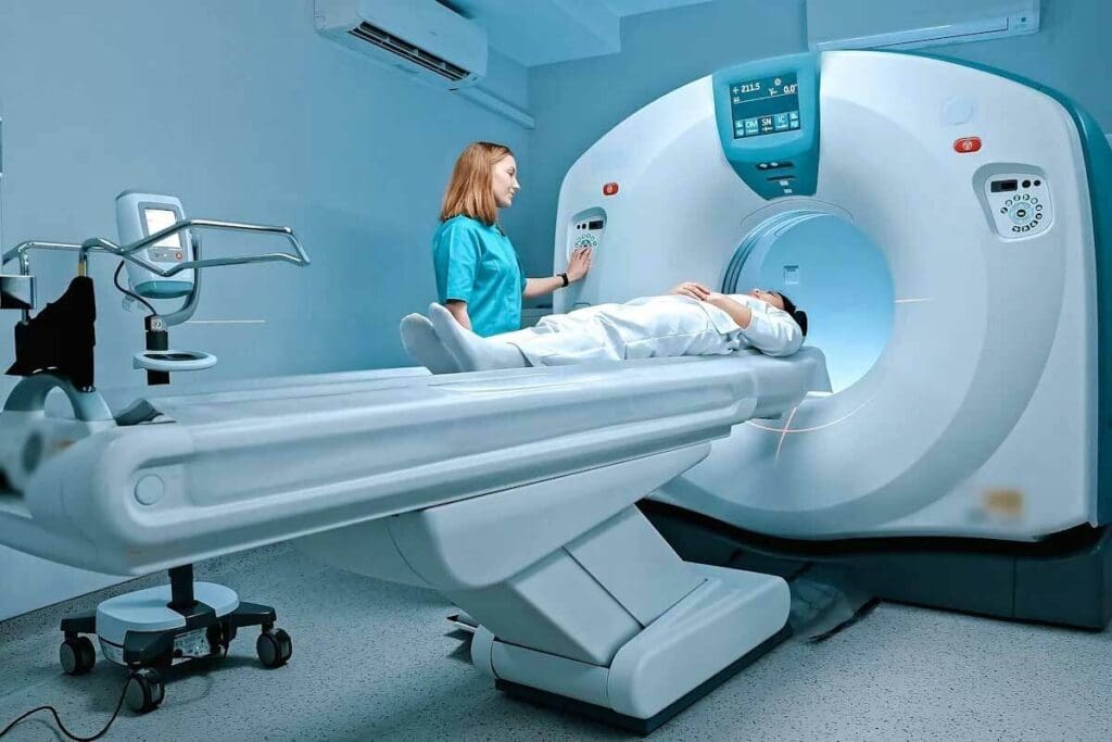
PET scanning works on the idea of positron emission and annihilation. A radiotracer is given to the body, which goes to active areas. Positrons from the radiotracer meet electrons, causing an event that makes two gamma rays.
Key aspects of the physics behind PET scanning include:
- The emission of positrons by the radiotracer
- The annihilation of positrons with electrons, producing gamma rays
- The detection of gamma rays by the PET scanner
From Data Collection to Medical Images
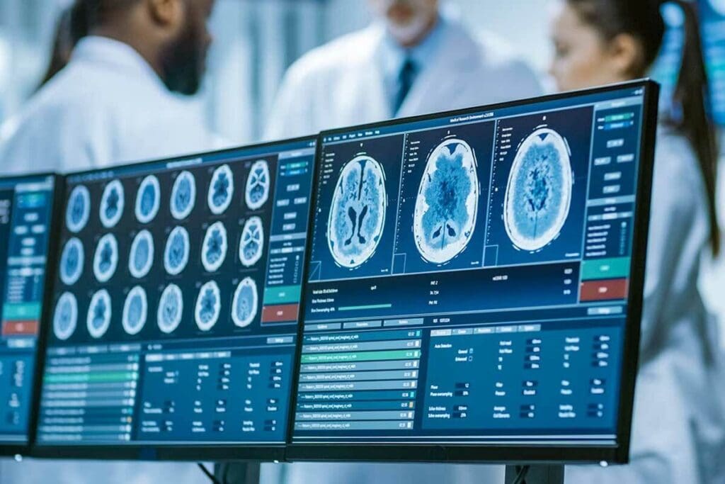
The PET scanner catches gamma rays and turns them into images. These images show how the body works, helping doctors find and treat diseases.
The steps to make medical images are:
- Data collection: detecting gamma rays emitted from the body
- Data reconstruction: using algorithms to reconstruct the data into images
- Image analysis: interpreting the reconstructed images to diagnose conditions
Understanding PET scanning shows its importance in medicine today.
Evolution of PET Technology Through the Decades
PET technology has grown a lot over the years. It has changed how we see inside the body. Each decade has brought big improvements.
Technological Milestones of the 1980s
The 1980s were key for PET technology. PET scanners became more common and were used in hospitals. Advancements in detector technology and image reconstruction algorithms have made images better and more accurate.
- Introduction of new scintillator materials
- Improvements in scanner design and sensitivity
- Development of more sophisticated image processing software
These changes helped PET technology become more popular in the next decades.
Breakthroughs in the 1990s
The 1990s saw a big leap with PET/CT hybrid scanners. This was thanks to David Townsend and Ronald Nutt. It mixed PET’s function with CT’s detail. A study on the National Center for Biotechnology Information showed that it improved diagnosis a lot.
PET/CT helped doctors plan treatments better, mainly for cancer. It lets them see how diseases affect the body more clearly.
21st Century Refinements and Improvements
In the 21st century, PET technology kept getting better. Scanners got more detailed and sensitive. This lets doctors spot smaller problems and see how treatments work better.
- Time-of-flight (TOF) PET, which improves image quality and reduces scan times
- Advanced reconstruction algorithms that enhance image resolution and accuracy
- Increased use of PET in neurology and cardiology, expanding beyond oncology
These updates made PET technology a key tool in medicine. It gives deep insights into how our bodies work.
The PET/CT Hybrid Revolution
PET/CT technology changed how we diagnose diseases. It combines two imaging types. This was thanks to David Townsend and Ronald Nutt’s work.
David Townsend and Ronald Nutt’s Innovative Vision
David Townsend and Ronald Nutt created the first PET/CT scanner. They wanted to mix PET’s function with CT’s detail. This helps doctors understand patients better.
Their work was based on earlier nuclear medicine research. They made a system that blends PET and CT images. This makes diagnosis more accurate, as noted in a related article, the combination of PET and CT has proven to be a perfect, enhancing diagnostic accuracy and treatment planning
How Combined Imaging Enhanced Diagnostic Capabilities
PET/CT technology has greatly improved diagnostics:
- Improved Accuracy: It combines two imaging types for better diagnoses.
- Enhanced Treatment Planning: Detailed images help in making better treatment plans.
- Better Patient Outcomes: It leads to better patient care by detecting diseases early and treating them precisely.
PET/CT technology is used in many medical fields. It has changed how we diagnose and treat diseases. The work of David Townsend and Ronald Nutt is key to this progress.
Radiopharmaceuticals: The Essential Components of PET Imaging
Radiopharmaceuticals are at the core of PET imaging. They help create detailed images of the body’s metabolic activities. These substances are key to diagnosing and monitoring diseases like cancer, neurological disorders, and heart diseases.
Development of FDG and Early Tracers
Fluorodeoxyglucose (FDG) is the most used radiopharmaceutical in PET imaging. It’s a glucose analog that cells absorb. This allows for the detection of tumors and checking how well treatments work.
Early tracers like FDG were made to target specific biological processes. Researchers keep innovating, making new tracers to identify conditions more accurately.
Modern Radiopharmaceuticals for Specific Conditions
Modern radiopharmaceuticals are made to target specific conditions. For example, tracers for Alzheimer’s disease bind to amyloid plaques. Others target low oxygen in tumors, helping guide treatment.
- Cardiac Imaging: Rubidium-82 is used for heart muscle blood flow imaging.
- Oncology: Tracers like Fluorothymidine (FLT) check tumor cell growth rates.
- Neurology: Tracers for tau proteins or amyloid help diagnose neurodegenerative diseases.
Production and Safety Considerations
Making radiopharmaceuticals involves complex steps like synthesis, purification, and quality control. These compounds have short half-lives, needing quick production and delivery. Keeping patients and healthcare workers safe is a top priority, with strict handling and administration protocols.
“The safe handling of radiopharmaceuticals is critical to prevent unnecessary radiation exposure to patients and staff. Strict guidelines and protocols are in place to ensure safety.”
We must follow these guidelines to keep PET imaging safe and effective.
Clinical Applications and Impact of PET Technology
PET technology lets us see how our bodies work at a detailed level. It’s a key tool in today’s medicine. It helps us understand and treat diseases better than ever before.
Revolutionizing Cancer Detection and Treatment
PET scans have changed how we fight cancer. They help find tumors early and track how well treatments work. FDG-PET is a big help because it spots cancer by showing where cells are most active.
The good things about PET in fighting cancer are:
- It finds cancer early
- It helps figure out how far cancer has spread
- It checks if treatments are working
- It guides doctors during biopsies and surgeries
| Cancer Type | PET Scan Application | Benefits |
| Lymphoma | Staging and monitoring treatment response | Improved accuracy in assessing disease progression |
| Lung Cancer | Evaluating solitary pulmonary nodules | Enhanced detection of malignant nodules |
| Breast Cancer | Assessing axillary lymph node involvement | Better staging and treatment planning |
Cardiac and Neurological Applications
PET scans also help in cardiology and neurology. In heart care, they check if the heart areas are working properly. This helps find problems early.
In brain health, PET scans are key for diagnosing diseases like Alzheimer’s. They show brain activity, helping doctors tell diseases apart and track how they change.
PET technology is very useful in today’s medicine. As we keep improving, PET scans will help us even more. They will open up new ways to diagnose and treat many diseases.
Global Adoption and Accessibility of PET Scanning
PET scanning is not equally available worldwide. This is due to different healthcare policies, economic conditions, and tech advancements. The use of PET technology varies greatly, influenced by many factors.
Regional Differences in PET Technology Implementation
PET technology is used differently in different places. In rich countries, PET scans are common in big hospitals and research centers. For example, in the U.S., they’re often used to find and track cancer.
But poor countries struggle to get PET scans. They face high costs, a lack of skilled people, and weak healthcare systems. This means not everyone gets the advanced care they need.
Economic Factors and Healthcare Integration
The cost of PET scanners and the drugs needed for them is very high. This makes it hard for many healthcare systems to afford. Also, making some drugs requires expensive cyclotrons.
How well a country’s healthcare works also matters. Places with strong healthcare and support for new tech tend to have more PET scans. Countries that focus on nuclear medicine and have clear rules usually do better.
In summary, many things affect how widely PET scanning is used around the world. These include healthcare policies, money, and new tech. As PET tech gets better, we must work to make sure everyone can get the care they need.
Conclusion: The Future Landscape of PET Technology
PET technology will keep being key in medical imaging and diagnostics. New research and development will bring better PET technology. This will help improve patient care.
New radiopharmaceuticals and tech will make PET scans better. They will help find diseases early and track them. This means doctors can give more precise treatments.
The future of PET technology looks bright. Breakthroughs in AI and machine learning could make PET scans even better. This will make getting scans easier and more efficient around the world.
We’re dedicated to using PET technology for top-notch healthcare. We’ll use the latest PET advancements to help patients get the best care possible.
FAQ
What is Positron Emission Tomography (PET) and how does it work?
PET is a medical imaging method that shows how the body works by detecting special particles. It uses a special medicine that emits these particles. The particles then create images of the body’s functions.
Who invented the PET scanner?
Professor Edward J. Hoffman and a medical expert created the first PET scanner. They worked at Washington University and made the first working PET scanner in 1974.
What is the significance of the PET/CT hybrid scanner?
The PET/CT scanner combines two imaging types. It uses PET to see how the body works and CT to see its structure. This combo helps doctors get more detailed images and make better diagnoses.
What are radiopharmaceuticals, and how are they used in PET imaging?
Radiopharmaceuticals are special medicines that help with PET imaging. They show where certain processes happen in the body. For example, FDG shows where tumors are because they use a lot of sugar.
How has PET technology evolved over the decades?
PET technology has grown a lot over the years. Big steps were made in the 1980s, 1990s, and later. These include better scanners, detectors, and ways to make images. New medicines have also been developed.
What are the clinical applications of PET scanning?
PET scanning helps find and treat cancer and diagnose heart and brain diseases. It shows how different parts of the body work. This makes it very useful in medicine.
How accessible is PET scanning globally?
PET scanning is not the same everywhere. It depends on the country’s healthcare, money, and technology. In some places, it’s hard to get because of cost and lack of technology.
What is the future of PET technology?
PET technology will keep getting better. New scanners, detectors, and medicines are coming. These will help doctors diagnose and treat diseases even better.
When was the PET scan invented?
The first PET scanner was made in 1974 by Professor Edward J. Hoffman and a medical expert.
What is PET imaging used for?
PET imaging is used for many things. It helps find and treat cancer, diagnose heart problems, and manage brain diseases. It shows how different parts of the body work.
References
- Portnow, L. H. (2013). The history of cerebral PET scanning: From physiology to imaging. Neurotherapeutics, 10(3), 404-415. Describes early work on PET by Michael Phelps and Edward Hoffman and subsequent developments. Retrieved from https://pmc.ncbi.nlm.nih.gov/articles/PMC3653214/


